Skull bones, features and markings II
•Download as PPTX, PDF•
17 likes•13,536 views

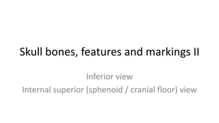
Inferior (mandible removed) and superior view (top of cranium removed - cranial floor / sphenoid view) skull anatomy warm-up for Anatomy and Physiology students. Bones, features and markings are shown
Report
Share
Report
Share

Recommended
03.18.09(c): Eye Histology
Slideshow is from the University of Michigan Medical
School's M1 CNS sequence
View additional course materials on Open.Michigan:
openmi.ch/med-M1CNS
Histology: Connective Tissues
A PowerPoint review of photomicrographs depicting the various features of the basic forms of connective tissues, not including bone tissue or blood. By Timothy Ballard, UNC Wilmington. Licensed under a Creative Commons License: Attribution Non-Commercial-NoDerivs. From http://www.lifescitrc.org/resource.cfm?submissionID=9054.
Appendicular skeletal system
The document discusses the appendicular skeleton, which includes the bones of the upper and lower limbs attached to the axial skeleton. It is divided into six major regions: the pectoral girdles, arms and forearms, hands, pelvis, thighs and legs, and feet and ankles. Each limb contains specific bones that make up its structure and allow for movement.
Appendicular skeleton
The document summarizes the main bones that make up the appendicular skeleton in humans. It describes the two pairs of girdles - the pectoral (shoulder) and pelvic (hip) girdles - that connect the upper and lower limbs to the axial skeleton. It then outlines the major bones that comprise each segmented limb, including the arm, forearm, hand, thigh, leg, and foot. For each bone, it identifies key anatomical features and points of articulation with other bones.
Aortic arches.pptx
The aortic arches develop from paired dorsal aortae that fuse to form the ascending aorta and arch. There are initially 6 pairs of aortic arches, which remodel such that the internal carotid, common carotid, subclavian and pulmonary arteries form from specific arches between the right and left sides. Anomalies can occur if the aortic arches do not regress properly, leading to conditions like patent ductus arteriosus, coarctation of the aorta, or interrupted aortic arch.
Neck Muscles
Neck muscles help support the cervical spine and contribute to movements of the head, neck, upper back, and shoulders
#neck #muscle #anatomy
Histology of cerebrum and cerebellum
The cerebellum is located in the posterior cranial fossa. It receives sensory data from the spinal cord and motor data from the cerebrum, and its output is motor in function. It controls the activity of the cerebrum and spinal cord, and dysfunctions result in clumsy, poorly coordinated movements. The cerebellum is responsible for equilibrium, eye and head movements, balancing, postural changes, muscle tone, smooth execution of movements, accuracy, and motor control. It has a cortex with molecular, purkinje, and granular layers containing various cell types like purkinje cells. The cerebrum has gray matter containing neuron types like pyramidal cells and white matter with nerve fibers. The cerebral cortex
Chapter 7&8
There are 206 bones in the human body grouped into the axial skeleton and appendicular skeleton. The axial skeleton includes 80 bones that make up the skull, vertebral column, ribs, and sternum. It forms the central core and foundation of the body. The appendicular skeleton includes 126 bones arranged in the upper and lower limbs, including their attaching girdles, forming the shoulders, arms, legs and allowing for movement.
Visual Apparatus
The visual apparatus contains several anatomical structures that work together to enable sight. These include the orbit, which houses the eyeball and extraocular muscles that control eye movement. The eyeball itself contains the retina, lens, and other structures surrounded by protective layers like the sclera. Nerves like the optic nerve carry visual signals from the eyes to the brain for processing.
Lacrimal apparatus & nose
The lacrimal apparatus consists of the lacrimal gland, conjunctival sac, lacrimal puncta and canaliculi, lacrimal sac, and nasolacrimal duct. The lacrimal gland secretes tears which drain through the puncta and canaliculi into the lacrimal sac and then nasolacrimal duct into the nose. Obstruction of this drainage system can cause epiphora or excessive tearing. The nose has functions of respiration, olfaction, air conditioning, and protection and is formed externally by skin, cartilage, and bone and internally by three nasal cavities with vestibules, respiratory and olfactory regions.
Surface anatomy
This document provides an overview of surface anatomy and the human skull. It defines surface anatomy as the study of external body features and notes that specific anatomical features include the head, ears, nose, mouth, and limbs. It then describes how the skeleton is composed of an endoskeleton for humans and defines the main parts of the human skeleton, including the axial and appendicular regions. Finally, it provides details on the parts of the human skull, listing the cranium, mandible, frontal bone, and other bones that make up the skull.
Skull and vertebrae
The skull is made up of 22 bones that form two sections - the neurocranium which encloses the brain, and the viscerocranium which forms the face. There are 8 cranial bones and 14 facial bones. The skull provides protection, support, and attachment sites for muscles. The vertebral column consists of 33 vertebrae including 9 fused bones in the sacrum and coccyx. It houses and protects the spinal cord while allowing for movement and curvature of the spine. Common spinal conditions addressed include spondylolisthesis, kyphosis, lordosis, and scoliosis.
Eye
The document describes the anatomy and structures of the eye. It discusses the three layers (tunics) that make up the eye - the outer fibrous layer, middle vascular layer, and inner retinal layer. It describes key optical components such as the cornea, pupil, iris, lens, vitreous humor, and aqueous humor. The document also outlines production and flow of aqueous humor, the six extrinsic eye muscles, and provides diagrams of eye anatomy including layers and muscles.
Applied and clinical anatomy of lower limb
The document describes the anatomy of the lower limb, including the pelvis, femur, patella, tibia, fibula, and hip joint. It discusses the bones that make up each part and their blood supply, fractures commonly seen in each bone, and movements at the hip joint. The lower limb consists of the gluteal region, thigh, leg, and foot and its main functions are to support body weight and enable locomotion.
Human anatomy
This material is prepared for the beginners of medical and Paramedical professionals. The books referred were Tortora, Gyton and Ross and Wilson for Human Anatomy and physiology. Leave a comment if you find it useful which would make me to upload more study materials for the beneficiaries.
Ophthalmic artery
ophthalmic artery, course and branches of ophthalmic artery, venous drainage of eye ball, superior and inferior ophthalmic vein and its drainage
T H E C R A N I A L N E R V E S
The document discusses the 12 pairs of cranial nerves:
- It describes the origin, function, and key characteristics of each cranial nerve. The cranial nerves are classified as sensory, motor, or mixed.
- The cranial nerves include the olfactory, optic, oculomotor, trochlear, trigeminal, abducens, facial, vestibulocochlear, glossopharyngeal, vagus, accessory, and hypoglossal nerves.
- Each cranial nerve is described in detail including its origin, function, and important clinical notes. Diagrams are included to illustrate some cranial nerves and their pathways.
An easy way to learn upper limb muscles
A summary for learning the muscles of the upper limb including their attachments, innervation, etc., without having to have too many books open. Resources: "Gray’s Anatomy", "Taschenatlas der Anatomie" and Wikipedia. Awaiting further proof-reading!
Ch 13 the peripheral nervous system and nervous activity
This document contains a list of figures and tables related to the peripheral nervous system and reflex activity from Lecture 13. There is no other text content, just a long list of figure and table references.
Anatomy of Radio ulnar joints
The document discusses the three types of radio-ulnar joints in the wrist: superior, middle, and inferior. It describes the ligaments that support these joints, including the fibrous capsule, annular ligament, quadrate band, interosseous membrane and oblique cord. The document also mentions the articular disc between the radius and ulna that allows for pronation and supination movements of the forearm. Finally, it lists some injuries to the radius, such as nurse maid's elbow, Colles fractures, and Smith's fractures.
Epithelium, cells,tissues & histology
This document discusses the four major types of adult tissues - epithelial, connective, muscle and nervous tissue. It provides details on the classification, structure and functions of each type of tissue. The key points are:
1) Tissues are classified based on structure, composition and function. The four major types are epithelial, connective, muscle and nervous tissue.
2) Epithelial tissues cover surfaces, line organs and form glands. They protect, absorb, secrete and transport. Connective tissues connect, support and protect other tissues. Muscle tissues contract to cause movement and pumping blood. Nervous tissues transmit electrical signals.
3) Each tissue contains different cell types and extracellular matrix to suit their functions.
Final apendicular skeleton (lower limbs).
This document provides information about the bones of the pelvic girdle and lower limbs. It begins by listing the objectives, which are to identify the bones and their markings, describe the divisions of the pelvis and differences between male and female pelvis. It then lists the bones of the lower limbs and provides details about the pelvic girdle bones including the ilium, ischium, pubis and acetabulum. It also describes the joints of the pelvic girdle and differences between male and female pelvis. Finally, it provides information about the lower limb bones including the femur, patella, tibia, fibula, tarsals, metatarsals and phalanges.
Histology quiz
The document lists different types of epithelial tissues and connective tissues, asking the reader to identify each one. It discusses pseudostratified columnar epithelium, simple columnar epithelium, stratified squamous epithelium, simple cuboidal epithelium, transitional epithelium, simple squamous epithelium, as well as adipose connective tissue, areolar connective tissue, cardiac muscle, compact bone, elastic cartilage, dense irregular connective tissue, dense regular connective tissue, fibrocartilage, hyaline cartilage, reticular connective tissue, skeletal muscle, and smooth muscle.
Appendicula skeleton
The appendicular skeleton includes the bones of the upper and lower limbs that are attached to the axial skeleton by shoulder and pelvic girdles. The pectoral girdle consists of bones that hold the upper limbs in place while the pelvic girdle consists of bones that hold the lower limbs in place. The adult pelvis is composed of four bones - the sacrum, coccyx, and left and right ossa coxae which protect the viscera and support the lower body. Each os coxa is formed from the ilium, ischium, and pubis bones. The pelvic brim divides the pelvis into the true pelvis and false pelvis. The foot has three longitudinal
cervical and brachial plexus
The cervical plexus and brachial plexus are formed by the branching and merging of spinal nerve roots in the neck and shoulder region. The cervical plexus is formed from cervical nerve roots C1-C4 and supplies nerves to the neck muscles and skin. The brachial plexus is formed from cervical nerve roots C5-T1 and supplies all nerves to the upper limb, including the axillary, musculocutaneous, radial, median, and ulnar nerves. Both plexuses demonstrate how spinal nerves branch and connect to innervate different regions of the body.
L2.11 liver pancreas peritoneum pdf
The document provides information on the structure and functions of the liver and pancreas. It discusses the liver's location, lobes, ligaments, vascular and biliary supply. The liver receives blood from the hepatic portal vein and hepatic arteries. It secretes bile into canaliculi between hepatocytes. The bile ducts drain into the right and left hepatic ducts. The pancreas is also mentioned. The peritoneum and its derivatives are briefly introduced.
Sense organs
This document discusses the primary sentient sense organs, including the eye. It describes the anatomy and histology of the eyeball and its tunics: sclera, choroid, and retina. In particular, it outlines the structures and functions of the cornea, iris, ciliary body, lens, vitreous body, and retina. The retina contains photoreceptor cells (rods and cones) and associative neurons that transmit visual signals via the optic nerve to the brain. The pigmented epithelium assists with light absorption and photoreceptor renewal.
09 [chapter 9 joints]
The document summarizes the key points about joints from chapter 9. It introduces the different types of joints in the body - fibrous, cartilaginous, and synovial joints. It describes their classification based on structure and function. Synovial joints are discussed in more detail, including their structure, types of movement they allow, and examples like the shoulder, hip, and knee joints. Factors that can affect the range of motion of synovial joints and joint replacement surgery are also covered.
The urinary system
Presentation used to introduce anatomy and physiology students to the urinary system anatomy and physiology. Glomerular filtration, tubular secretion and tubular reabsorption are shown.
Oogenesis female reproductive system hormone signaling in female
This presentation is used for high school anatomy and physiology students to (re)introduce oogenesis and female reproductive anatomy and physiology.
More Related Content
What's hot
Visual Apparatus
The visual apparatus contains several anatomical structures that work together to enable sight. These include the orbit, which houses the eyeball and extraocular muscles that control eye movement. The eyeball itself contains the retina, lens, and other structures surrounded by protective layers like the sclera. Nerves like the optic nerve carry visual signals from the eyes to the brain for processing.
Lacrimal apparatus & nose
The lacrimal apparatus consists of the lacrimal gland, conjunctival sac, lacrimal puncta and canaliculi, lacrimal sac, and nasolacrimal duct. The lacrimal gland secretes tears which drain through the puncta and canaliculi into the lacrimal sac and then nasolacrimal duct into the nose. Obstruction of this drainage system can cause epiphora or excessive tearing. The nose has functions of respiration, olfaction, air conditioning, and protection and is formed externally by skin, cartilage, and bone and internally by three nasal cavities with vestibules, respiratory and olfactory regions.
Surface anatomy
This document provides an overview of surface anatomy and the human skull. It defines surface anatomy as the study of external body features and notes that specific anatomical features include the head, ears, nose, mouth, and limbs. It then describes how the skeleton is composed of an endoskeleton for humans and defines the main parts of the human skeleton, including the axial and appendicular regions. Finally, it provides details on the parts of the human skull, listing the cranium, mandible, frontal bone, and other bones that make up the skull.
Skull and vertebrae
The skull is made up of 22 bones that form two sections - the neurocranium which encloses the brain, and the viscerocranium which forms the face. There are 8 cranial bones and 14 facial bones. The skull provides protection, support, and attachment sites for muscles. The vertebral column consists of 33 vertebrae including 9 fused bones in the sacrum and coccyx. It houses and protects the spinal cord while allowing for movement and curvature of the spine. Common spinal conditions addressed include spondylolisthesis, kyphosis, lordosis, and scoliosis.
Eye
The document describes the anatomy and structures of the eye. It discusses the three layers (tunics) that make up the eye - the outer fibrous layer, middle vascular layer, and inner retinal layer. It describes key optical components such as the cornea, pupil, iris, lens, vitreous humor, and aqueous humor. The document also outlines production and flow of aqueous humor, the six extrinsic eye muscles, and provides diagrams of eye anatomy including layers and muscles.
Applied and clinical anatomy of lower limb
The document describes the anatomy of the lower limb, including the pelvis, femur, patella, tibia, fibula, and hip joint. It discusses the bones that make up each part and their blood supply, fractures commonly seen in each bone, and movements at the hip joint. The lower limb consists of the gluteal region, thigh, leg, and foot and its main functions are to support body weight and enable locomotion.
Human anatomy
This material is prepared for the beginners of medical and Paramedical professionals. The books referred were Tortora, Gyton and Ross and Wilson for Human Anatomy and physiology. Leave a comment if you find it useful which would make me to upload more study materials for the beneficiaries.
Ophthalmic artery
ophthalmic artery, course and branches of ophthalmic artery, venous drainage of eye ball, superior and inferior ophthalmic vein and its drainage
T H E C R A N I A L N E R V E S
The document discusses the 12 pairs of cranial nerves:
- It describes the origin, function, and key characteristics of each cranial nerve. The cranial nerves are classified as sensory, motor, or mixed.
- The cranial nerves include the olfactory, optic, oculomotor, trochlear, trigeminal, abducens, facial, vestibulocochlear, glossopharyngeal, vagus, accessory, and hypoglossal nerves.
- Each cranial nerve is described in detail including its origin, function, and important clinical notes. Diagrams are included to illustrate some cranial nerves and their pathways.
An easy way to learn upper limb muscles
A summary for learning the muscles of the upper limb including their attachments, innervation, etc., without having to have too many books open. Resources: "Gray’s Anatomy", "Taschenatlas der Anatomie" and Wikipedia. Awaiting further proof-reading!
Ch 13 the peripheral nervous system and nervous activity
This document contains a list of figures and tables related to the peripheral nervous system and reflex activity from Lecture 13. There is no other text content, just a long list of figure and table references.
Anatomy of Radio ulnar joints
The document discusses the three types of radio-ulnar joints in the wrist: superior, middle, and inferior. It describes the ligaments that support these joints, including the fibrous capsule, annular ligament, quadrate band, interosseous membrane and oblique cord. The document also mentions the articular disc between the radius and ulna that allows for pronation and supination movements of the forearm. Finally, it lists some injuries to the radius, such as nurse maid's elbow, Colles fractures, and Smith's fractures.
Epithelium, cells,tissues & histology
This document discusses the four major types of adult tissues - epithelial, connective, muscle and nervous tissue. It provides details on the classification, structure and functions of each type of tissue. The key points are:
1) Tissues are classified based on structure, composition and function. The four major types are epithelial, connective, muscle and nervous tissue.
2) Epithelial tissues cover surfaces, line organs and form glands. They protect, absorb, secrete and transport. Connective tissues connect, support and protect other tissues. Muscle tissues contract to cause movement and pumping blood. Nervous tissues transmit electrical signals.
3) Each tissue contains different cell types and extracellular matrix to suit their functions.
Final apendicular skeleton (lower limbs).
This document provides information about the bones of the pelvic girdle and lower limbs. It begins by listing the objectives, which are to identify the bones and their markings, describe the divisions of the pelvis and differences between male and female pelvis. It then lists the bones of the lower limbs and provides details about the pelvic girdle bones including the ilium, ischium, pubis and acetabulum. It also describes the joints of the pelvic girdle and differences between male and female pelvis. Finally, it provides information about the lower limb bones including the femur, patella, tibia, fibula, tarsals, metatarsals and phalanges.
Histology quiz
The document lists different types of epithelial tissues and connective tissues, asking the reader to identify each one. It discusses pseudostratified columnar epithelium, simple columnar epithelium, stratified squamous epithelium, simple cuboidal epithelium, transitional epithelium, simple squamous epithelium, as well as adipose connective tissue, areolar connective tissue, cardiac muscle, compact bone, elastic cartilage, dense irregular connective tissue, dense regular connective tissue, fibrocartilage, hyaline cartilage, reticular connective tissue, skeletal muscle, and smooth muscle.
Appendicula skeleton
The appendicular skeleton includes the bones of the upper and lower limbs that are attached to the axial skeleton by shoulder and pelvic girdles. The pectoral girdle consists of bones that hold the upper limbs in place while the pelvic girdle consists of bones that hold the lower limbs in place. The adult pelvis is composed of four bones - the sacrum, coccyx, and left and right ossa coxae which protect the viscera and support the lower body. Each os coxa is formed from the ilium, ischium, and pubis bones. The pelvic brim divides the pelvis into the true pelvis and false pelvis. The foot has three longitudinal
cervical and brachial plexus
The cervical plexus and brachial plexus are formed by the branching and merging of spinal nerve roots in the neck and shoulder region. The cervical plexus is formed from cervical nerve roots C1-C4 and supplies nerves to the neck muscles and skin. The brachial plexus is formed from cervical nerve roots C5-T1 and supplies all nerves to the upper limb, including the axillary, musculocutaneous, radial, median, and ulnar nerves. Both plexuses demonstrate how spinal nerves branch and connect to innervate different regions of the body.
L2.11 liver pancreas peritoneum pdf
The document provides information on the structure and functions of the liver and pancreas. It discusses the liver's location, lobes, ligaments, vascular and biliary supply. The liver receives blood from the hepatic portal vein and hepatic arteries. It secretes bile into canaliculi between hepatocytes. The bile ducts drain into the right and left hepatic ducts. The pancreas is also mentioned. The peritoneum and its derivatives are briefly introduced.
Sense organs
This document discusses the primary sentient sense organs, including the eye. It describes the anatomy and histology of the eyeball and its tunics: sclera, choroid, and retina. In particular, it outlines the structures and functions of the cornea, iris, ciliary body, lens, vitreous body, and retina. The retina contains photoreceptor cells (rods and cones) and associative neurons that transmit visual signals via the optic nerve to the brain. The pigmented epithelium assists with light absorption and photoreceptor renewal.
09 [chapter 9 joints]
The document summarizes the key points about joints from chapter 9. It introduces the different types of joints in the body - fibrous, cartilaginous, and synovial joints. It describes their classification based on structure and function. Synovial joints are discussed in more detail, including their structure, types of movement they allow, and examples like the shoulder, hip, and knee joints. Factors that can affect the range of motion of synovial joints and joint replacement surgery are also covered.
What's hot (20)
Ch 13 the peripheral nervous system and nervous activity
Ch 13 the peripheral nervous system and nervous activity
More from James H. Workman
The urinary system
Presentation used to introduce anatomy and physiology students to the urinary system anatomy and physiology. Glomerular filtration, tubular secretion and tubular reabsorption are shown.
Oogenesis female reproductive system hormone signaling in female
This presentation is used for high school anatomy and physiology students to (re)introduce oogenesis and female reproductive anatomy and physiology.
Meiosis and the male reproductive system
Used to (re)ntroduce high school anatomy and physiology students to the process of meiosis / gametogenesis. More specifically, this presentation focuses on spermatogenesis and the male reproductive system anatomy.
Spinal cord and spinal nerves
The spinal cord is protected within the vertebrae and meninges. It acts as a highway for sensory and motor nerve impulses between the brain and spinal nerves. The spinal cord contains 31 pairs of spinal nerves that emerge at different levels to innervate different parts of the body. Sensory information enters the spinal cord via ascending tracts in the posterior region while motor commands exit via descending tracts in the anterior region, allowing for integration of reflexes. Spinal nerves branch into plexuses at certain levels to further distribute nerve signals.
Nervous system 3; Synapses and Neurotransmitters
Lecture notes and diagrams for Anatomy and Physiology students describing / showing the connections between nerve cells (synapses) and how neurotransmitters work. Video of animation that shows how drugs affect neurotransmitters is included, although it will not show in slideshare.
Nervous tissue 1
Lecture notes and diagrams to help high school anatomy and physiology students learn the general functions of the nervous system and types of glial support nerve cells, types of neurons and anatomy of typical neurons.
Nervous tissue 2
The document discusses nerve impulses and nerve fiber types. It explains that nerve fibers maintain a resting potential of around -70 mV due to ion transport mechanisms. When an impulse is initiated, this potential rapidly changes and then repolarizes. Impulse frequency is limited by the sodium-potassium pump recovery time. Impulse speed is affected by factors like temperature, fiber diameter, and myelination. There are three main nerve fiber types - type A fibers are large and myelinated with fast conduction, type B are medium and myelinated, and type C are small and unmyelinated with slow conduction.
Muscular system
This document provides an overview of the muscular system, including muscle types, actions, and examples of lever systems. It describes how muscle attachments determine their actions and lists common movement terms. Finally, it details the major skeletal muscles in the head, upper arm, forearm, hand, respiratory system, abdomen, pectoral girdle, leg, ankle, and foot. It concludes with a brief overview of common muscular system diseases.
Muscle tissue 3
Variation in fast and slow twitch muscle fibers, wave summation, incomplete and complete tetanus and treppe phenomena
Muscle tissue 2
This document summarizes the key components of muscle tissue and muscle contraction. It discusses nerve impulses, motor units, neuromuscular junctions, and the sliding filament theory of muscle contraction. The main components that allow for contraction are described, including myosin, actin, tropomyosin, troponin, and the calcium release initiated by a nerve impulse that allows for crossbridge binding and sarcomere shortening. Regions of the sarcomere such as the Z-line, A-band, I-band, H-zone, and M-line are also outlined.
Muscle tissue 1
Types of muscle and functional characteristics of muscle. Levels of organization of muscle, fascicles, myofibrils, and myofibers. Microanatomy and functional unit of contraction, the sarcomere. Used as basis for lecture to high school science students
Articulations and movement
Functional and structural classification of articulations (joints) and directional terms of movement for Anatomy and Physiology students
Appendicular skeleton bones and features 3
Bones of the forearm and hand (ulna radius and carpals, metacarpals and phalanges) are described and depicted. For anatomy and phsyiology students.
Appendicular skeleton and bone features 1
The document discusses the appendicular skeleton and bone features, specifically focusing on the pectoral girdle and upper arm bones which include the clavicle, scapula, and humerus. It provides information on each of these three bones that make up the shoulder region.
Axial skeleton, vertebrae and thorax
The axial skeleton consists of the vertebral column, which includes the bones that form the spine, as well as the thorax, which contains the ribs and sternum. The vertebral column provides structure and protection to the spinal cord and supports the weight of the head and upper body. The thorax protects the lungs and heart by the rib cage and sternum.
Skull bones, features and markings 3
Introduction to bones and features of the infant human skull and the the features and markings of the mandible for Anatomy and Physiology students.
Skull bones and features 1
The skull is composed of 22 bones, including 8 cranial bones and 14 facial bones. The cranial bones are the frontal, occipital, ethmoid, sphenoid, and two parietal and temporal bones each. The facial bones include the vomer, mandible, two maxillae, two zygomatic, two nasal, two nasal concha, two lacrimal, and two palatine bones. Key skull features include the sagittal, coronal, lambdoidal, and squamosal sutures, as well as the orbit.
Human skeleton bone name and numbers warm up
Used as a quick learning check // check for understanding warm up in my Anatomy and Physiology class
Introduction to 206 bones of the human body
Introductory categorization and display of diagrams of the 206 bones in the human body.
For anatomy and physiology students.
Skeletal tissue
The skeletal system includes bones and cartilage that provide structure, allow for movement, and protect organs. The medical specialty of orthopedics focuses on treating the skeleton and joints. Skeletal tissue includes cartilage and bone, which are types of dense connective tissue. Bones have important functions like support, protection, movement, mineral storage, and blood cell production. Bone is made up of osteogenic cells, osteoblasts that form bone, osteocytes embedded in bone matrix, and osteoclasts that resorb bone. There are different types of bones like long bones with a diaphysis, epiphyses, and metaphysis. Bone formation occurs through intramembranous ossification or endochondral ossification. Common bone
More from James H. Workman (20)
Oogenesis female reproductive system hormone signaling in female
Oogenesis female reproductive system hormone signaling in female
Recently uploaded
Reimagining Your Library Space: How to Increase the Vibes in Your Library No ...
Librarians are leading the way in creating future-ready citizens – now we need to update our spaces to match. In this session, attendees will get inspiration for transforming their library spaces. You’ll learn how to survey students and patrons, create a focus group, and use design thinking to brainstorm ideas for your space. We’ll discuss budget friendly ways to change your space as well as how to find funding. No matter where you’re at, you’ll find ideas for reimagining your space in this session.
Leveraging Generative AI to Drive Nonprofit Innovation
In this webinar, participants learned how to utilize Generative AI to streamline operations and elevate member engagement. Amazon Web Service experts provided a customer specific use cases and dived into low/no-code tools that are quick and easy to deploy through Amazon Web Service (AWS.)
What is Digital Literacy? A guest blog from Andy McLaughlin, University of Ab...
What is Digital Literacy? A guest blog from Andy McLaughlin, University of Aberdeen
PCOS corelations and management through Ayurveda.
This presentation includes basic of PCOS their pathology and treatment and also Ayurveda correlation of PCOS and Ayurvedic line of treatment mentioned in classics.
South African Journal of Science: Writing with integrity workshop (2024)
South African Journal of Science: Writing with integrity workshop (2024)Academy of Science of South Africa
A workshop hosted by the South African Journal of Science aimed at postgraduate students and early career researchers with little or no experience in writing and publishing journal articles.BÀI TẬP BỔ TRỢ TIẾNG ANH 8 CẢ NĂM - GLOBAL SUCCESS - NĂM HỌC 2023-2024 (CÓ FI...
BÀI TẬP BỔ TRỢ TIẾNG ANH 8 CẢ NĂM - GLOBAL SUCCESS - NĂM HỌC 2023-2024 (CÓ FI...Nguyen Thanh Tu Collection
https://app.box.com/s/y977uz6bpd3af4qsebv7r9b7s21935vdLAND USE LAND COVER AND NDVI OF MIRZAPUR DISTRICT, UP
This Dissertation explores the particular circumstances of Mirzapur, a region located in the
core of India. Mirzapur, with its varied terrains and abundant biodiversity, offers an optimal
environment for investigating the changes in vegetation cover dynamics. Our study utilizes
advanced technologies such as GIS (Geographic Information Systems) and Remote sensing to
analyze the transformations that have taken place over the course of a decade.
The complex relationship between human activities and the environment has been the focus
of extensive research and worry. As the global community grapples with swift urbanization,
population expansion, and economic progress, the effects on natural ecosystems are becoming
more evident. A crucial element of this impact is the alteration of vegetation cover, which plays a
significant role in maintaining the ecological equilibrium of our planet.Land serves as the foundation for all human activities and provides the necessary materials for
these activities. As the most crucial natural resource, its utilization by humans results in different
'Land uses,' which are determined by both human activities and the physical characteristics of the
land.
The utilization of land is impacted by human needs and environmental factors. In countries
like India, rapid population growth and the emphasis on extensive resource exploitation can lead
to significant land degradation, adversely affecting the region's land cover.
Therefore, human intervention has significantly influenced land use patterns over many
centuries, evolving its structure over time and space. In the present era, these changes have
accelerated due to factors such as agriculture and urbanization. Information regarding land use and
cover is essential for various planning and management tasks related to the Earth's surface,
providing crucial environmental data for scientific, resource management, policy purposes, and
diverse human activities.
Accurate understanding of land use and cover is imperative for the development planning
of any area. Consequently, a wide range of professionals, including earth system scientists, land
and water managers, and urban planners, are interested in obtaining data on land use and cover
changes, conversion trends, and other related patterns. The spatial dimensions of land use and
cover support policymakers and scientists in making well-informed decisions, as alterations in
these patterns indicate shifts in economic and social conditions. Monitoring such changes with the
help of Advanced technologies like Remote Sensing and Geographic Information Systems is
crucial for coordinated efforts across different administrative levels. Advanced technologies like
Remote Sensing and Geographic Information Systems
9
Changes in vegetation cover refer to variations in the distribution, composition, and overall
structure of plant communities across different temporal and spatial scales. These changes can
occur natural.
বাংলাদেশ অর্থনৈতিক সমীক্ষা (Economic Review) ২০২৪ UJS App.pdf
বাংলাদেশের অর্থনৈতিক সমীক্ষা ২০২৪ [Bangladesh Economic Review 2024 Bangla.pdf] কম্পিউটার , ট্যাব ও স্মার্ট ফোন ভার্সন সহ সম্পূর্ণ বাংলা ই-বুক বা pdf বই " সুচিপত্র ...বুকমার্ক মেনু 🔖 ও হাইপার লিংক মেনু 📝👆 যুক্ত ..
আমাদের সবার জন্য খুব খুব গুরুত্বপূর্ণ একটি বই ..বিসিএস, ব্যাংক, ইউনিভার্সিটি ভর্তি ও যে কোন প্রতিযোগিতা মূলক পরীক্ষার জন্য এর খুব ইম্পরট্যান্ট একটি বিষয় ...তাছাড়া বাংলাদেশের সাম্প্রতিক যে কোন ডাটা বা তথ্য এই বইতে পাবেন ...
তাই একজন নাগরিক হিসাবে এই তথ্য গুলো আপনার জানা প্রয়োজন ...।
বিসিএস ও ব্যাংক এর লিখিত পরীক্ষা ...+এছাড়া মাধ্যমিক ও উচ্চমাধ্যমিকের স্টুডেন্টদের জন্য অনেক কাজে আসবে ...
Traditional Musical Instruments of Arunachal Pradesh and Uttar Pradesh - RAYH...
Traditional Musical Instruments of Arunachal Pradesh and Uttar Pradesh
How to Make a Field Mandatory in Odoo 17
In Odoo, making a field required can be done through both Python code and XML views. When you set the required attribute to True in Python code, it makes the field required across all views where it's used. Conversely, when you set the required attribute in XML views, it makes the field required only in the context of that particular view.
Wound healing PPT
This document provides an overview of wound healing, its functions, stages, mechanisms, factors affecting it, and complications.
A wound is a break in the integrity of the skin or tissues, which may be associated with disruption of the structure and function.
Healing is the body’s response to injury in an attempt to restore normal structure and functions.
Healing can occur in two ways: Regeneration and Repair
There are 4 phases of wound healing: hemostasis, inflammation, proliferation, and remodeling. This document also describes the mechanism of wound healing. Factors that affect healing include infection, uncontrolled diabetes, poor nutrition, age, anemia, the presence of foreign bodies, etc.
Complications of wound healing like infection, hyperpigmentation of scar, contractures, and keloid formation.
How to Fix the Import Error in the Odoo 17
An import error occurs when a program fails to import a module or library, disrupting its execution. In languages like Python, this issue arises when the specified module cannot be found or accessed, hindering the program's functionality. Resolving import errors is crucial for maintaining smooth software operation and uninterrupted development processes.
Advanced Java[Extra Concepts, Not Difficult].docx
This is part 2 of my Java Learning Journey. This contains Hashing, ArrayList, LinkedList, Date and Time Classes, Calendar Class and more.
BÀI TẬP DẠY THÊM TIẾNG ANH LỚP 7 CẢ NĂM FRIENDS PLUS SÁCH CHÂN TRỜI SÁNG TẠO ...
BÀI TẬP DẠY THÊM TIẾNG ANH LỚP 7 CẢ NĂM FRIENDS PLUS SÁCH CHÂN TRỜI SÁNG TẠO ...Nguyen Thanh Tu Collection
https://app.box.com/s/qhtvq32h4ybf9t49ku85x0n3xl4jhr15The Diamonds of 2023-2024 in the IGRA collection
A review of the growth of the Israel Genealogy Research Association Database Collection for the last 12 months. Our collection is now passed the 3 million mark and still growing. See which archives have contributed the most. See the different types of records we have, and which years have had records added. You can also see what we have for the future.
Recently uploaded (20)
Reimagining Your Library Space: How to Increase the Vibes in Your Library No ...
Reimagining Your Library Space: How to Increase the Vibes in Your Library No ...
Leveraging Generative AI to Drive Nonprofit Innovation
Leveraging Generative AI to Drive Nonprofit Innovation
What is Digital Literacy? A guest blog from Andy McLaughlin, University of Ab...
What is Digital Literacy? A guest blog from Andy McLaughlin, University of Ab...
South African Journal of Science: Writing with integrity workshop (2024)
South African Journal of Science: Writing with integrity workshop (2024)
BÀI TẬP BỔ TRỢ TIẾNG ANH 8 CẢ NĂM - GLOBAL SUCCESS - NĂM HỌC 2023-2024 (CÓ FI...
BÀI TẬP BỔ TRỢ TIẾNG ANH 8 CẢ NĂM - GLOBAL SUCCESS - NĂM HỌC 2023-2024 (CÓ FI...
LAND USE LAND COVER AND NDVI OF MIRZAPUR DISTRICT, UP
LAND USE LAND COVER AND NDVI OF MIRZAPUR DISTRICT, UP
Digital Artefact 1 - Tiny Home Environmental Design
Digital Artefact 1 - Tiny Home Environmental Design
বাংলাদেশ অর্থনৈতিক সমীক্ষা (Economic Review) ২০২৪ UJS App.pdf
বাংলাদেশ অর্থনৈতিক সমীক্ষা (Economic Review) ২০২৪ UJS App.pdf
Traditional Musical Instruments of Arunachal Pradesh and Uttar Pradesh - RAYH...
Traditional Musical Instruments of Arunachal Pradesh and Uttar Pradesh - RAYH...
BÀI TẬP DẠY THÊM TIẾNG ANH LỚP 7 CẢ NĂM FRIENDS PLUS SÁCH CHÂN TRỜI SÁNG TẠO ...
BÀI TẬP DẠY THÊM TIẾNG ANH LỚP 7 CẢ NĂM FRIENDS PLUS SÁCH CHÂN TRỜI SÁNG TẠO ...
Skull bones, features and markings II
- 1. Skull bones, features and markings II Inferior view Internal superior (sphenoid / cranial floor) view
- 4. Practice – and check you work!
- 10. Practice – and check you work!
- 12. Sphenoid and Occipital bones (detail)