Histology of Renal Tubule and its Variation in relation to Function
•Download as PPTX, PDF•
3 likes•305 views

The structural and functional unit of kidney, the nephron is highly specialized in view of its function. Here we look at the various histological variations/modification in relation to its function of the Renal Tubule

Recommended
Recommended
More Related Content
What's hot
What's hot (20)
Similar to Histology of Renal Tubule and its Variation in relation to Function
Similar to Histology of Renal Tubule and its Variation in relation to Function (20)
More from Saran A K
More from Saran A K (20)
Recently uploaded
Recently uploaded (20)
Histology of Renal Tubule and its Variation in relation to Function
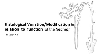
- 1. Histological Variation/Modification in relation to function of the Nephron Dr. Saran A K 1
- 2. Nephron Tubular Segments Common Histological Features Histological Features • 1. Renal Corpuscle • 2. Proximal Convoluted Tubule • 3. Loop of Henle • 4. Distal Convoluted Tubule • 5. Collecting Duct Applied Aspects References 2
- 3. • The nephron is the structural and functional unit of the kidney. • Each kidney contains approximately 1.3 million nephrons. • The nephron consists of the • Renal corpuscle • Glomerulus • Bowman’s Capsule • Tubule system Coronal Section Through a Unipapillary Kidney Fig 1.1 Comprehensive Clinical Nephrology 3
- 4. Nephron Divisions Length Bowman’s Capsule Proximal Tubule PCT and PST(pars recta) 15 mm, 55µm in diameter Loop of Henle Descending Thin Segment Ascending Thin Segment Thick Ascending Limb Length variable up to 25mm Cortical Nephrons Juxta Medullary (15%) Distal Convoluted Tubule 5 mm Collecting Duct Cortical Collecting Duct Medullary Collecting Duct 20 mm 45-65 mm (Total Length) 4
- 5. General Histological Features 1. Single-layer epithelium 2. Basolateral Membrane invaginated to form basal spaces 3. Cytoplasmic processes from lateral and basal surfaces of the cells interdigitate with the similar processes of adjacent cells, forming Lateral Intercellular space. Lateral space continuous with peritubular space functionally Diagram of a Juxtamedullary Nephron Fig 38-1 Ganong’s Review of Medical Physiology 26th Edition 5
- 6. 2. Tight Junctions • Adjacent cells united by apical tight junctions consisting of a tight junction (zonula occludens), an adherens junction, and, rarely, a desmosome • Leaky tight junctions • Tight tight junctions Fig 38-8 Ganong’s Review of Medical Physiology 26th Edition Peritubular Space 6
- 7. 1. Paracellular transport : depends on the type of tight junction 2. Transcellular transport depends on specific channels, carriers, and transporters included in the apical and basolateral cell membranes. Figure 1.10. Tubular epithelia Comprehensive Clinical Nephrology Transport Systems 7
- 8. • Glomerulus • Capillary Endothelium (E) • Bowman’s Capsule • Parietal Layer (PE) • Visceral Layer • Podocytes (PO) • Glomerular Membrane Renal Corpuscle Figure 1.4. Comprehensive Clinical Nephrology 8 1. Renal Corpuscle
- 9. Glomerular Membrane/ Filtration Barrier 1. Glomerular Endothelium : numerous fenestrae - pore size 70-90 nm 2. Glomerular Basement Membrane (GBM): acts as a physical barrier and an ion-selective filter 3. Visceral layer of Bowman’s capsule: podocytes -foot processes or pedicels. Slits closed by filtration slit diaphragm acts as a size-selective filter. Three Layers of Filtration Barrier Fig 22.13 Medical Physiology Rhodes and Rhotney 4th Edition 9
- 10. Scanning Electron Micrograph of a Glomerulus EM of Glomerular Capillary and Podocyte Fig 20.12, Fig 20.13 Histology A Text and Atlas, Michael H Ross GM permits the free passage of neutral substances up to 4nm in diameter and almost totally excludes those with diameters greater than 8nm. 10
- 11. • Mesangial Cells • Intra glomerular • Structural Support • Regulate Blood Flow • Phagocytic Activity • Extra glomerular (lacis cells) • Juxtaglomerular Cells • Macula Densa Juxta Glomerular Apparatus • feedback control of GFR and Blood flow in same nephron Structure of Renal Corpuscle Fig 20.7 Histology A Text and Atlas, Michael H Ross 11
- 12. 2. Proximal Convoluted Tubule • The epithelial cells are cuboidal in shape • A layer of closely packed microvilli covers the luminal surface forming a brush border • The cells have prominent basal and lateral processes and interdigitate extensively Drawing of proximal convoluted tubule cells Fig 20.17 Histology A Text and Atlas, Michael H Ross 12
- 13. Electron Micrograph of Proximal Tubule Cell Fig 20.18 Histology A Text and Atlas, Michael H Ross • Prominent Gogli Apparatus with ER and vacolues for synthesizing membrane components, sorting and targeting them to surface sites • Numerous mitochondria concentrated in the basal processes created by the invagination of basal membrane. 13
- 14. Cellular Ultrastructure and Primary Transport in PCT Fig 27-6 Guyton and Hall Medical Physiology • Glucose, Sodium Chloride and Sodium Bicarbonate actively absorbed • Transporters present in plasmalemma membrane (SGLT1 and NHE3) • Na+-K+-ATPase in basolateral membrane • Abundant mitochondria supplies energy • Water : 66% of all the water absorption • well adapted for the processes of reabsorption and secretion brush border and basal processes • Aquaporin1 abundance (Obligatory) 14
- 15. • Proteins and large peptides are endocytosed • Prominent Endosomal-Lysosomal system present towards the apex • Organic acids and bases, such as drugs and drug metabolites are secreted. Figure 1.11. PCT Comprehensive Clinical Nephrology 15
- 16. • Consist of the Thin Descending Limb and the Thick Ascending Limb • Long Looped Nephrons have a Thin Ascending Limb • The Thin Descending Limb is permeable to water and few solutes while the Thick Ascending Limb is impermeable to water but not to solutes. 3. Loop of Henle Thin Descending Limb Thick Ascending Limb • 2-14mm,30 µ • 12mm, 60µ • Flat squamous cell • Leaky Tight Junctions • Cuboidal cell • Tight Tight Junctions 16
- 17. Thin Descending Limb Thick Ascending Limb • Few mitochondria • Large number of mitochondria • Poorly developed luminal and basolateral surface • Extensive invagination of basal membrane • Passive Diffusion of Substances • Water 15-20% • Active Reabsorption • Na+-K+-2Cl- Co-transporter • Na+-H+ Counter transport • Na+-K+ ATPase 17
- 18. • Thin Descending Limb helps in the absorption of water and increases tubular fluid osmolality • Leaky Tight Junctions • low level of metabolic activity • designed for the passive diffusion of water and urea • Thick Ascending Limb forms Diluting Segment of the kidney along with DCT • Impermeable to water (tight tight junctions) • Metabolically active and designed to perform active reabsorption of solutes Cellular Ultrastructure and Primary Transport in LoH Fig 27-8 Guyton and Hall Medical Physiology 18
- 19. Distal Nephron • Made up of • Distal Convoluted Tubule • Connecting Tubule • The Collecting Duct • The junction between the cortical thick ascending limb and the distal nephron contains JG Apparatus 19
- 20. 4. Distal Convoluted Tubule Electron Microscope of a Distal Convoluted Tubule Cell Fig 20.18 Histology A Text and Atlas, Michael H Ross • Cuboidal Epithelium • Extensive basal and lateral IP • Greatest numerical density of Mitochondria • Lack a brush border but has Microvilli 20
- 21. • Reabsorption of Na • Early DCT: Transporters present in plasmalemma membrane (Na+-Cl- Co transporter) • Late DCT: ENaC (P Cells) • Regulated by Aldosterone – Na+ Balance • Na+-K+-ATPase in basolateral membrane • Abundant Mitochondria • 5% filtered water reabsorbed • Early DCT : impermeable • Late DCT : Controlled by ADH ( V2 Receptors and Aquaporin 2 in P Cells) and by Na+ absorption by aldosterone 21 Cellular Ultrastructure and Primary Transport DCT Fig 27-9Guyton and Hall Medical Physiology
- 22. 5. Collecting Duct • Collecting Duct is divided into • Cortical Collecting Duct • Medullary Collecting Duct • Cells appear like in DCT but taller and more granular • Contains two distinct type of cells • Principal Cells (P) • Intercalated Cells (I) 22
- 23. Principal (P) or the Light Cells Intercalated (I) or Dark Cells • More in number • Less in number • Relative paucity of cell organelles • Abundant mitochondria • Basally located interdigitations with neighboring cells. • Few short microvilli • Have more microvilli • Numerous vesicles are present in the apical cytoplasm 23
- 24. • Water Reabsorption : Fine Control of Urine Conc. • 8-10% • P Cell • Antidiuretic hormone (ADH) acts on V2R • aquaporin-2 (AQP-2) to luminal surface, AQP3 and AQP4 to interstitium • Facultative Reabsorption • Sodium Reabsorption and Potassium Secretion • P Cells • through ENaC • ENaC respond to Aldosterone - RAAS 24 Cortical Collecting Duct
- 25. • Secretion of H+ : Acidification of Urine • The intercalated cells are involved in the Reabsorption of HCO3 - with concomitant secretion of H+ leading to further acidification in Late DCT and CD • In Acidosis number of H+ pumps increases by insertion of the tubulovesicles into apical cell membrane 25 Primary Active Secretion of H+ Intercalated Cells (Late DCT and CD) Fig 30-6 Guyton and Hall Medical Physiology
- 26. • Urea Transport : Concentrating Mechanism • The terminal portions of CD express the urea transport system (UTB1) - recycling of Urea • H+-ATPase Transporter • H2O Reabsorption depending on the level of ADH 26 Medullary Collecting Duct Cellular Ultrastructure and Primary Transport Medullary CD Fig 27-13 Guyton and Hall Medical Physiology
- 27. • Filtration Membrane 1. Alport’s syndrome (Hereditary Glomerulonephritis) Mutation in 5th chain of type IV collagen 2. Autoimmune response to 3rd chain of type IV collagen Goodpasture syndrome • Mesangial Cells 1. IgA nephropathy (Berger disease) 2. Diabetic Nephropathy Applied Aspects 27
- 28. • Diabetes Insipidus 1. Central Diabetes Insipidus (CDI) 2. Nephrogenic Diabetes Insipidus defective AQP-2 and ADH receptor proteins • Diuretics 28 Type Action Examples Loop Diuretics Inhibit Na+-K+-2Cl- in Thick Ascending Limb of LoH Furosemide Thiazide Diuretics Inhibit Na+Cl- symporter in DCT Chlorothiazide K+ Sparing Diuretics • Aldosterone antagonist • Direct ENaC Blocker Inhibit ENaC in late DCT and Cortical CD Spironolactone Amiloride Carbonic Anhydrase Inhibitor Prevent HCO3 - Reabsorption in PCT and secretion of H+ Na+-H+ Antiport blocked Acetazolamide Dorzolamide Osmotic Diuretics Increases fluid osmolality LoH Mannitol
- 29. 29 • Renal Tubular Syndromes 1. Fanconi Syndrome : Generalized reabsorption defect in PCT 2. Bartter Syndrome : Reabsorption Defect in Thick Ascending Loop of Henle Na+-K+-2Cl- Co-transporter (mimics Loop Diuretic) 3. Gitelman Syndrome : Reabsorption Defect of NaCl in DCT (mimics thiazide Diuretic) 4. Liddle Syndrome : Gain of function mutation of ENaC Channels in Collecting Tubule
- 30. 30
- 31. References 1. Ganong. Review of Medical Physiology 22nd Edition McGraw Hill 2. Best and Taylor’s Physiological Basis of Medical Practice 12th Edition Williams & Wilkins 3. Michael H Ross. Histology : A Text and Atlas 6th Edition Williams & Wilkins 4. Samson Wright’s Applied Physiology 13th Edition Oxford University Press 5. Guyton & Hall. Textbook of Medical Physiology 11th Edition Saunders Elsevier 6. Ganong. Review of Medical Physiology 26th Edition McGraw Hill 31
- 32. Thank You! 32
Editor's Notes
- Basolateral Membrane of PCT, DCT and TAL of LoH is invaginated to form basal spaces also bringing abundant mitochondria in contact to plasma membrane in contrast to TDL and CD
- Lamina Rara Interna Lamina Rara Externa Lamina Densa
- Plasma Osmolality Increases. ADH Secretion Incereases