10. Introduction
• A. arteriole – carries blood to glomerulus.
• Glomerulus – filters protein free plasma into
tubular component.
• E. arteriole – carries blood from glomerulus.
• Peritubular capillary – supply the renal tissue,
involved in exchanges with fluid in T. lumen.
• JGA – produces substances involved in control
of kidney function.
11. Cont..
• Bowman's capsule – collect G. filtrate.
• Proximal Tubules – uncontrolled reabsorption
and secretion of selected substances occur
here.
• Loop of Henle – establishes an osmotic
gradient in renal medulla. Important in
kidney’s ability to produce urine of varying
concentration.
12. Cont..
• Distal tubule and Collecting duct – variable,
controlled reabsorption of Na⁺ and H₂O and
secretion of K⁺ and H⁺ occur here.
• Fluid leaving the collecting duct is urine, which
enters the renal pelvis.
13. Function of Kidney
• 1) Role in Homeostasis.
• Excreting the end product of bodily metabolism.
• Maintaining water balance in the body.
• Maintaining the Electrolyte balance.
• Maintain the acid base balance by adjusting
urinary output of H⁺ and HCO₃-.
• 2) Producing erythropoietin.
• 3) Regulation of blood pressure
• 4) Converting vitamin D into its active form.
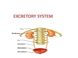
14. Kidney
• Bean – shaped,
• Location – retroperitoneally on the posterior
abdominal wall, one on each side of V. column at
the level of T₁₂ to L₁ vertebra.
• Weight – 150 gm, 10 cm x 5cm x 2.5 cm.
• Rt. Kidney is slightly lower than left kidney
• Outer cortex and inner medulla.
• Medulla contains 10 to 15 pyramids, which
terminate medially in renal papillae.
15. Cont..
• Papillae projects into calyces, 10 to 15 minor
calyces join to form 2 major calyces.
• Which come out through the pelvis of kidney
to widened end of kidney.
• Renal hilus – blood vessels, lymphatic's and
nerves enter into or exit from kidney via hilus.
• Renal pelvis – flattened, funnel – shaped
expansion of superior end of ureter.
16. Microscopic structure of kidney
• Cortex and medulla of kidney are composed of
nephron, blood vessels, lymphatic's and nerves.
Nephron
• Functional unit of kidney.
• Each kidney consists of 1.3 million nephron.
• Each nephron is capable of forming urine.
• Nephron consists of 2 major part – renal corpuscle and
renal tubules.
• The kidney cannot regenerate new nephron. Therefore,
with renal injury, disease, or normal aging, there is a
gradual decrease in nephron number.
17. Renal corpuscle
Glomerulus
• Each nephron contains a tuft of glomerular
capillaries called the glomerulus, through
which large amounts of fluid are filtered from
the blood,
Bowman's capsule
• Encloses the glomerulus.
• Formed of two layers - visceral layer and
parietal layer.
18. Cont..
• Space between visceral and parietal layer is called
bowman's space or urinary space.
Ultra structure of glomerular membrane
• It separates blood of glomerular capillaries from
the fluid present in bowman's space.
• Also called filtration barrier.
Layer of the membrane.
• Capillary endothelium - basement membrane -
bowman's visceral epithelium,
19. Structure of glomerular membrane
• 1) It is thin membrane and made up of five layers
• Layer 1.- foot process of podocytes.
• The final part of the membrane is a layer of
epithelial cells (podocytes) that encircle the outer
surface of the capillaries.
• The foot processes are separated by gaps called
slit pores 25nm through glomerular filtrate
moves.
• The epithelial cells also have negative charges,
provide additional restriction to filtration of
plasma proteins.
20. Cont..
• Layer 2.- Lamina rara externa- overlying foot
processes of podocytes.
• Layer 3.- lamina densa- dense structural portion
of basement membrane.
• Layer 4 - Lamina rara interna – provide bed for
capillary endothelium.
• Layer 5.- Endothelial cell layer- it is fenestrated,
contains pores with diameter 70 to 90nm. And
freely permeable to water, small solutes and even
to small proteins.
21.
22.
23.
24. Cont..
• 2. Glomerular capillaries.
• Each glomerulus contain 6 lobules and each of
these consists of 3- 6 capillary loops.
• Anastomoses occur between the capillaries
within 1 lobule.
• Arrangement of both arterioles within glomerulus
allows the maintenance of much higher pressure
60 mmhg in G. capillary.
• High capillary pressure is well adapted for
filtration.
25. Cont.
• 3. Major function of G. membrane is to produce
an ultra filtrate.
• Filtrate contains all constituents of plasma except
protein.
• Permits the free passage of neutral substances up
to 4 nm in diameter and excludes those
diameters greater than 8 nm.
• The total area of glomerular capillary
endothelium across which filtration occurs in
humans is about 0.8 m2.
26. Epithelium lining Renal Tubule
• Tubules are lining by cuboidal except thin
segment these are flat or squamous type.
• Apical surface of cuboidal cells have microvilli,
present in proximal tubule.
• Basolateral membrane of PCT cells, thick
ascending segment cells and DCT cells contain
mitochondria ( transport function ).
• Descending and ascending thin limb have poorly
developed basolateral surface contain few
mitochondria.
27. Cont..
• Lateral surface of cells form 2 type of tight
junction.
Leaky tight junction
• Permit H₂O and solutes to diffuse across them.
• Present in proximal tubule.
Tight junction
• Do not permit H₂O, solutes to diffuse across them
easily.
• Present in Distal tubule.
29. Proximal Convoluted Tubule
• P.C.T. is about 15 mm long and 55 m in
diameter.
• Single layer of cells with curved outline and
brush border formed by microvilli.
• Tubule cells are rich in mitochondria, they are
responsible for active transport of 80% of Na⁺
filtered, out of the tubular fluid into the
peritubular capillary blood.
30. Cont..
• Tubular cells are united at the apex by tight
junction while between base of the cells have
extensions into extracellular space called
lateral intercellular space.
31. Loop of Henle
• The descending portion of the loop and the
proximal portion of the ascending limb are
made up of thin, permeable cells.
• Thick portion of the ascending limb is formed
by low cuboidal epithelium, containing many
mitochondria.
• In humans, only 15% of the nephron have long
loops.
32. Cortical nephron
• Short loops of
Henle
juxtamedullary
nephron
• long loops of Henle
extending down
into the medullary
pyramids.
33. Cont..
• The thick end of the ascending limb of the
loop of Henle reaches the glomerulus of the
nephron from which the tubule arose and
nestles between its afferent and efferent
arterioles.
• Specialized cells at the end form the macula
densa, which is close to the efferent and
particularly the afferent arteriole
34.
35. Cont..
• The macula, the neighboring lacis cells, and
the renin-secreting juxtaglomerular cells in
the afferent arteriole form the JGA.
36. Cont..
• Descending thin limb – H₂O passive
reabsorption through tight junction.
• Ascending limb – impermeable to H₂O.
• Early part of DCT – impermeable to H₂O,
active reabsorption of NACL.
• Later part of DCT and CT – H⁺ secretion, Na⁺
reabsorption ( 8% ), and K⁺ secretion and H₂O
reabsorption ( 10 to 15% ).
37. Distal convoluted tubule
• D.C.T. starts at the macula densa, is about 5
mm long.
• Low cuboidal epithelium is lower than that of
the proximal tubule, and although a few
microvilli are present,
• There is no distinct brush border.
• Macula densa maintain the NACL
concentration in DCT.
38. Collecting Ducts
• DCT join to form collecting ducts, 20 mm long
and lined by clear cuboidal epithelium.
• Pass through the renal cortex and medulla to
empty into the pelvis of the kidney at the
apexes of the medullary pyramids.
• The collecting ducts epithelium consist of 2
types of cells principal cells (P cells) and
intercalated cells (I cells).
39. Cont..
Principal cell
• Increase the permeability of CT to water in the
presence of ADH by increasing pore size
through Aquaporine-2.
Intercalated cells
• Secrete acids, help transport of bicarbonate
and are responsible for acidic urine.
• The total length of the nephron, including the
collecting ducts, ranges from 45 to 65 mm
40. Juxta Glomerular Apparatus
• Combination of specialized tubular and
vascular cells located at where afferent and
efferent arterioles enter and leave glomerulus
JGA composed of 3 types of cells.
• Juxtaglomerular cells. JG cells
• Macula Densa cells
• Mesangial cells of Lacis cell.
41.
42. Juxtaglomerular cells
• Specialized myoepithelial cells ( modified
vascular smooth muscle cells ).
• Located in the media of afferent arteriole.
• Have well developed G. apparatus, and E.
reticulum, abundant mitochondria and
ribosomes.
• They synthesize, store and release a P. enzyme
called Renin stored in granules of JG cells.
43. Cont..
• It acts as baroreceptor and respond to change
in Transmural Pressure Gradient between
afferent arteriole and interstitium.
• Innervated by sympathetic nerve fiber and
release Renin in response to sympathetic
discharge.
• These vascular volume receptor monitor Renal
Perfusion Pressure and stimulated by
hypovolemia or ↓ed Renal perfusion pressure
44. Macula Densa Cells
• Specialized Renal Tubular Epithelial cells.
• Located at site where thick segment of
ascending limb of LOH continued as DCT.
• Cells are in direct contact with Mesangial cells,
in close contact with JG cells and adjoining
with both A and E arterioles.
• Cells have prominent nuclei and function as
Chemoreceptor.
45. Cont..
• Stimulated by↓ed Na⁺ conc. (NACL) load
causing ↑ed renin release.
• They are not innervated.
• They are not well adapted for reabsorption
• They do not show signs of secretory activity.
• Cells maintain the NACL concentration in DCT.
46. Mesangial Cells
• Supporting cells of JGA and found between
capillary loops.
• They are contact with both JG cells and Macula
Densa cells
• They are contractile and play role in regulation of
glomerular filtration.
• They show granulation to secrete renin in
conditions of extreme hyperactivity.
• Also secrete verious substances and take up
immune complexes.
47. JGA regulate Renin secretion into
blood stream
Factors inhibit renin secretion
Inhibition of JG cells by
• Stretch due to ↑ in afferent arterial pressure.
• Angiotensin ii - arteriole constrictor.
• Vasopressin ( ADH ) - strong vasoconstrictor,
affect H₂O reabsorption in R. tubule. After severe
hemorrhage its concentration rises to high level
and shows vasoconstrictor effect.
↑ Na⁺ and CL⁻ reabsorption across macula densa.
48. Cont..
Factors stimulate Renin secretion
Stimulation of JG cells by
• Increase in sympathetic activity due to
hypovolemia, hypotension.
• Increase in circulating catecholamine's
• Prostaglandins – vasodilator
Sodium depletion, diuretics, congestive
cardiac failure.
49. Renin Angiotensin System
Operate during
• Regulation of blood pressure
• Regulation of extracellular fluid volume.
Renin secretion and Angiotensin formation
• Renin secretion stimulated by decrease in BP
• Renin catalyzes the conversion of
angiotensinogen ( plasma ) to Angiotensin I.
• Angiotensin I converted into Angiotensin II by
Angiotensin converting enzyme.
50. Cont
• ACE present in endothelium of blood vessels
entire the body especially in lung and kidney.
• Angiotensin persist in blood until inactivated
by multiple blood and tissue enzymes called
angiotensinase.
51. Action of Angiotensin II
Vasoconstriction – arteriole constriction
increase both systolic and diastolic BP.
Decrease in salt and water excretion by kidney
By direct action on the kidney
• Angiotensin II constrict E. arteriole cause
diminishes blood flow through P. capillaries
allowing rapid osmotic reabsorption from
tubule.
52. Cont..
• Angiotensin II directly stimulate epithelial cells of
tubule to increase reabsorption of sodium and
water.
By stimulating secretion of aldosterone.
• Angiotensin II stimulate adrenal gland to secrete
aldosterone cause increase salt and water
reabsorption by epithelial cell.
Stimulation of thirst.
• Angiotensin II is powerful stimulator for thrust
lead to increase water intake and increase blood
volume.
53. Renal Blood Supply
• Blood flow to the two kidneys is normally about
22 % of the cardiac output, or 1100 ml/min.
• The renal artery enters the kidney through the
hilum and branches to form the interlobar
arteries, arcuate arteries, interlobular arteries (
radial arteries) and afferent arterioles.
• Lead to glomerular capillaries, where large
amounts of fluid and solutes are filtered to begin
urine formation.
54.
55. • The distal ends of the capillaries of each
glomerulus coalesce to form the efferent
arteriole, leads to the peritubular capillaries, that
surrounds the renal tubules.
• The renal circulation has two capillary beds, the
glomerular and peritubular capillaries, arranged
in series and separated by the efferent arterioles,
• Which help regulate the hydrostatic pressure in
both sets of capillaries.
56. Cont..
• High hydrostatic pressure in the glomerular
capillaries (about60 mm Hg) causes rapid fluid
filtration.
• Lower hydrostatic pressure in the peritubular
capillaries (about 13 mm Hg) permits rapid fluid
reabsorption.
• By adjusting the resistance of the Afferent and
Efferent arterioles, the kidneys can regulate the
hydrostatic pressure in both the glomerular and
peritubular capillaries.
57. Cont..
• Change in glomerular filtration, tubular
reabsorption in response to body
homeostatic demands.
• The peritubular capillaries empty into the
vessels of the venous system, which run
parallel to the arteriolar vessels.
• To form the interlobular vein, arcuate vein,
interlobar vein, and renal vein and leaves the
kidney beside the renal artery and ureter.
58. Cont..
• For the cortical nephron, - the entire tubular
system is surrounded by an extensive network of
peritubular capillaries.
• For the juxtamedullary néphrons, - long efférent
artérioles extend from the glomeruli down into
the outer medulla.
• Then divide into specialized peritubular
capillaries called vasa recta that extend
downward into the medulla, lying side by side
with the loops of Henle.
59. Cont..
• The vasa recta return toward the cortex and
empty into the cortical veins.
• This specialized network of capillaries in the
medulla plays an essential role in the formation
of a concentrated urine.
• Total surface of renal capillaries is approx = total
surface area of tubules, both being about 12 m2.
• The volume of blood in the renal capillaries at any
given time is 30 to 40 mL.
60. Factors affecting blood supply to
kidney
Catecholamine's ( NE ) - ↓ RBF – constriction
Dopamine - ↑ RBF – vasodilatation.
Angiotensin II - ↓ RBF
• Low concentration cause constrict E arterioles.
• High concentration cause constrict both
arterioles.
Prostaglandins - ↑ RBF – vasodilatation.
Exercise - ↓ RBF – release of NE and Angiotensin
II by increases sympathetic activity.
61. Cont..
Haemorrhage - ↓ RBF – NE and Angiotensin II
Change of posture - ↓ RBF
Increase in renal arteriole BP – ↑ RBF
decrease in sympathetic tone lead to arteriole
dilation.
Acetylcholine - ↑ RBF by producing
vasodilatation.
High protein diet - ↑RBF- by increasing
glomerular capillary pressure.
62. Characteristics of renal blood flow
Under basal condition RBF is 1.2 to 1.3L/M ie.
(300 – 400 ml/ 100gm /M ).
• RBF is very high compare to other body organs
example –
- Coronary blood flow – 60-80ml/100gm/m
- Brain blood flow – 55ml/100gm/m
- Skeletal muscle blood flow – 3-4ml/100gm/m
• High RBF produce high GFR for excretion of
metabolic waste products.