
Recommended
Recommended
More Related Content
What's hot
What's hot (20)
Viewers also liked
Viewers also liked (12)
Similar to 237299274 1case-report-11-new
Similar to 237299274 1case-report-11-new (17)
More from homeworkping3
More from homeworkping3 (20)
Recently uploaded
Recently uploaded (20)
237299274 1case-report-11-new
- 1. Get Homework/Assignment Done Homeworkping.com Homework Help https://www.homeworkping.com/ Research Paper help https://www.homeworkping.com/ Online Tutoring https://www.homeworkping.com/ click here for freelancing tutoring sites Case report Primary Granulocytic sarcoma Lip in a nonleukemic patient - A rare presentation Srivastava kirti, Paul Sayan, Gupta Deepak,Tripathi A K*,Goel Madhu Mati**Pant MC,Verma Jitendra. Department of Radiotherapy ,*Department of Medicine ,** Department of Pathology. CSMMU (formerly KGMU),lucknow Abstract- A sixty year old male presented with a swelling in the upper lip. On cytological examination patient was diagnosed as a case of Granulocytic Sarcoma. His bone marrow examination was normal suggesting no systemic disease. Patient achieved complete response by radiotherapy alone. Eighteen months following treatment patient is completely asymptomatic. Key Words: Granulocytic Sarcoma, Radiotherapy Introduction Granulocytic sarcoma (GS) also called myeloblastoma or myeloid sarcoma is a rare solid tumor composed of immature cells. It was first described by A. Burns in 1811 ( 5 ). GS is a rare occurrence with an estimated incidence of .7 / million 1
- 2. in children and 2/ million in adults this tumor occurs in about 5% of adults and 13% of children with myeloid Leukemia . In autopsy series GS occurs in 2-8% of patients with acute and chronic myelogenous leukemia. Such tumors often display a greenish color due to the enzymatic action of myeloperoxidase in the tumor cells , hence the term “Chloroma” was given to this lesion in 1853.More frequently the term Extra –medullary myeloid tumor (EMMT ) has been proposed to include all the forms of extramedullary myeloid leukemia infiltrates.. They occur most commonly in bones, periostium , soft tissues, Lymph Nodes & skin but can occur virtually anywhere. It is extremely rare in Oral cavity and often present as a lump in the gingiva, palate and extraction sockets. In view of the rarity of its occurrence and diagnostic importance we report here a case of primary GS presenting as a solitary lump in the lip with out bone marrow involvement. Case History A sixty year old male patient presented to our OPD with a history of gradually progressive swelling in right upper lip for one month which was not associated with bleeding or pain and no difficulty in chewing. On examination a lumpy swelling was noted in right upper lip with redness. ( fig:1 )The lump was non tender and there was no bleeding from the lump it was soft to firm in consistency. The lump was mainly present on right side while it had crossed the midline to involve medial 2/3rd of left side of upper lip. There were some necrotic areas on the right side of buccal mucosa with surrounding odema with mild tenderness without any bleeding .Cytological examination of the swelling was done suggesting granulocytic sarcoma featuring scattered single large cells with high N:C ratio, oval to round to irregular concentrically placed nuclei with granular chromatic & prominent single to multiple nucleoli. The cells have scanty to moderate amount of basophilic cytoplasm. Some of the cells contain cytoplasmic granule, background is of RBCs & neutrophils and lymphocytes ( Fig 2 ). A second opinion was taken from the Pathology department of our Institution where it was confirmed to 2
- 3. be granulocytic sarcoma .After discussing the case in tumor board patient received local radiotherapy 30 Gy in 15# @ 200 cGys / # by Co-60 by parallel and opposed lat fields. Patient achieved complete response following completion of radiotherapy he was then advised for chemotherapy. Due to some financial constraints patient did not receive chemotherapy but was on regular follow up. Eighteen months following completion of radiotherapy patient is completely asymptomatic ( Fig 3. ). His bone marrow and General blood picture is normal till date. Discussion: Granulocytic sarcomas often pose a diagnostic dilemma for the oncologists. Granulocytic sarcoma (also called myeloblastoma or myeloid sarcoma) is a rare solid tumor composed of immature myeloid cells. It was first described in 1811.1 Such tumors often display a greenish color due to the enzymatic action of myeloperoxidase in the tumor cells; hence, the term "chloroma" was given to this lesion in 1853.2,3 The WHO classification separates these tumors into myeloid or monocytic sarcomas. Most tumors have been observed in patients with acute myelogenous leukemia and myeloproliferative disorders3 ; however, they have been described in patients with myelodysplasia4 and chronic lymphocytic leukemia.5,6 Granulocytic sarcomas may also occur in the absence of leukemia,7 as notedin our patient. The most common sites of involvement are bone, periosteum, soft tissue, lymph node, and skin.3 Rare occurrences in muscle,8 meninges,9 breast,10 mediastinum, and ovary11 have also been reported. The present case demonstrates the unusual extramedullary presentation with lip involvement. This often predates involvement of the marrow but in our case eighteen months following treatment patient is absolutely normal. Though Granulocytic sarcoma is treated as acute myelogenous leukemia with systemic chemotherapy with or without local radiotherapy , Local radiation therapy may be necessary in selected cases, like the case we have reported . 3
- 4. References: 1. Menasce LP, Banerjee SS, Beckett E,Harris M. Extra-medullary myeloid tumour ( granulocytoma ) is often misdiagonosed: A study of 26 cases.Histopathology 1999;34:391-8. 2. Yamauchi K,Yasuda M comparison in treatments of nonleukemic granulocytic sarcoma:report of two cases and a review of 72 cases in the literature.Cancer 2002 Mar 15;94(6):1739-46. 3. Byrd JC, Edenfield JW,Shields DJ,et al: Extramedullary myeloid tumors in acute non lymphocytic myeloid leukemia: A Clinical review.J Clin Oncol 13: 1800, 1995. 4. 5.Burns A: Observations of Surgical Anatomy, Head and Neck. Edinburgh, United Kingdom, Thomas Royce and Co, 1811, pp 364-366 6.. King A: A case of chloroma. Monthly J Med 17:97, 1853 7. Neiman RS, Barcos M, Berard C, et al: Granulocytic sarcoma: A clinicopathologic study of 61 biopsied cases. Cancer 48:1426-1437, 1981[CrossRef][Medline] 8. List AF, Gonzalez-Osete G, Kummet T, et al: Granulocytic sarcoma in myelodysplastic syndromes: Clinical marker of disease acceleration. Am J Med 90:274- 276, 1991[Medline] 9. Majumdar G, Singh AK: Cord compression: A rare complication of chronic lymphocytic leukaemia. J Clin Pathol 45:258-259, 1992[Abstract/Free Full Text] 10. Michalevicz R, Burstein A, Razon N, et al: Spinal epidural compression in chronic lymphocytic leukemia. Cancer 64:1961-1964, 1989[Medline] 11. Buckland ME, Scolyer RA, Donellan MB, et al: Spinal chloroma presenting with triplegia in an aleukaemic patient. Pathology 33:386-389, 2001[Medline] 12. Bassichis B, McClay J, Wiatrak B: Chloroma of the masseteric muscle. Int J Pediatr Otorhinolaryngol 53:57-61, 2000[Medline] 13. Binder C, Tiemann M, Haase D, et al: Isolated meningeal chloroma (granulocytic sarcoma): A case report and review of the literature. Ann Hematol 79:459-462, 2000[Medline] 14. Ngu IWY, Sinclair EC, Greenaway S, et al: Unusual presentation of granulocytic sarcoma in the breast: A case report and review of the literature. Diagn Cytopathol 24:53- 57, 2001[Medline] 4
- 5. 15. Sreejith G, Gangadharan VP, Elizabeth K, et al: Primary Granulocytic sarcoma of the ovary. Am J Clin Oncol 23:239-240, 2000[Medline Fig 2. Microphotograph from cytological smear of Fine needle aspirate from the lip swelling smear showing immature myeloid cells including blasts. ( May Grunwald Giemsa X 400 ) 5
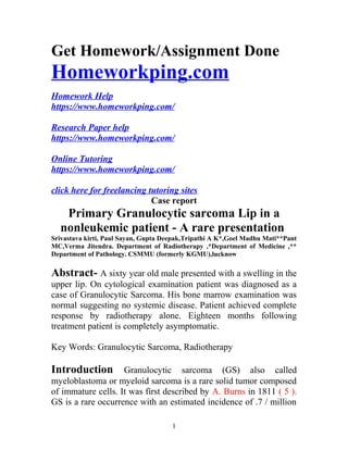
- 6. Figure: 1 Pretreatment lesion involving right upper lip 6
- 7. Figure: 3 Post treatment picture showing complete disappearance of the lesion 7