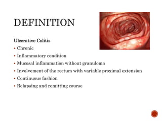
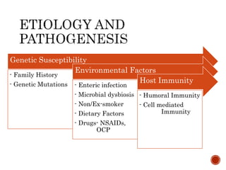
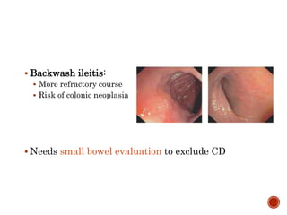
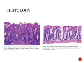
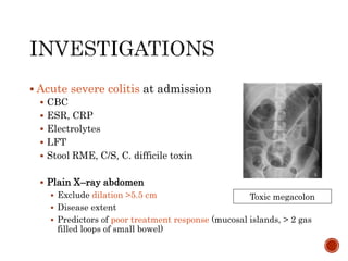
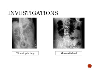
Dr. S M Ali Hasan
MD Phase-B resident
Dept. of Gastroenterology
 Definition
 Epidemiology
 Etiology and Pathogenesis
 Pathology
 Diagnosis
 Assessment
 Management
 Complication and extra-intestinal manifestation
Ulcerative Colitis
 Chronic
 Inflammatory condition
 Mucosal inflammation without granuloma
 Involvement of the rectum with variable proximal extension
 Continuous fashion
 Relapsing and remitting course
Prevalence
 Europe (Norway- 505/100000)
 North America (USA- 286/100000)
 Southern Asia (India- 44/100000)
Incidence
 Europe (Faroe Islands- 58/100000/yr)
 North America (Canada- 23/100000/yr)
 Southern Asia (India- 6/100000/yr)
Ng, Shi et al. 2018
Genetic Susceptibility
- Family History
- Genetic Mutations
Environmental Factors
- Enteric infection
- Microbial dysbiosis
- Non/Ex-smoker
- Dietary Factors
- Drugs- NSAIDs,
OCP
Host Immunity
- Humoral Immunity
- Cell mediated
Immunity
 Increased risks
 Family history of UC
 NSAID (short term
COX2 inhibitor may be
safe)
 Decreased risks
 Appendicitis/ mesenteric
lymphadenitis in
childhood/ adolescence
 Smoking
 Protect PSC and
pouchitis
 Always involves the rectum
 Variable proximal extension
 Most severe distally
 Progressively less severe proximally
 Continuous and symmetrical
 Sharp transition between diseased and healthy segment of the
colon
 Limited to mucosa and superficial submucosa
 Rectal sparing:
 Topical therapy
 Untreated children
 Cecal patch
 Peri-appendiceal skip lesions (75%)
 More responsive course
 Risk of pouchitis after IPAA
 Backwash ileitis:
 More refractory course
 Risk of colonic neoplasia
 Needs small bowel evaluation to exclude CD
 According to disease extent (Montreal
classification), Silverberg et al.,
 E1 – Proctitis: Distal to rectosigmoid junction
 E2 – Left-sided: Limited distal to splenic flexure
 E3 – Pancolitis/Extensive: involvement proximal to splenic
flexure.
 Proximal extension of proctitis – 20-30%
 Importance:
 Choice of treatment and drug delivery system
 Selection for surveillance
According to disease severity
 Remission
 Active
 Mild
 Moderate
 Severe active disease
 Diagnosis depends on
 Clinical features
 Laboratory parameter
 Imaging
 Colonoscopy
 Histopathology
 Exclusion of infective cause
 Clinical features:
Depends on extent and severity
 Rectal bleeding (> 90%)
 Loose stool (> 6 weeks)
 Rectal urgency
 Tenesmus
 Mucopurulent exudate
 Nocturnal defecation
 Crampy abdominal pain
 Proctitis:
 Rectal bleeding
 Rectal urgency
 Mucus discharge
 Tenesmus
 Severe constipation
 Systemic symptoms:
 Weight loss
 Fever
 Tachycardia
 Nausea
 Vomiting
 Extra-intestinal manifestation
 Arthropathy
 Ocular
 Dermatological
 Hepato-biliary
 History
 Symptoms
 Family history
 Travel history
 Drug history
 Examination
 Anemia
 Pulse
 Blood pressure
 Height, weight
 Abdomen: Distension,
tenderness, Bowel sound
 Perianal: inspection,
DRE.
Differential diagnosis
 Crohn’s colitis
 Infectious colitis
 Ischemic colitis
 Radiation colitis
 SRUS
 Amoebic colitis
 Schistosomiasis
 Colon cancer
 NSAID enteropathy
 HSP, Behcet’s disease
Initial
 CBC – Hb- , PC-
 Electrolytes
 Liver function test
 Renal function test
 Iron profile
 Vitamin-D level
 CRP
 F. Calprotectin
 Exclusion of infective diarrhoea including C. difficile
 Stool RME
 Stool culture
 Toxin A & B
 Colonoscopy/Sigmoidoscopy
 Histopathology
In relapse or treatment refractory UC
 Test for C. difficile and CMV
 C. difficile infection
 Stool for c. difficile toxin
 Pseudo membrane are usually absent (0-13%)
 CMV
 Biopsy
 Histopathology:
multiple inclusion body
 IHC (> 2 CMV +ve cells)
 PCR in blood
 Biomarker
 pANCA limited sensitivity, routine use for Dx
 ASCA therapeutic decision is not justified
 F. calprotectin – most sensitive in activity, Dx & treatment
response
 Elastase
 Lactoferrin
 Acute severe colitis at admission
 CBC
 ESR, CRP
 Electrolytes
 LFT
 Stool RME, C/S, C. difficile toxin
 Plain X–ray abdomen
 Exclude dilation >5.5 cm
 Disease extent
 Predictors of poor treatment response (mucosal islands, > 2 gas
filled loops of small bowel)
Toxic megacolon
Thumb printing Mucosal island
Acute severe colitis at admission
 Flexible sigmoidoscopy
 Confirm diagnosis
 Exclude infection – CMV
 Preparation: unprepared bowel. Phosphate enema can be
used
 Full colonoscopy is not recommended
 After active disease has been controlled in newly diagnosed
patient full colonoscopy should carried out to see extent and
to exclude CD
 Clinical assessment
 Endoscopic assessment
 Histological assessment
 Radiological assessment
 Biomarkers
 Quality of life (QoL)
Clinical assessment
 About 17 indices
 For assessment of mild to moderate disease
 Simple colitis clinical activity index (SCCAI)
 Partial Mayo clinic index (PMI)
 For severe disease
 Truelove and Witts criteria
SCCAI
Partial Mayo clinic
Index (PMI)
Mild Moderate Severe
Bloody stool < 4 ≥ 4 if ≥ 6 and
Pulse < 90 ≤ 90 > 90
Temperature < 37.5 ≤ 37.5 > 37.5
Hemoglobin > 11.5 ≥ 10.5 < 10.5
ESR < 20 ≤ 30 > 30
CRP Normal ≤ 30 > 30
Truelove & Witts
Endoscopic assessment of severity
 Ulcerative colitis endoscopic index of severity
(UCEIS)
 Mayo clinic endoscopic subscore
 UCEIS_ UC endoscopic index of severity
UCEIS_ UC endoscopic index of
severity
Vascular pattern Normal = 0
Patchy obliteration = 1
Complete obliteration = 2
Bleeding None = 0
Mucosal = 1
Luminal mild = 2
Luminal moderate or severe = 3
Erosions and ulcers None = 0
Erosions = 1
Superficial ulcers = 2
Deep ulcers = 3
 Mayo score (3 days)
Mayo index 0 1 2 3
Stool
frequency
Norma
l
1-2/day
> Normal
3-4/day
> Normal
≥ 5/day
> Normal
Rectal
bleeding
None Streaks Obvious Mostly blood
Mucosa Norma
l
Mild friability
Erythema
Decreased
vascular
pattern
Moderate
friability
Marked erythema
Erosion
Absent vascular
pattern
Spontaneous
bleeding
Ulceration
Physician’s
global
assessment
Norma
l
Mild Moderate Severe
Histological marker
 Remission
 Persistent architectural distortion to normalization
 Active
 Transmucosal inflammatory infiltrate
 Basal plasmacytosis
 Cryptitis
 Crypt abscess
 Histological inflammation can present in
endoscopically inactive disease
 Is a risk factor for CRC in long standing case
Histological assessment of severity
 26 indices, 2 are validated
 Nancy index mostly recommended
 Robarts histology index (RHI)
Ulceration
Yes
Grade- 4: Severe
Active disease
Acute inflammatory
cell infiltrate
No
No
Yes
Moderate
or Severe
Mild
Grade- 3:
Moderately Active
disease
Grade- 2: Mildly
Active diseaseChronic inflammatory
cell infiltrate
Moderate or
marked
increased
No or
mild
increase
Grade- 1: No active
inflammatory
infiltrate
Grade- 0: No
histologic
significance disease
NANCY INDEX
ROBERTS HISTOLOGY INDEX
Radiological assessment
 Plain X-ray
 Used in Acute severe UC
 Toxic dilation (> 5.5 cm)
 Also to diagnose proximal constipation in refractory distal
UC
 Cross-sectional imaging
 To assess complication
 Exclude small bowel inflammation and Crohn’s disease
 Severe stenosis or comorbidity limits colonoscopy
 MRI/USG
 Disease activity
 Disease extent
 Simplified MR colonography index (MRC-S)
 Bowel ultrasound severity score
Simplified MR colonography index (MRC-S)
Bowel ultrasound severity score
Laboratory marker of severity
 CRP
 ESR
 Procalcitonin
 Low albumin
 F. Calprotectin
 Lactoferrin
 Elastase
 CRP
Oxford Criteria in acute severe colitis
Quality of Life (QoL)
 IBD-Control
 IBDQ
 SIBDQ
 CUCQ-8
 Short form SF 36v2
 Short form SF 12v2
 EuroQol (EQ-5D)
Based on
 Distribution
 Proctitis
 Left-sided colitis
 Extensive colitis
 Age of onset
 Disease duration
 Pattern of disease
 Infrequent (≤ 1 year)
 Frequent (≥ 2/year)
 Continuous (no
remission)
 Disease course
 Response to previous
medication
 Side effect of medication
 Extraintestinal
manifestation
 Proctitis (Mild to moderate)
 1st line:
 Mesalamine 1 g suppository once daily
 Alternative: Mesalamine foam or enema
 Topical mesalamine >> more effective than topical steroid
 Combined topical Mesalamine with oral Mesalamine or
topical steroid is more effective
Topical Mesalamine
Topical Mesalamine +
Topical steroid
(Budesonide 2g foam)
Oral 5-ASA + Topical
Mesalamine / topical
steroid
Refractory proctitis
 May require
 Systemic steroid
 Immunomodulators and /or
 Biologics
 1st line: Mesalamine enema ≥ 1g/day plus oral Mesalamine ≥
2.4 g/day
 Topical Mesalamine is more effective than topical steroid
 Once daily Mesalamine is equally effective as divided dose.
 Mild to moderate disease refractory or intolerant to 5-ASA
may be treated with Budesonide 9mg/day
 Moderate to severe disease or mild disease not
responding to 5-ASA
 Severe disease require Hospitalization
 Systemic steroid: Prednisolone 40mg/day or Beclomethasone
5mg/day
Think about…
 Poor adherence
 Delivery of inadequate conc. Of active drug
 Unrecognized complications (Proximal constipation, infection)
 Inappropriate diagnosis (CD, CRC etc.)
Stool C/S, endoscopy and biopsy, X-ray abdomen
Oral steroid + oral
and rectal 5-ASA
1. IV steroid
2. Salvage therapy
- Oral/rectal ciclosporin
- Oral/rectal tacrolimus
- Infliximab
Colectomy
Refractory proctitis or distal colitis
Review of symptoms, treatment history, medication
adherence
Proximal constipation
affect drug delivery
Laxative
Failed
Mild to moderate
 1st line: Mesalamine enema ≥ 1 g/day plus oral Mesalamine ≥
2.4 g/day (more benefit at 4.8g/day)
Moderate to severe disease
or mild disease refractory to 5-ASA (already on ≥ 2g /day
Mesalamine with relapse):
 Systemic steroid
 Oral Prednisolone 40 mg/day then taper 5mg weekly total 8
weeks
 2nd generation steroid:
 Oral Beclomethasone 5 mg/day – 4 weeks then alternate weekly
4 weeks (Similar efficacy to prednisolone 40mg)
 Diagnosis:
 Bloody diarrhoea ≥ 6 times/day and
 Any sign of systemic toxicity
 Pulse > 90 bpm
 Temperature >37.5 °C
 Hb <10.5 g/dl
 ESR > 30 mm/hr or CRP > 30 mg/L
 Hospitalization
 IV corticosteroid
 Methyl prednisolone 60 mg/day or
 Hydrocortisone 100 mg 4 times daily for 7-10 days
 Alternative: IV cyclosporine monotherapy if steroid contra-
indicated
 IV fluid & electrolyte replacement:
 Potassium supplementation at least 60 mmol/day
 Hypokalemia and hypomagnesaemia promote toxic
megacolon
 Appropriate investigations to diagnose and exclude
infection
 Unprepared Sigmoidoscopy with biopsy ( Dx and exclusion
of CMV)
 Stool culture and C. difficile toxin
 If C. difficile found positive then
 Oral vancomycin
 Fecal transplantation (FT) can be considered
 If possible, immunosuppressive drug should be stopped
 Subcutaneous LMWH to minimize risk of
thromboembolism
 Nutritional support:
 Enteral nutrition is most appropriate
 Bowel rest through IV nutrition does not alter outcomes
 Withdrawal of anti cholinergic, anti-diarrheal,
NSAID, opioids (risk of toxic megacolon)
 Antibiotic:
 Only if infection is considered
 Immediately prior to surgery
 Blood transfusion to maintain Hb conc. 8-10 g/dl
 IV steroid refractory UC
 Response should be assessed by 3rd day
 Prediction of IV steroid failure:
 Clinical marker:
 Stool frequency > 12 at Day-2, > 8/day or 3-8/day + CRP
>45mg/L at Day-3
 Biochemical marker: ESR >75 mm/hr, temp > 38°C
 Radiological/Endoscopic:
 Abdominal X-ray: dilatation > 5.5 cm or mucosal island
 Endoscopy: deep ulcerations
 Salvage therapy:
 Ciclosporin – 2 mg /kg/day for 8 days followed by 4 mg/kg/day
oral
 Tacrolimus
 Infliximab – 5 mg/kg on 0,2,6 weeks
 All responders
 Start Azathioprine at day 7
 Taper steroid from day 8
 Non responders following 4-7 days of salvage therapy:
 Colectomy
Complications
 Toxic megacolon
 Total or segmental non obstructive dilatation ≥ 5.5 cm plus
systemic toxicity
 Risk factors –
 Hypokalemia
 Hypomagnesaemia
 Bowel preparation
 Use of anti diarrheal therapy
 Management –
 i.v steroid
 Vancomycin until stool negative for C. difficile
 Consultation with colorectal surgeon
 If not improved - colectomy
 Perforation
 Inappropriate total colonoscopy
 Toxic megacolon
 Thromboembolism
• Thiopurines (EL-2)
• Anti TNF (EL-1)
preferably with
thiopurines
• Vedolizumab (EL-2)
• Methotrexate (EL-2)
Steroid dependent active UC
• Different Anti TNF
• Vedolizumab (EL-2)
• Colectomy
• IV steroid (EL-4)
• Anti TNF (EL-1)
preferably with
thiopurines
• Vedolizumab (EL-2)
• Tacrolimus (EL-2)
Oral Steroid refractory active UC
• Different Anti TNF
• Vedolizumab (EL-2)
• Colectomy
• Anti TNF (EL-1)
preferably with
thiopurines
• Vedolizumab (EL-2)
Immunomodulator refractory UC
• Different Anti TNF
• Vedolizumab (EL-2)
• Colectomy
 Goals –
 Steroid free remission
 Clinical and endoscopic remission
 Long term therapy
 Intermittent therapy may be acceptable for some
patients with proctitis
 Choice of maintenance therapy depends on
 Disease extent
 Disease course
 Previous medication failure and adverse event
 Severity of most recent flare
 Treatment used to induce remission
 Safety of therapy
 Cancer prevention
 Stepwise escalation of therapy
Add Anti-
TNF/
vedolizumab
Add thiopurines
Dose escalation of oral
/rectal 5-ASA
 5-ASA
 1st line maintenance therapy after induction with 5-ASA/
oral or rectal steroid
 Rectal 5-ASA: for proctitis or left-sided colitis
 Combination of oral and rectal 5-ASA – 2nd line maintenance
therapy
 Oral mesalamine 2 g/day once daily administration is
preferred
 Sulfasalazine is equally or slightly more effective but more
toxic.
 Thiopurines
 Mild to moderate disease
 Indications
 Early/ frequent relapse on Mesalamine optimum dose
 Mesalamine intolerant
 Steroid dependent
 Induction with Ciclosporin or Tacrolimus
 Reduce risk of colectomy after induction with ciclosporin
• Oral Ciclosporin for 6
months
• Continue Azathioprine
Start Oral ciclosporin 4
mg/kg/day and Azathioprine
Induction with i.v Ciclosporin
2mg/kg/day – 8 days
• Vedolizumab and
maintenance with
Vedolizumab (EL2)
Continue Anti TNF ±
thiopurines (EL1)
Induction with Anti TNF
Alternative:
thiopurines (EL3)
 Anti TNF & anti adhesion therapy
Anaemia (21%)
 Iron deficiency
 Anaemia of chronic disease
 Both
 Investigation
 CBC, PBF
 Ferritin, T. SAT
 CRP
Anaemia
Without active
inflammation
Active disease/inflammation
Ferritin <30 µg/L Ferritin upto 100
µg/L
Ferritin > 100 µg/L,
TSAT < 20%
ACD
IDA
 Atypical case:
 Vitamin B12
 Folate
 Haptoglobin
 LDH
 Coombs test
 Treatment
 Iron supplementation (oral >> parenteral)
 Blood transfusion (Hb <8, Ferritin <15)
 Erythropoetin (refractory case)
 Arthropathy (20%) – 2nd most common
Arthropathy
Axial Peripheral
Type – I Type – II
• Pauci-articular (< 5)
• Large joint
• Asymmetrical
• Acute self limiting
• Related to
intestinal disease
activity
• Poly-articular (>5)
• Small joint
• Symmetrical
• Chronic persistent
• Independent of
disease activity
• Inflammatory
LBP
• MRI/Radiological
evidence of
sacroilitis
Exclusion of
other form
 Treatment:
 Target:
 Reduce inflammation
 Pain relief
 Reduce disability
 Type-I:
 Effective treatment of UC flare
 Sulfasalazine
 Rest
 Physiotherapy
 Type – II
 NSAID/systemic steroid
 Axial/ AS
 Consultation with rheumatologist
 Sulfasalazine, MTX, Azathioprine are ineffective
 Choice
 NSAID
 Anti-TNF – Infliximab, Adalimumab, Golimumab
Metabolic bone disease
 Diagnosis: BMD
 Persistently active disease
 Repeated exposure to steroid
 Long duration disease
 Treatment:
 Calcium 500-1000 mg/day
 Vitamin-D 800-1000 IU/day
 Prophylactic Calcium/Vit-D with steroid
 Bisphosphonates: post menopausal women, spontaneous
fracture.
Cutaneous manifestations
 Erythema nodosum:
 Diagnosis: Clinical,
atypical case – skin biopsy
 Treatment:
 Treatment of underlying UC
 Systemic steroid (severe case)
 Immunomodulators/Anti-TNF (refractory case)
 Pyoderma gangrenosum:
 Dermatological consultation
 Systemic steroid
 Infliximab/Adalimumab
 Topical CNI
Ocular manifestation
 Episcleritis:
 Topical steroid and NSAID
 Uveitis:
 Topical/ Systemic steroid
 Urgent referral to ophthalmologist
 Resistant case – Anti TNF
Hepatobiliary
 PSC – increased risk of CRC
 Diagnosis: MRCP
 Treatment: UDCA
 Pericholangitis
 Steatosis
 Cirrhosis
 Chronic hepatitis
 Gall stone
 UC doesn’t affect fertility
 If pregnancy occurs at remission >> risk of relapse
is equal as non pregnant
 If pregnancy occurs at active disease >> Risk of
persistent disease activity
 Should advised to conceive during remission
 Disease activity is associate with preterm labor,
LBW
 No specific risk for congenital malformation
 Medications are safe except MTX
 All patient should be tested for HBV and HCV
infection at diagnosis
 If HBV found negative >> vaccination
 If HBsAg Positive, before, during and 12 months
after IM therapy >> Antiviral drug irrespective of
HBV-DNA level.(EL2)
 Or monitoring HBV-DNA 2-3 months interval
without antiviral. (EL5)
 All patient should screen for latent or active TB at
diagnosis and before starting of Anti-TNF
 History
 Chest X ray
 MT/IGRA
 UC & Latent TB:
 Anti TB at least for 3 weeks before Anti-TNF
 UC & Active TB:
 Anti TB at least 2 months before Anti-TNF
Risk factors for CRC
 Disease duration
 Disease extent
 More severe / persistent inflammation
 PSC
 Family history of CRC
 Male gender
 Raised CRP level
All UC patients
With PSC Without PSC
No further
surveillance
• Rectum without
• Past/current
• Endoscopic/Histolo
gic inflammation
proximal to rectum
• Others without only
rectum involvement
After Dx of PSC
annually irrespective
of activity, extent,
duration
All patients at 8 year of Dx
To reassess extent & exclude dysplasia
• High risk
• Intermediate risk
• No risk
Intermediate risk No riskHigh risk
• Stricture or dysplasia
detected within 5 year
• PSC
• Extensive colitis with
severe active
inflammation
• Extensive colitis with
mild to moderate active
inflammation
• Post inflammatory polyp
• F/H of CRC in 1°
relative ≥ 50 years
Annually 2-3 yearly 5 yearly
Surveillance colonoscopy
 Should be done at remission
 Chromoendoscopy with targeted biopsy
 High detection rate
 White light endoscopy
 Random biopsy every 10 cm &
 Targeted biopsy of any visible lesion
 Mesalamine – decrease incidence of CRC
 Other medication having no significant evidence of
chemoprevention
 LGD/HGD should be confirmed by independent GI
specialist pathologist
Endoscopically visible
dysplasia
Polypoid dysplasia Non-polypoid dysplasia
Polypectomy 1 Endoscopic resection possible
2. No evidence of non-polypoid or
invisible dysplasia elsewhere
Other cases
Colectomy
regardless of
Grade of dysplasiaEndoscopic resection and
continued surveillance
Endoscopically Non-
visible dysplasia
High grade dysplasia Low grade dysplasia
Colectomy Colectomy or
Annual surveillance
Referral to expert IBD
surveillance endoscopist
Repeat with high definition
chromo-endoscope
Controversial
Discuss with the
patients, GI specialist
and surgeon
Goals:
 To remove the disease
 With little alteration of normal physiology and life style
 Indications
Acute indications Chronic indications
• Fulminant colitis
• Toxic megacolon
• Perforation
• Massive hemorrhage
• Unresponsive to medical
Mx
• Poor quality of life and
fear of carcinoma
• Steroid dependency,
toxicity or refractory
• High grade dysplasia
• Carcinoma
• Severe EIM
• Growth retardation
• Chronic bleeding
requiring transfusion
• Large bowel obstruction
 The surgical choices are:
Restorative
proctocolectomy with IPAA
Total colectomy with IRA
Total proctocolectomy with
permanent end ileostomy
Restorative proctocolectomy with IPAA
 Resection of colon and rectum
 Formation of ileal pouch
 Pouch anal anastomosis
Indication:
 Procedure of choice
Contraindications:
 Poor sphincter control
 Crohn’s disease
 Low rectal cancer
Complications
 Pelvic sepsis
 Small bowel
obstruction
 Anastomotic stricture
 Pouchitis
 Pouch failure
Diagnosis:
Symptoms:
 Increased frequency
 Liquid stool
 Abdominal cramping
 Urgency
 Tenesmus
 Pelvic discomfort
 Rectal bleeding
 Fever
 EIM
Endoscopy:
 Defuse erythema
 Edema
 Granularity
 Friability
 Spontaneous or contact
bleeding
 Loss of vascular pattern
 Erosion
 Ulceration
Histological evidence of
pouchitis
 Differential diagnosis
 Ischemia
 Crohn’s disease
 CMV or C. difficile infection
 Collagenous pouchitis
 Risk factors
 Extensive UC
 PSC
 Being non-smoker
 pANCA positive serology
 NSAID use
 Management
Pouchitis
Acute pouchitis (< 4 weeks) Chronic Pouchitis (> 4 weeks)
Metronidazole or Ciprofloxacin
Antidiarrheal drug
Combination of 2 antibiotic
Oral steroid
Topical tacrolimus
Infliximab (refractory)
Adalimumab
Maintenance of remission: Probiotics (VSL#3)
 ECCO 2017: Third European evidence-based
consensus on diagnosis and management of
ulcerative colitis
 Sleisenger and Fordtran’s gastrointestinal and liver
disease: Pathophysiology/diagnosis/management
(10th edition)
 AIG IBD Manual: A clinician’s guide for day-to-day
practice
Ulcerative colitis by Dr. Ali Hasan

Ulcerative colitis by Dr. Ali Hasan

  • 1.
    Dr. S MAli Hasan MD Phase-B resident Dept. of Gastroenterology
  • 2.
     Definition  Epidemiology Etiology and Pathogenesis  Pathology  Diagnosis  Assessment  Management  Complication and extra-intestinal manifestation
  • 3.
    Ulcerative Colitis  Chronic Inflammatory condition  Mucosal inflammation without granuloma  Involvement of the rectum with variable proximal extension  Continuous fashion  Relapsing and remitting course
  • 4.
    Prevalence  Europe (Norway-505/100000)  North America (USA- 286/100000)  Southern Asia (India- 44/100000) Incidence  Europe (Faroe Islands- 58/100000/yr)  North America (Canada- 23/100000/yr)  Southern Asia (India- 6/100000/yr) Ng, Shi et al. 2018
  • 7.
    Genetic Susceptibility - FamilyHistory - Genetic Mutations Environmental Factors - Enteric infection - Microbial dysbiosis - Non/Ex-smoker - Dietary Factors - Drugs- NSAIDs, OCP Host Immunity - Humoral Immunity - Cell mediated Immunity
  • 8.
     Increased risks Family history of UC  NSAID (short term COX2 inhibitor may be safe)  Decreased risks  Appendicitis/ mesenteric lymphadenitis in childhood/ adolescence  Smoking  Protect PSC and pouchitis
  • 9.
     Always involvesthe rectum  Variable proximal extension  Most severe distally  Progressively less severe proximally  Continuous and symmetrical  Sharp transition between diseased and healthy segment of the colon  Limited to mucosa and superficial submucosa
  • 10.
     Rectal sparing: Topical therapy  Untreated children  Cecal patch  Peri-appendiceal skip lesions (75%)  More responsive course  Risk of pouchitis after IPAA
  • 11.
     Backwash ileitis: More refractory course  Risk of colonic neoplasia  Needs small bowel evaluation to exclude CD
  • 12.
     According todisease extent (Montreal classification), Silverberg et al.,  E1 – Proctitis: Distal to rectosigmoid junction  E2 – Left-sided: Limited distal to splenic flexure  E3 – Pancolitis/Extensive: involvement proximal to splenic flexure.
  • 13.
     Proximal extensionof proctitis – 20-30%  Importance:  Choice of treatment and drug delivery system  Selection for surveillance
  • 14.
    According to diseaseseverity  Remission  Active  Mild  Moderate  Severe active disease
  • 15.
     Diagnosis dependson  Clinical features  Laboratory parameter  Imaging  Colonoscopy  Histopathology  Exclusion of infective cause
  • 16.
     Clinical features: Dependson extent and severity  Rectal bleeding (> 90%)  Loose stool (> 6 weeks)  Rectal urgency  Tenesmus  Mucopurulent exudate  Nocturnal defecation  Crampy abdominal pain
  • 17.
     Proctitis:  Rectalbleeding  Rectal urgency  Mucus discharge  Tenesmus  Severe constipation  Systemic symptoms:  Weight loss  Fever  Tachycardia  Nausea  Vomiting  Extra-intestinal manifestation  Arthropathy  Ocular  Dermatological  Hepato-biliary
  • 18.
     History  Symptoms Family history  Travel history  Drug history  Examination  Anemia  Pulse  Blood pressure  Height, weight  Abdomen: Distension, tenderness, Bowel sound  Perianal: inspection, DRE.
  • 19.
    Differential diagnosis  Crohn’scolitis  Infectious colitis  Ischemic colitis  Radiation colitis  SRUS  Amoebic colitis  Schistosomiasis  Colon cancer  NSAID enteropathy  HSP, Behcet’s disease
  • 20.
    Initial  CBC –Hb- , PC-  Electrolytes  Liver function test  Renal function test  Iron profile  Vitamin-D level  CRP  F. Calprotectin
  • 21.
     Exclusion ofinfective diarrhoea including C. difficile  Stool RME  Stool culture  Toxin A & B  Colonoscopy/Sigmoidoscopy  Histopathology
  • 23.
    In relapse ortreatment refractory UC  Test for C. difficile and CMV  C. difficile infection  Stool for c. difficile toxin  Pseudo membrane are usually absent (0-13%)  CMV  Biopsy  Histopathology: multiple inclusion body  IHC (> 2 CMV +ve cells)  PCR in blood
  • 25.
     Biomarker  pANCAlimited sensitivity, routine use for Dx  ASCA therapeutic decision is not justified  F. calprotectin – most sensitive in activity, Dx & treatment response  Elastase  Lactoferrin
  • 26.
     Acute severecolitis at admission  CBC  ESR, CRP  Electrolytes  LFT  Stool RME, C/S, C. difficile toxin  Plain X–ray abdomen  Exclude dilation >5.5 cm  Disease extent  Predictors of poor treatment response (mucosal islands, > 2 gas filled loops of small bowel) Toxic megacolon
  • 27.
  • 28.
    Acute severe colitisat admission  Flexible sigmoidoscopy  Confirm diagnosis  Exclude infection – CMV  Preparation: unprepared bowel. Phosphate enema can be used  Full colonoscopy is not recommended  After active disease has been controlled in newly diagnosed patient full colonoscopy should carried out to see extent and to exclude CD
  • 29.
     Clinical assessment Endoscopic assessment  Histological assessment  Radiological assessment  Biomarkers  Quality of life (QoL)
  • 30.
    Clinical assessment  About17 indices  For assessment of mild to moderate disease  Simple colitis clinical activity index (SCCAI)  Partial Mayo clinic index (PMI)  For severe disease  Truelove and Witts criteria
  • 31.
  • 32.
  • 33.
    Mild Moderate Severe Bloodystool < 4 ≥ 4 if ≥ 6 and Pulse < 90 ≤ 90 > 90 Temperature < 37.5 ≤ 37.5 > 37.5 Hemoglobin > 11.5 ≥ 10.5 < 10.5 ESR < 20 ≤ 30 > 30 CRP Normal ≤ 30 > 30 Truelove & Witts
  • 34.
    Endoscopic assessment ofseverity  Ulcerative colitis endoscopic index of severity (UCEIS)  Mayo clinic endoscopic subscore
  • 35.
     UCEIS_ UCendoscopic index of severity UCEIS_ UC endoscopic index of severity Vascular pattern Normal = 0 Patchy obliteration = 1 Complete obliteration = 2 Bleeding None = 0 Mucosal = 1 Luminal mild = 2 Luminal moderate or severe = 3 Erosions and ulcers None = 0 Erosions = 1 Superficial ulcers = 2 Deep ulcers = 3
  • 36.
     Mayo score(3 days) Mayo index 0 1 2 3 Stool frequency Norma l 1-2/day > Normal 3-4/day > Normal ≥ 5/day > Normal Rectal bleeding None Streaks Obvious Mostly blood Mucosa Norma l Mild friability Erythema Decreased vascular pattern Moderate friability Marked erythema Erosion Absent vascular pattern Spontaneous bleeding Ulceration Physician’s global assessment Norma l Mild Moderate Severe
  • 37.
    Histological marker  Remission Persistent architectural distortion to normalization  Active  Transmucosal inflammatory infiltrate  Basal plasmacytosis  Cryptitis  Crypt abscess  Histological inflammation can present in endoscopically inactive disease  Is a risk factor for CRC in long standing case
  • 38.
    Histological assessment ofseverity  26 indices, 2 are validated  Nancy index mostly recommended  Robarts histology index (RHI)
  • 39.
    Ulceration Yes Grade- 4: Severe Activedisease Acute inflammatory cell infiltrate No No Yes Moderate or Severe Mild Grade- 3: Moderately Active disease Grade- 2: Mildly Active diseaseChronic inflammatory cell infiltrate Moderate or marked increased No or mild increase Grade- 1: No active inflammatory infiltrate Grade- 0: No histologic significance disease NANCY INDEX
  • 40.
  • 41.
    Radiological assessment  PlainX-ray  Used in Acute severe UC  Toxic dilation (> 5.5 cm)  Also to diagnose proximal constipation in refractory distal UC
  • 42.
     Cross-sectional imaging To assess complication  Exclude small bowel inflammation and Crohn’s disease  Severe stenosis or comorbidity limits colonoscopy  MRI/USG  Disease activity  Disease extent  Simplified MR colonography index (MRC-S)  Bowel ultrasound severity score
  • 43.
  • 44.
  • 45.
    Laboratory marker ofseverity  CRP  ESR  Procalcitonin  Low albumin  F. Calprotectin  Lactoferrin  Elastase
  • 46.
     CRP Oxford Criteriain acute severe colitis
  • 47.
    Quality of Life(QoL)  IBD-Control  IBDQ  SIBDQ  CUCQ-8  Short form SF 36v2  Short form SF 12v2  EuroQol (EQ-5D)
  • 49.
    Based on  Distribution Proctitis  Left-sided colitis  Extensive colitis  Age of onset  Disease duration  Pattern of disease  Infrequent (≤ 1 year)  Frequent (≥ 2/year)  Continuous (no remission)  Disease course  Response to previous medication  Side effect of medication  Extraintestinal manifestation
  • 50.
     Proctitis (Mildto moderate)  1st line:  Mesalamine 1 g suppository once daily  Alternative: Mesalamine foam or enema  Topical mesalamine >> more effective than topical steroid  Combined topical Mesalamine with oral Mesalamine or topical steroid is more effective
  • 51.
    Topical Mesalamine Topical Mesalamine+ Topical steroid (Budesonide 2g foam) Oral 5-ASA + Topical Mesalamine / topical steroid Refractory proctitis
  • 52.
     May require Systemic steroid  Immunomodulators and /or  Biologics
  • 53.
     1st line:Mesalamine enema ≥ 1g/day plus oral Mesalamine ≥ 2.4 g/day  Topical Mesalamine is more effective than topical steroid  Once daily Mesalamine is equally effective as divided dose.  Mild to moderate disease refractory or intolerant to 5-ASA may be treated with Budesonide 9mg/day
  • 54.
     Moderate tosevere disease or mild disease not responding to 5-ASA  Severe disease require Hospitalization  Systemic steroid: Prednisolone 40mg/day or Beclomethasone 5mg/day
  • 55.
    Think about…  Pooradherence  Delivery of inadequate conc. Of active drug  Unrecognized complications (Proximal constipation, infection)  Inappropriate diagnosis (CD, CRC etc.)
  • 56.
    Stool C/S, endoscopyand biopsy, X-ray abdomen Oral steroid + oral and rectal 5-ASA 1. IV steroid 2. Salvage therapy - Oral/rectal ciclosporin - Oral/rectal tacrolimus - Infliximab Colectomy Refractory proctitis or distal colitis Review of symptoms, treatment history, medication adherence Proximal constipation affect drug delivery Laxative Failed
  • 57.
    Mild to moderate 1st line: Mesalamine enema ≥ 1 g/day plus oral Mesalamine ≥ 2.4 g/day (more benefit at 4.8g/day) Moderate to severe disease or mild disease refractory to 5-ASA (already on ≥ 2g /day Mesalamine with relapse):  Systemic steroid  Oral Prednisolone 40 mg/day then taper 5mg weekly total 8 weeks  2nd generation steroid:  Oral Beclomethasone 5 mg/day – 4 weeks then alternate weekly 4 weeks (Similar efficacy to prednisolone 40mg)
  • 58.
     Diagnosis:  Bloodydiarrhoea ≥ 6 times/day and  Any sign of systemic toxicity  Pulse > 90 bpm  Temperature >37.5 °C  Hb <10.5 g/dl  ESR > 30 mm/hr or CRP > 30 mg/L
  • 59.
     Hospitalization  IVcorticosteroid  Methyl prednisolone 60 mg/day or  Hydrocortisone 100 mg 4 times daily for 7-10 days  Alternative: IV cyclosporine monotherapy if steroid contra- indicated  IV fluid & electrolyte replacement:  Potassium supplementation at least 60 mmol/day  Hypokalemia and hypomagnesaemia promote toxic megacolon
  • 60.
     Appropriate investigationsto diagnose and exclude infection  Unprepared Sigmoidoscopy with biopsy ( Dx and exclusion of CMV)  Stool culture and C. difficile toxin  If C. difficile found positive then  Oral vancomycin  Fecal transplantation (FT) can be considered  If possible, immunosuppressive drug should be stopped
  • 61.
     Subcutaneous LMWHto minimize risk of thromboembolism  Nutritional support:  Enteral nutrition is most appropriate  Bowel rest through IV nutrition does not alter outcomes  Withdrawal of anti cholinergic, anti-diarrheal, NSAID, opioids (risk of toxic megacolon)
  • 62.
     Antibiotic:  Onlyif infection is considered  Immediately prior to surgery  Blood transfusion to maintain Hb conc. 8-10 g/dl
  • 63.
     IV steroidrefractory UC  Response should be assessed by 3rd day  Prediction of IV steroid failure:  Clinical marker:  Stool frequency > 12 at Day-2, > 8/day or 3-8/day + CRP >45mg/L at Day-3  Biochemical marker: ESR >75 mm/hr, temp > 38°C  Radiological/Endoscopic:  Abdominal X-ray: dilatation > 5.5 cm or mucosal island  Endoscopy: deep ulcerations
  • 64.
     Salvage therapy: Ciclosporin – 2 mg /kg/day for 8 days followed by 4 mg/kg/day oral  Tacrolimus  Infliximab – 5 mg/kg on 0,2,6 weeks  All responders  Start Azathioprine at day 7  Taper steroid from day 8  Non responders following 4-7 days of salvage therapy:  Colectomy
  • 65.
    Complications  Toxic megacolon Total or segmental non obstructive dilatation ≥ 5.5 cm plus systemic toxicity  Risk factors –  Hypokalemia  Hypomagnesaemia  Bowel preparation  Use of anti diarrheal therapy  Management –  i.v steroid  Vancomycin until stool negative for C. difficile  Consultation with colorectal surgeon  If not improved - colectomy
  • 66.
     Perforation  Inappropriatetotal colonoscopy  Toxic megacolon  Thromboembolism
  • 67.
    • Thiopurines (EL-2) •Anti TNF (EL-1) preferably with thiopurines • Vedolizumab (EL-2) • Methotrexate (EL-2) Steroid dependent active UC • Different Anti TNF • Vedolizumab (EL-2) • Colectomy
  • 68.
    • IV steroid(EL-4) • Anti TNF (EL-1) preferably with thiopurines • Vedolizumab (EL-2) • Tacrolimus (EL-2) Oral Steroid refractory active UC • Different Anti TNF • Vedolizumab (EL-2) • Colectomy
  • 69.
    • Anti TNF(EL-1) preferably with thiopurines • Vedolizumab (EL-2) Immunomodulator refractory UC • Different Anti TNF • Vedolizumab (EL-2) • Colectomy
  • 70.
     Goals – Steroid free remission  Clinical and endoscopic remission  Long term therapy  Intermittent therapy may be acceptable for some patients with proctitis
  • 71.
     Choice ofmaintenance therapy depends on  Disease extent  Disease course  Previous medication failure and adverse event  Severity of most recent flare  Treatment used to induce remission  Safety of therapy  Cancer prevention
  • 72.
     Stepwise escalationof therapy Add Anti- TNF/ vedolizumab Add thiopurines Dose escalation of oral /rectal 5-ASA
  • 73.
     5-ASA  1stline maintenance therapy after induction with 5-ASA/ oral or rectal steroid  Rectal 5-ASA: for proctitis or left-sided colitis  Combination of oral and rectal 5-ASA – 2nd line maintenance therapy  Oral mesalamine 2 g/day once daily administration is preferred  Sulfasalazine is equally or slightly more effective but more toxic.
  • 74.
     Thiopurines  Mildto moderate disease  Indications  Early/ frequent relapse on Mesalamine optimum dose  Mesalamine intolerant  Steroid dependent  Induction with Ciclosporin or Tacrolimus  Reduce risk of colectomy after induction with ciclosporin
  • 75.
    • Oral Ciclosporinfor 6 months • Continue Azathioprine Start Oral ciclosporin 4 mg/kg/day and Azathioprine Induction with i.v Ciclosporin 2mg/kg/day – 8 days
  • 76.
    • Vedolizumab and maintenancewith Vedolizumab (EL2) Continue Anti TNF ± thiopurines (EL1) Induction with Anti TNF Alternative: thiopurines (EL3)  Anti TNF & anti adhesion therapy
  • 77.
    Anaemia (21%)  Irondeficiency  Anaemia of chronic disease  Both  Investigation  CBC, PBF  Ferritin, T. SAT  CRP
  • 78.
    Anaemia Without active inflammation Active disease/inflammation Ferritin<30 µg/L Ferritin upto 100 µg/L Ferritin > 100 µg/L, TSAT < 20% ACD IDA
  • 79.
     Atypical case: Vitamin B12  Folate  Haptoglobin  LDH  Coombs test  Treatment  Iron supplementation (oral >> parenteral)  Blood transfusion (Hb <8, Ferritin <15)  Erythropoetin (refractory case)
  • 80.
     Arthropathy (20%)– 2nd most common Arthropathy Axial Peripheral Type – I Type – II • Pauci-articular (< 5) • Large joint • Asymmetrical • Acute self limiting • Related to intestinal disease activity • Poly-articular (>5) • Small joint • Symmetrical • Chronic persistent • Independent of disease activity • Inflammatory LBP • MRI/Radiological evidence of sacroilitis Exclusion of other form
  • 81.
     Treatment:  Target: Reduce inflammation  Pain relief  Reduce disability  Type-I:  Effective treatment of UC flare  Sulfasalazine  Rest  Physiotherapy
  • 82.
     Type –II  NSAID/systemic steroid  Axial/ AS  Consultation with rheumatologist  Sulfasalazine, MTX, Azathioprine are ineffective  Choice  NSAID  Anti-TNF – Infliximab, Adalimumab, Golimumab
  • 83.
    Metabolic bone disease Diagnosis: BMD  Persistently active disease  Repeated exposure to steroid  Long duration disease  Treatment:  Calcium 500-1000 mg/day  Vitamin-D 800-1000 IU/day  Prophylactic Calcium/Vit-D with steroid  Bisphosphonates: post menopausal women, spontaneous fracture.
  • 84.
    Cutaneous manifestations  Erythemanodosum:  Diagnosis: Clinical, atypical case – skin biopsy  Treatment:  Treatment of underlying UC  Systemic steroid (severe case)  Immunomodulators/Anti-TNF (refractory case)
  • 85.
     Pyoderma gangrenosum: Dermatological consultation  Systemic steroid  Infliximab/Adalimumab  Topical CNI
  • 86.
    Ocular manifestation  Episcleritis: Topical steroid and NSAID  Uveitis:  Topical/ Systemic steroid  Urgent referral to ophthalmologist  Resistant case – Anti TNF
  • 87.
    Hepatobiliary  PSC –increased risk of CRC  Diagnosis: MRCP  Treatment: UDCA  Pericholangitis  Steatosis  Cirrhosis  Chronic hepatitis  Gall stone
  • 88.
     UC doesn’taffect fertility  If pregnancy occurs at remission >> risk of relapse is equal as non pregnant  If pregnancy occurs at active disease >> Risk of persistent disease activity  Should advised to conceive during remission
  • 89.
     Disease activityis associate with preterm labor, LBW  No specific risk for congenital malformation  Medications are safe except MTX
  • 90.
     All patientshould be tested for HBV and HCV infection at diagnosis  If HBV found negative >> vaccination  If HBsAg Positive, before, during and 12 months after IM therapy >> Antiviral drug irrespective of HBV-DNA level.(EL2)  Or monitoring HBV-DNA 2-3 months interval without antiviral. (EL5)
  • 91.
     All patientshould screen for latent or active TB at diagnosis and before starting of Anti-TNF  History  Chest X ray  MT/IGRA  UC & Latent TB:  Anti TB at least for 3 weeks before Anti-TNF  UC & Active TB:  Anti TB at least 2 months before Anti-TNF
  • 92.
    Risk factors forCRC  Disease duration  Disease extent  More severe / persistent inflammation  PSC  Family history of CRC  Male gender  Raised CRP level
  • 93.
    All UC patients WithPSC Without PSC No further surveillance • Rectum without • Past/current • Endoscopic/Histolo gic inflammation proximal to rectum • Others without only rectum involvement After Dx of PSC annually irrespective of activity, extent, duration All patients at 8 year of Dx To reassess extent & exclude dysplasia • High risk • Intermediate risk • No risk
  • 94.
    Intermediate risk NoriskHigh risk • Stricture or dysplasia detected within 5 year • PSC • Extensive colitis with severe active inflammation • Extensive colitis with mild to moderate active inflammation • Post inflammatory polyp • F/H of CRC in 1° relative ≥ 50 years Annually 2-3 yearly 5 yearly
  • 95.
    Surveillance colonoscopy  Shouldbe done at remission  Chromoendoscopy with targeted biopsy  High detection rate  White light endoscopy  Random biopsy every 10 cm &  Targeted biopsy of any visible lesion
  • 96.
     Mesalamine –decrease incidence of CRC  Other medication having no significant evidence of chemoprevention
  • 97.
     LGD/HGD shouldbe confirmed by independent GI specialist pathologist
  • 98.
    Endoscopically visible dysplasia Polypoid dysplasiaNon-polypoid dysplasia Polypectomy 1 Endoscopic resection possible 2. No evidence of non-polypoid or invisible dysplasia elsewhere Other cases Colectomy regardless of Grade of dysplasiaEndoscopic resection and continued surveillance
  • 99.
    Endoscopically Non- visible dysplasia Highgrade dysplasia Low grade dysplasia Colectomy Colectomy or Annual surveillance Referral to expert IBD surveillance endoscopist Repeat with high definition chromo-endoscope Controversial Discuss with the patients, GI specialist and surgeon
  • 100.
    Goals:  To removethe disease  With little alteration of normal physiology and life style
  • 101.
     Indications Acute indicationsChronic indications • Fulminant colitis • Toxic megacolon • Perforation • Massive hemorrhage • Unresponsive to medical Mx • Poor quality of life and fear of carcinoma • Steroid dependency, toxicity or refractory • High grade dysplasia • Carcinoma • Severe EIM • Growth retardation • Chronic bleeding requiring transfusion • Large bowel obstruction
  • 102.
     The surgicalchoices are: Restorative proctocolectomy with IPAA Total colectomy with IRA Total proctocolectomy with permanent end ileostomy
  • 103.
    Restorative proctocolectomy withIPAA  Resection of colon and rectum  Formation of ileal pouch  Pouch anal anastomosis Indication:  Procedure of choice
  • 104.
    Contraindications:  Poor sphinctercontrol  Crohn’s disease  Low rectal cancer Complications  Pelvic sepsis  Small bowel obstruction  Anastomotic stricture  Pouchitis  Pouch failure
  • 105.
    Diagnosis: Symptoms:  Increased frequency Liquid stool  Abdominal cramping  Urgency  Tenesmus  Pelvic discomfort  Rectal bleeding  Fever  EIM
  • 106.
    Endoscopy:  Defuse erythema Edema  Granularity  Friability  Spontaneous or contact bleeding  Loss of vascular pattern  Erosion  Ulceration Histological evidence of pouchitis
  • 107.
     Differential diagnosis Ischemia  Crohn’s disease  CMV or C. difficile infection  Collagenous pouchitis
  • 108.
     Risk factors Extensive UC  PSC  Being non-smoker  pANCA positive serology  NSAID use
  • 109.
     Management Pouchitis Acute pouchitis(< 4 weeks) Chronic Pouchitis (> 4 weeks) Metronidazole or Ciprofloxacin Antidiarrheal drug Combination of 2 antibiotic Oral steroid Topical tacrolimus Infliximab (refractory) Adalimumab Maintenance of remission: Probiotics (VSL#3)
  • 110.
     ECCO 2017:Third European evidence-based consensus on diagnosis and management of ulcerative colitis  Sleisenger and Fordtran’s gastrointestinal and liver disease: Pathophysiology/diagnosis/management (10th edition)  AIG IBD Manual: A clinician’s guide for day-to-day practice