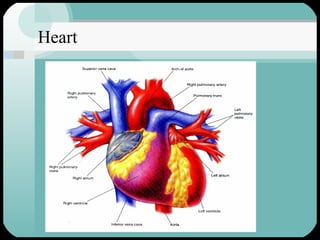
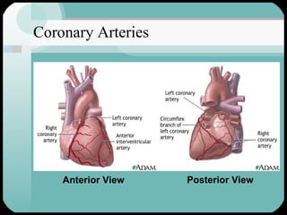
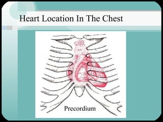
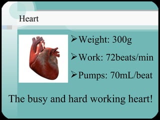
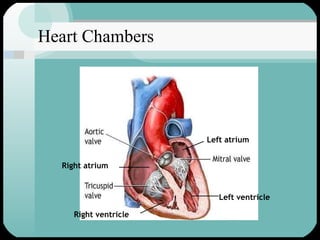
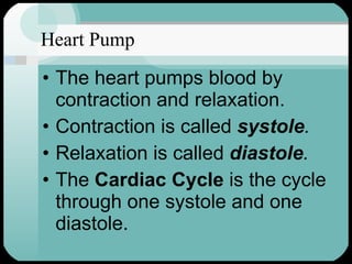
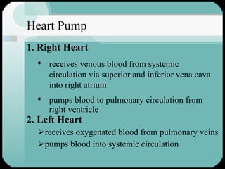
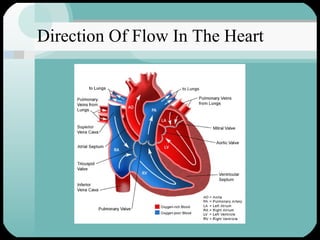
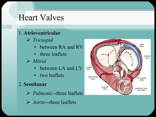
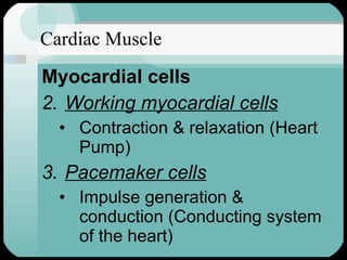
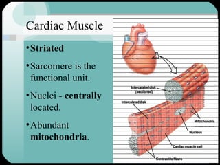
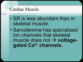
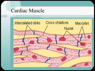
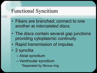
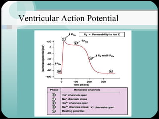
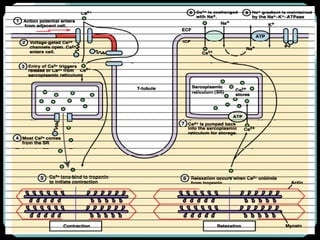
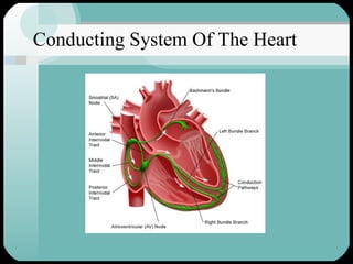
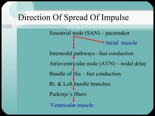
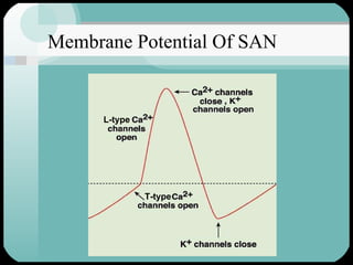
Cardiac Muscle Dr. Mohammed Abdul Hannan Hazari Assistant Professor, Department of Physiology, Deccan College of Medical Sciences, Hyderabad
Heart
Coronary Arteries Anterior View Posterior View
Heart Location In The Chest Precordium
Weight: 300g Work: 72beats/min Pumps: 70mL/beat The busy and hard working heart! Heart
Heart Chambers Left atrium Right atrium Left ventricle Right ventricle
Heart Pump The heart pumps blood by contraction and relaxation. Contraction is called  systole . Relaxation is called  diastole . The  Cardiac Cycle  is the cycle through one systole and one diastole.
Heart Pump 2. Left Heart receives oxygenated blood from pulmonary veins pumps blood into systemic circulation 1. Right Heart receives venous blood from systemic circulation via superior and inferior vena cava into right atrium pumps blood to pulmonary circulation from right ventricle
Direction Of Flow In The Heart
Heart Valves 1.  Atrioventricular Tricuspid between RA and RV three leaflets Mitral between LA and LV two leaflets 2.  Semilunar Pulmonic --three leaflets Aortic --three leaflets
Cardiac Muscle Myocardial cells Working myocardial cells Contraction & relaxation (Heart Pump) Pacemaker cells Impulse generation & conduction (Conducting system of the heart)
Cardiac Muscle Striated Sarcomere is the functional unit. Nuclei -  centrally  located. Abundant  mitochondria .
Cardiac Muscle SR is less abundant than in skeletal muscle. Sarcolemma has specialized ion channels that skeletal muscle does not     voltage-gated Ca 2+  channels.
The Sarcomere 1.6 – 1.7  μ
Cardiac Muscle
Functional Syncitium Fibers are branched; connect to one another at  intercalated discs . The discs contain several gap junctions providing cytoplasmic continuity. Rapid transmission of impulse. 2 syncitia Atrial syncitium Ventricular syncitium *Separated by fibrous ring
Ventricular Action Potential
 
Conducting System Of The Heart
Direction Of Spread Of Impulse Sinoatrial node (SAN) – pacemaker   Internodal pathways –fast conduction Atrioventricular node (AVN) – nodal delay Bundle of His – fast conduction Rt. & Left bundle branches Purkinje’s fibers Atrial  muscle Ventricular muscle
Membrane Potential Of SAN L-type T-type
Conduction Speeds In Cardiac Tissue 1 Ventricular muscle 4 Purkinje system 1 Bundle of His 0.05 AV node 1 Atrial pathways 0.05 SA node Conduction Rate (mts/s) Tissue
Questions?
Thank You

Cardiac muscle

  • 1.
    Cardiac Muscle Dr.Mohammed Abdul Hannan Hazari Assistant Professor, Department of Physiology, Deccan College of Medical Sciences, Hyderabad
  • 2.
  • 3.
    Coronary Arteries AnteriorView Posterior View
  • 4.
    Heart Location InThe Chest Precordium
  • 5.
    Weight: 300g Work:72beats/min Pumps: 70mL/beat The busy and hard working heart! Heart
  • 6.
    Heart Chambers Leftatrium Right atrium Left ventricle Right ventricle
  • 7.
    Heart Pump Theheart pumps blood by contraction and relaxation. Contraction is called systole . Relaxation is called diastole . The Cardiac Cycle is the cycle through one systole and one diastole.
  • 8.
    Heart Pump 2.Left Heart receives oxygenated blood from pulmonary veins pumps blood into systemic circulation 1. Right Heart receives venous blood from systemic circulation via superior and inferior vena cava into right atrium pumps blood to pulmonary circulation from right ventricle
  • 9.
    Direction Of FlowIn The Heart
  • 10.
    Heart Valves 1. Atrioventricular Tricuspid between RA and RV three leaflets Mitral between LA and LV two leaflets 2. Semilunar Pulmonic --three leaflets Aortic --three leaflets
  • 11.
    Cardiac Muscle Myocardialcells Working myocardial cells Contraction & relaxation (Heart Pump) Pacemaker cells Impulse generation & conduction (Conducting system of the heart)
  • 12.
    Cardiac Muscle StriatedSarcomere is the functional unit. Nuclei - centrally located. Abundant mitochondria .
  • 13.
    Cardiac Muscle SRis less abundant than in skeletal muscle. Sarcolemma has specialized ion channels that skeletal muscle does not  voltage-gated Ca 2+ channels.
  • 14.
  • 15.
  • 16.
    Functional Syncitium Fibersare branched; connect to one another at intercalated discs . The discs contain several gap junctions providing cytoplasmic continuity. Rapid transmission of impulse. 2 syncitia Atrial syncitium Ventricular syncitium *Separated by fibrous ring
  • 17.
  • 18.
  • 19.
  • 20.
    Direction Of SpreadOf Impulse Sinoatrial node (SAN) – pacemaker Internodal pathways –fast conduction Atrioventricular node (AVN) – nodal delay Bundle of His – fast conduction Rt. & Left bundle branches Purkinje’s fibers Atrial muscle Ventricular muscle
  • 21.
    Membrane Potential OfSAN L-type T-type
  • 22.
    Conduction Speeds InCardiac Tissue 1 Ventricular muscle 4 Purkinje system 1 Bundle of His 0.05 AV node 1 Atrial pathways 0.05 SA node Conduction Rate (mts/s) Tissue
  • 23.
  • 24.