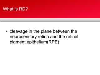
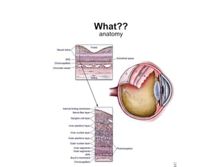
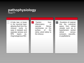
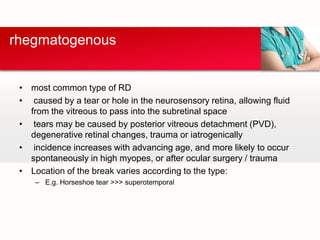
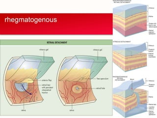
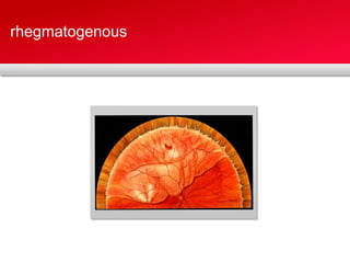
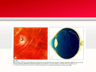
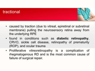
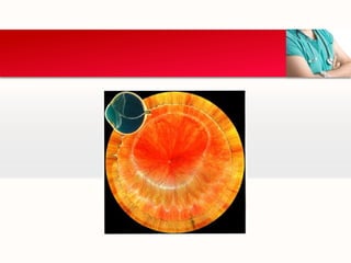
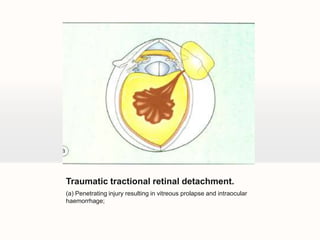
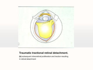
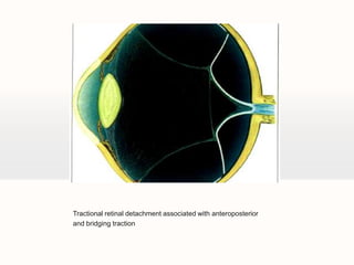
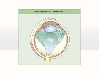
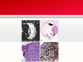
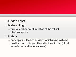
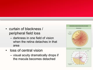
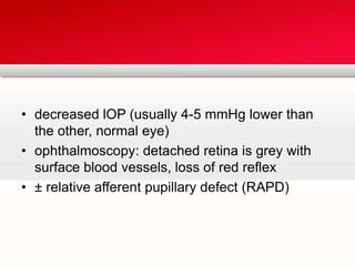
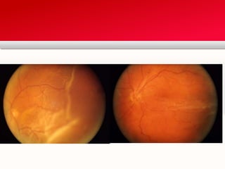
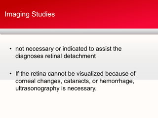
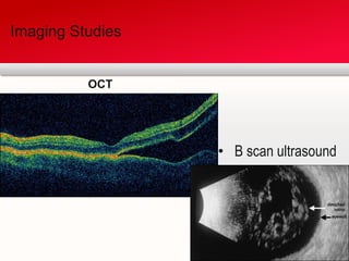
Retinal Detachment (RD)By: Hamza J. AlGhamdi
Objectives✓1What??types2rhegmatogenous34tractional5exudative6Clinical Features7cont…… with Salem
What is RD?cleavage in the plane between the neurosensory retina and the retinal pigment epithelium(RPE)
What??anatomy
pathophysiologyHow??tTraction from inflammatory or vascular fibrous membranes on the surface of the retina, which tether to the vitreoustExudation of material into the subretinal space from retinal vessels such as in hypertension, central retinal venous occlusion, vasculitis, or papilledemaA hole, tear, or break in the neuronal layer allowing fluid from the vitreous cavity to seep in between and separate sensory and RPE layers (ie, rhegmatogenous RD)321
Types
most common type of RDcaused by a tear or hole in the neurosensory retina, allowing fluid from the vitreous to pass into the subretinalspace tears may be caused by posterior vitreous detachment (PVD), degenerative retinal changes, trauma or iatrogenicallyincidence increases with advancing age, and more likely to occur spontaneously in high myopes, or after ocular surgery / traumaLocation of the break varies according to the type:E.g. Horseshoe tear >>> superotemporalrhegmatogenous
rhegmatogenous
rhegmatogenous
Your own footerYour LogoPathogenesis of ocular damage by blunt trauma;
Types
caused by traction (due to vitreal, epiretinal or subretinal membrane) pulling the neurosensory retina away from the underlying RPEfound in conditions such as diabetic retinopathy, CRVO, sickle cell disease, retinopathy of prematurity (ROP), and ocular traumaProliferative vitreoretinopathy is a complication of rhegmatogenous RD and is the most common cause of failure of surgical repair.tractional
Traumatic tractional retinal detachment.(a) Penetrating injury resulting in vitreous prolapse and intraocular haemorrhage;
Traumatic tractional retinal detachment.(b) subsequent vitreoretinal proliferation and traction resultingin retinal detachment
Tractional retinal detachment associated with anteroposteriorand bridging traction
Types
caused by damage to the RPE resulting in fluid accumulation in the subretinal spacemain causes are intraocular tumours, posterior uveitis, central serous retinopathyexudative
Clinical features
sudden onsetflashes of lightdue to mechanical stimulation of the retinal photoreceptorsfloatershazy spots in the line of vision which move with eye position, due to drops of blood in the vitreous (blood vessels tear as the retina tears)
curtain of blackness / peripheral field lossdarkness in one field of vision when the retina detaches in that arealoss of central visionvisual acuity dramatically drops if the macula becomes detached
decreased lOP (usually 4-5 mmHg lower than the other, normal eye)ophthalmoscopy: detached retina is grey with surface blood vessels, loss of red reflex± relative afferent pupillary defect (RAPD)
not necessary or indicated to assist the diagnoses retinal detachmentIf the retina cannot be visualized because of corneal changes, cataracts, or hemorrhage, ultrasonography is necessary.Imaging Studies
Imaging StudiesOCT B scan ultrasound
THANK YOU!

Retinal detachment

  • 1.
  • 2.
  • 3.
    What is RD?cleavagein the plane between the neurosensory retina and the retinal pigment epithelium(RPE)
  • 4.
  • 5.
    pathophysiologyHow??tTraction from inflammatoryor vascular fibrous membranes on the surface of the retina, which tether to the vitreoustExudation of material into the subretinal space from retinal vessels such as in hypertension, central retinal venous occlusion, vasculitis, or papilledemaA hole, tear, or break in the neuronal layer allowing fluid from the vitreous cavity to seep in between and separate sensory and RPE layers (ie, rhegmatogenous RD)321
  • 6.
  • 7.
    most common typeof RDcaused by a tear or hole in the neurosensory retina, allowing fluid from the vitreous to pass into the subretinalspace tears may be caused by posterior vitreous detachment (PVD), degenerative retinal changes, trauma or iatrogenicallyincidence increases with advancing age, and more likely to occur spontaneously in high myopes, or after ocular surgery / traumaLocation of the break varies according to the type:E.g. Horseshoe tear >>> superotemporalrhegmatogenous
  • 8.
  • 9.
  • 11.
    Your own footerYourLogoPathogenesis of ocular damage by blunt trauma;
  • 12.
  • 13.
    caused by traction(due to vitreal, epiretinal or subretinal membrane) pulling the neurosensory retina away from the underlying RPEfound in conditions such as diabetic retinopathy, CRVO, sickle cell disease, retinopathy of prematurity (ROP), and ocular traumaProliferative vitreoretinopathy is a complication of rhegmatogenous RD and is the most common cause of failure of surgical repair.tractional
  • 15.
    Traumatic tractional retinaldetachment.(a) Penetrating injury resulting in vitreous prolapse and intraocular haemorrhage;
  • 16.
    Traumatic tractional retinaldetachment.(b) subsequent vitreoretinal proliferation and traction resultingin retinal detachment
  • 17.
    Tractional retinal detachmentassociated with anteroposteriorand bridging traction
  • 19.
  • 20.
    caused by damageto the RPE resulting in fluid accumulation in the subretinal spacemain causes are intraocular tumours, posterior uveitis, central serous retinopathyexudative
  • 22.
  • 23.
    sudden onsetflashes oflightdue to mechanical stimulation of the retinal photoreceptorsfloatershazy spots in the line of vision which move with eye position, due to drops of blood in the vitreous (blood vessels tear as the retina tears)
  • 24.
    curtain of blackness/ peripheral field lossdarkness in one field of vision when the retina detaches in that arealoss of central visionvisual acuity dramatically drops if the macula becomes detached
  • 25.
    decreased lOP (usually4-5 mmHg lower than the other, normal eye)ophthalmoscopy: detached retina is grey with surface blood vessels, loss of red reflex± relative afferent pupillary defect (RAPD)
  • 27.
    not necessary orindicated to assist the diagnoses retinal detachmentIf the retina cannot be visualized because of corneal changes, cataracts, or hemorrhage, ultrasonography is necessary.Imaging Studies
  • 28.
    Imaging StudiesOCT Bscan ultrasound
  • 29.