SlideShare a Scribd company logo
1 of 8
X-Ray CRYSTALLOGRAPHY : Bragg’s law and applications
Diffraction refers to various phenomena which occur when a wave encounters an obstacle
or a slit.
It is defined as the bending of light around the corners of an obstacle or aperture into the
region of geometrical shadow of the obstacle.
What is the Significance of the Diffracted Pattern?
First we need to understand how the diffracted pattern is actually constructed and what
figure out what it tells us. Only then will we fully understand how and why it is a valuable
tool to understanding the molecular arrangements of atoms.
DESTRUCTIVE INTERFERENCE Destructive interference occurs when there is a collision of
photons in such a way that their intensities cancel out.
CONSTRUCTIVE INTERFERENCE: Constructive interference occurs when there is a collision
of protons in a way that their intensities combine.
After we observe the reflections of the photons on the detector, we change the angle at
which the x ray beam hits the crystal.
The crystal should be as perfect as possible! Why does the crystal have to be perfect?
Think of it this way. Atoms of the same type and that are in the same place within the
structure will result in an additive diffraction, which would definitely be easier to detect!
Therefore, the more perfect a crystal is, the more atoms that will be present in the same
places in the structure, which would be easier to detect.
Imperfect crystals usually result in fuzzy patterns that are hard to interpret.
We would then have to combine all the reflections together to find the three-dimensional
diffraction pattern. But why do we need to look at it at different angles????
What is Bragg’s Law and why is it Important? This equation explains why the faces of
crystals appear to reflect (diffract) X-ray beams at certain angles of incidence θ.
nλ = 2d sinθ
d : lattice interplanar spacing of the crystal θ : x-ray incidence angle (Bragg angle)
λ : wavelength of the characteristic x-rays
Deriving Bragg’s Law: Bragg's Law can easily be derived by considering the conditions
necessary to make the phases of the beams coincide when the incident angle equals and
reflecting angle.
The rays of the incident beam are always in phase and parallel up to the point at which
the top beam strikes the top layer at atom z.
The second beam continues to the next layer where it is scattered by atom B.
The second beam must travel the extra distance AB + BC if the two beams are to continue
traveling adjacent and parallel.
This extra distance must be an integral (n) multiple of the wavelength (λ) for the phases
of the two beams to be the same:
nλ = AB +BC (2).
Isomorphous Replacement (SIR, MIR)
To solve the phase of a protein crystal the complexity of the problem (i.e. the number of
variables) has to be reduced.
One way of doing that is to soak the crystal in a heavy atom solution and compare the
diffraction pattern of this derivative crystal to the diffraction pattern of the native crystal.
The heavy atoms in the structure contribute enormously to the diffraction which makes
their contribution easy to detect and the difference of the two diffraction patterns can be
taken
With direct methods the position of the heavy atoms (i.e. the heavy atom substructure) can
be determined.
Knowing these positions it is possible calculate the heavy atom contribution to each
structure factor.
This in turn means that for each structure factor one can determine the structure factors
of the heavy atoms (FH) and the amplitudes of the native protein (|FP|) and the heavy
atom derivative (|FPH|).
Drawing this relationship in the space of complex numbers is called the Harker
construction gives two possible solutions for the phase of each structure factor.
Solving the structure this way is called single isomorphous replacement (SIR).
By using at least one other heavy atom derivative this phase ambiguity can be solved
which is then called multiple isomporphous replacement (MIR).
The difference between a native structure factor (FP) and the derivative structure factor
(FPH) equals the heavy atom structure factor (FH) (left figure).
With direct methods we can solve the heavy atom structure factors using the amplitudes of
from the native and the derivative crystal.
Drawing a circle with the amplitudes of the native crystal as radius around one end of the
heavy atom structure factor and another circle with the amplitude of the derivative crystal
as radius we get two possible phases for the native crystal structure factor.
Considerations for Selecting GC/MS or LC/MS
Gas chromatography and mass spectrometry (GC/MS) are an effective combination for the
analysis of volatile chemicals.
Gas chromatography uses a carrier gas to move analytes through acoated, fused silica
capillary.
Separation occurs based on differential partition between the gas phase and the coating
inside the capillary.
GC/MS requires the analyte to be vaporized in order for migration through the capillary to
occur.
Analytes, therefore, must be volatile or amenable to chemical derivatization to render them
volatile.
Electron ionization (EI) is the most commonly used GC/MS ionization technique.
It is very robust and reproducible. It does not suffer from ion Suppression.
Electron ionization causes characteristic mass spectral fragmentation patterns.
Thus, EI spectra from unknowns can be searched against libraries of spectra to achieve
identification.
Liquid chromatography can separate metabolites that are not volatile and have not been
derivatized. As a result, LC/MS can analyze a much wider range of chemical species than
GC/MS. Electrospray ionization (ESI) is most commonly used in LC/MS.
Unlike EI for GC/MS, ion suppression can occur so co-eluting compounds may be underestimated or not
detected at all.
Therefore, for complex samples, greater separation is necessary for the reliable LC/MS
results.
The Importance of Animals in Biomedical Research
Throughout history, scientists have been solving medical problems, developing new
techniques and treatments, and curing diseases – all by using animals in biomedical
research
Why Use Animal Models?
 Very limited number of studies can be done on humans
 Allows for controlled experiments
 Environmental variables can be controlled
 Dosage/route of exposures can be controlled/varied
 Experiments can be replicated
 Physiology/anatomy can be matched to humans
Why are animals used in Research?
 organs and body systems similar to humans and other animals
 susceptible to the same diseases that affect humans
 short life span allows animals to be studied throughout their entire life
 environment easily controllable to keep experimental variables to a minimum
What animals are used in Research?
 Laboratory mice are used in research more often than any other animal species;
 These mice, plus other rodents such as rats and hamsters, make up more than 90% of
the total number of animals used; and
 Other animal species, including dogs, cats, rabbits, farm animals, fish, frogs, birds,
nonhuman primates, and many others, make up the remaining 10% of animals used
in research.
“Natural Models”
Good - Most related to the natural disease, most likely to portray all aspects of the disease
Bad - Restrictive in terms of time and types of animals used, also in terms of animals
developing illnesses (in aging studies).
1)Expensive 2) Long term studies 3) Restrictive at an experimental level
“Semi-Natural Models”:
Good – Usually good models to study disease because they are based on phenotypical
presentations of the disease.
Bad – Often lack appropriate controls because they are bred for a phenotype and the
genetics are unknown.
Lesion Models
Good - to study the function of a structure and how the loss of that structure affects other
cell populations
Bad - because most diseases are not specific to a structure or a cell population – High
mortality & variability
Chemical Models
Good - to study selective depletion of specific cell populations and examine the role of
transmitters / inducers in disease, more specific than lesions and sometimes leads to full
phenotype expression
Bad - because it is artificially induced and is incomplete with regards to phenotype
(pathology/not pathology), it does not always translate to the real population
Transgenic Models
Good - excellent to study the role of genes and genetic mutations in disease
Bad - because diseases like all biological events are a mix of environment and genetics so a
lot of times the genetic mutations do not recapitulate the disease.
Also, while some of the genetics are highly conserved a mouse/fish/fly/worm do not
respond the same way than humans do nor do animals of the same species!!!
(BACKGROUNDS!!!)
Alternative to Animals in Research
Cell culture and computer-generated models Cell Culture
 Preliminary screen to check the toxicity of substances
 Can answer fundamental questions about biology
 Cannot provide information about potential impacts on entire living organism
Alternatives to Animal Use in Research: A suitable replacement for animals should:
 Reliably predict biological phenomena
 Be at least as good (or better!) and a consistent model for risk assessment in humans
as well as in animals
 Be extensively validated, tested and accepted by regulatory agencies as suitable
substitutes
Model organisms are found among Prokaryotes, Protists , Fungi, Plants and Animals.
 Among the prokaryotes : Escherichia coli is the classical model of molecular biology,
the favorite organism to study transcription, translation, recombination, DNA repair
and the regulation of gene activity.
 The human parasite Plasmodium, the causative agent of malaria, and the genetic
model organisms such as Dictyostelium and Volvox are the most prominent in the
protists
 In the fungi group several models of great importance for genetics like
Saccharomyces cerevisiae Schizosaccharomyces pombe and Neurospora crassa.
 Yeasts, which are unicellular ascomycotan fungi, have proved to be extremely useful
organisms for the study of basic eukaryotic phenomena (genetic recombination,
macromolecular synthesis, metabolic regulation, cell cycle and general cell biology)
 The most prominent animal model organisms are fruit flies (Drosophila
melanogaster) , mouse (Mus musculus), nematode (Caenorhabditis elegans) .
 The regenerative capacity of stem cells offers unprecedented opportunities for
developing medical therapies for debilitating diseases and represents a source of
new, healthy tissue to treat or replace diseased or injured human organs.
 They are an excellent model that will help us to understand how they transform into
the myriad of specialized cells that make us what we are.
Flow Cytometry
Applications: The uses of flow in research has boomed since the mid-1980s, and is now the
gold standard for a variety of applications.
Immunophenotyping DNA cell cycle/tumor ploidy Membrane potential
Ion flux Cell viability Intracellular protein staining
pH changes Cell tracking and proliferation Sorting
Redox state Chromatin structure Total protein
Lipids Surface charge Membrane fusion/runover
Enzyme activity Oxidative metabolism Sulfhydryl groups/glutathione
DNA synthesis DNA degradation Gene expression
Phagocytosis Microparticle analysis
What Is Flow Cytometry? Flow ~ motion, Cyto ~ cell Metry ~ measure
Measuring both intrinsic and extrinsic properties of cells while in a moving fluid stream
What Happens in a Flow Cytometer?
• Cells in suspension flow single file
• through a focused laser where they scatter light and emit fluorescence that is
filtered,
• measured, then converted to digitized values that are stored in a file
• which can then be analyzed and interpreted within specialized software.
Fluidics Interrogation Electronics Interpretation
• The Fluidics System “Cells in suspension flow single file”
• Cells must flow one-by-one into the Cytometer to do single cell analysis
• Accomplished through a pressurized laminar flow system.
• The sample is injected into a sheath fluid as it passes through a small orifice (50µm-
300µm)
Interrogation : Light source needs to be focused on the same point where cells are focused.
Light source 99%=Lasers
Lasers : Light amplification by stimulated emission of radiation
• Lasers provide a single wavelength of light (monochromatic)
• They can provide milliwatts to watts of power
• Low divergence
• Provide coherent light
• Gas, dye, or solid state
Coherent: all emitting photons have same wavelength, phase and direction as stimulation
photons
END
If you want Word/PDF???
Soft copy of Word/PDF = 0.013$ (US dollar) (5Rs Indian rupee)
Contact :(I will share ppt via Email/WhatsApp/Telegram
Email: gnccmysore@gmail.com
Telegram:+919738137533(only for Chat)
*Once you done payment, Please intimate me.

More Related Content

What's hot

X ray crystallography and X ray Diffraction
X ray crystallography and X ray DiffractionX ray crystallography and X ray Diffraction
X ray crystallography and X ray DiffractionFaisal Hussain
 
Methods of study of protein by kk sahu
Methods of study of protein by kk sahuMethods of study of protein by kk sahu
Methods of study of protein by kk sahuKAUSHAL SAHU
 
X ray crystallography
X ray crystallography X ray crystallography
X ray crystallography ArchanaKR11
 
X ray crystallography
X ray crystallographyX ray crystallography
X ray crystallographymirzausman555
 
X-ray Crystallography
X-ray CrystallographyX-ray Crystallography
X-ray CrystallographyAkansh Goel
 
X - RAY CRYSTALLOGRAPHY TECHNIQUE
X - RAY CRYSTALLOGRAPHY TECHNIQUEX - RAY CRYSTALLOGRAPHY TECHNIQUE
X - RAY CRYSTALLOGRAPHY TECHNIQUEAYESHA KABEER
 
X ray Crystallography Manik
X ray Crystallography ManikX ray Crystallography Manik
X ray Crystallography ManikImran Nur Manik
 
Radio active labeling
Radio active labelingRadio active labeling
Radio active labelingAfra Fathima
 
Protein structure determination and our software tools
Protein structure determinationand our software toolsProtein structure determinationand our software tools
Protein structure determination and our software toolsMark Berjanskii
 
X ray powder diffraction
X ray powder diffractionX ray powder diffraction
X ray powder diffractionchiranjibi68
 
Crystallography and XRD.pptx
Crystallography and XRD.pptxCrystallography and XRD.pptx
Crystallography and XRD.pptxssusere0ab90
 

What's hot (20)

Autoradiography
AutoradiographyAutoradiography
Autoradiography
 
X ray crystallography
X ray crystallographyX ray crystallography
X ray crystallography
 
X ray crystallography and X ray Diffraction
X ray crystallography and X ray DiffractionX ray crystallography and X ray Diffraction
X ray crystallography and X ray Diffraction
 
Methods of study of protein by kk sahu
Methods of study of protein by kk sahuMethods of study of protein by kk sahu
Methods of study of protein by kk sahu
 
Autoradiography
AutoradiographyAutoradiography
Autoradiography
 
X ray crystallography
X ray crystallographyX ray crystallography
X ray crystallography
 
X ray crystallography
X ray crystallography X ray crystallography
X ray crystallography
 
X ray crystallography
X ray crystallographyX ray crystallography
X ray crystallography
 
X-ray Crystallography
X-ray CrystallographyX-ray Crystallography
X-ray Crystallography
 
X ray diffraction
X ray diffractionX ray diffraction
X ray diffraction
 
X - RAY CRYSTALLOGRAPHY TECHNIQUE
X - RAY CRYSTALLOGRAPHY TECHNIQUEX - RAY CRYSTALLOGRAPHY TECHNIQUE
X - RAY CRYSTALLOGRAPHY TECHNIQUE
 
X ray Crystallography Manik
X ray Crystallography ManikX ray Crystallography Manik
X ray Crystallography Manik
 
"X ray crystallography" - Tathagata Pradhan , Department of Pharmaceutical Ch...
"X ray crystallography" - Tathagata Pradhan , Department of Pharmaceutical Ch..."X ray crystallography" - Tathagata Pradhan , Department of Pharmaceutical Ch...
"X ray crystallography" - Tathagata Pradhan , Department of Pharmaceutical Ch...
 
Radio active labeling
Radio active labelingRadio active labeling
Radio active labeling
 
Protein structure determination and our software tools
Protein structure determinationand our software toolsProtein structure determinationand our software tools
Protein structure determination and our software tools
 
X ray crystallography
X ray crystallographyX ray crystallography
X ray crystallography
 
X ray powder diffraction
X ray powder diffractionX ray powder diffraction
X ray powder diffraction
 
X ray crystallography slideshare
X ray crystallography slideshareX ray crystallography slideshare
X ray crystallography slideshare
 
Crystallography and XRD.pptx
Crystallography and XRD.pptxCrystallography and XRD.pptx
Crystallography and XRD.pptx
 
X ray
X rayX ray
X ray
 

Similar to X ray crystallography

BT631-11-x-ray_crystallography_introduction
BT631-11-x-ray_crystallography_introductionBT631-11-x-ray_crystallography_introduction
BT631-11-x-ray_crystallography_introductionRajesh G
 
structural biology-Protein structure function relationship
structural biology-Protein structure function relationshipstructural biology-Protein structure function relationship
structural biology-Protein structure function relationshipMSCW Mysore
 
Infrared-Spectroscopy-ppt.pptx
Infrared-Spectroscopy-ppt.pptxInfrared-Spectroscopy-ppt.pptx
Infrared-Spectroscopy-ppt.pptxNagen87
 
Drug-Polymer Compatibility Studies By XRD
Drug-Polymer Compatibility Studies By XRDDrug-Polymer Compatibility Studies By XRD
Drug-Polymer Compatibility Studies By XRDVenkatesh Goli
 
Pigments and Colors:Extraction,Characterization
Pigments and Colors:Extraction,CharacterizationPigments and Colors:Extraction,Characterization
Pigments and Colors:Extraction,CharacterizationPRUTHVIRAJ K
 
Biological effects of radiation
Biological effects of radiationBiological effects of radiation
Biological effects of radiationDR.URVASHI NIKTE
 
organic-spectroscopic-analysis
organic-spectroscopic-analysisorganic-spectroscopic-analysis
organic-spectroscopic-analysisAlexandra Elena
 
Ir absorption spectroscopy
Ir absorption spectroscopyIr absorption spectroscopy
Ir absorption spectroscopyYogesh Mhadgut
 
Dipti_X ray crystallography (1).pptx
Dipti_X ray crystallography (1).pptxDipti_X ray crystallography (1).pptx
Dipti_X ray crystallography (1).pptxSourabYadav1
 
Int.+Spectroscopy+&+UV.ppt
Int.+Spectroscopy+&+UV.pptInt.+Spectroscopy+&+UV.ppt
Int.+Spectroscopy+&+UV.pptTahsinAhmed32
 
Quantification of Fluorophore Concentrations in Turbid Media by Spatial Frequ...
Quantification of Fluorophore Concentrations in Turbid Media by Spatial Frequ...Quantification of Fluorophore Concentrations in Turbid Media by Spatial Frequ...
Quantification of Fluorophore Concentrations in Turbid Media by Spatial Frequ...sela shefy
 
B.Sc Biotech II BAT Unit 1 Spectroscopy
B.Sc Biotech II BAT Unit 1 SpectroscopyB.Sc Biotech II BAT Unit 1 Spectroscopy
B.Sc Biotech II BAT Unit 1 SpectroscopyRai University
 
Radiobiology- Basic Guide
Radiobiology- Basic Guide Radiobiology- Basic Guide
Radiobiology- Basic Guide Aaditya Sinha
 
Microwave and infrared spectroscopy of polyatomic molecules
Microwave and infrared spectroscopy of polyatomic moleculesMicrowave and infrared spectroscopy of polyatomic molecules
Microwave and infrared spectroscopy of polyatomic moleculesAreebaWarraich1
 
Davey l1 macromolec-struc-anlys(1) lec 1
Davey l1 macromolec-struc-anlys(1) lec 1Davey l1 macromolec-struc-anlys(1) lec 1
Davey l1 macromolec-struc-anlys(1) lec 1RANJANI001
 

Similar to X ray crystallography (20)

BT631-11-x-ray_crystallography_introduction
BT631-11-x-ray_crystallography_introductionBT631-11-x-ray_crystallography_introduction
BT631-11-x-ray_crystallography_introduction
 
structural biology-Protein structure function relationship
structural biology-Protein structure function relationshipstructural biology-Protein structure function relationship
structural biology-Protein structure function relationship
 
Infrared-Spectroscopy-ppt.pptx
Infrared-Spectroscopy-ppt.pptxInfrared-Spectroscopy-ppt.pptx
Infrared-Spectroscopy-ppt.pptx
 
Drug-Polymer Compatibility Studies By XRD
Drug-Polymer Compatibility Studies By XRDDrug-Polymer Compatibility Studies By XRD
Drug-Polymer Compatibility Studies By XRD
 
Pigments and Colors:Extraction,Characterization
Pigments and Colors:Extraction,CharacterizationPigments and Colors:Extraction,Characterization
Pigments and Colors:Extraction,Characterization
 
Biological effects of radiation
Biological effects of radiationBiological effects of radiation
Biological effects of radiation
 
organic-spectroscopic-analysis
organic-spectroscopic-analysisorganic-spectroscopic-analysis
organic-spectroscopic-analysis
 
Ir absorption spectroscopy
Ir absorption spectroscopyIr absorption spectroscopy
Ir absorption spectroscopy
 
Ir spectroscopy
Ir spectroscopyIr spectroscopy
Ir spectroscopy
 
principles of xrd
principles of xrdprinciples of xrd
principles of xrd
 
Dipti_X ray crystallography (1).pptx
Dipti_X ray crystallography (1).pptxDipti_X ray crystallography (1).pptx
Dipti_X ray crystallography (1).pptx
 
Int.+Spectroscopy+&+UV.ppt
Int.+Spectroscopy+&+UV.pptInt.+Spectroscopy+&+UV.ppt
Int.+Spectroscopy+&+UV.ppt
 
Quantification of Fluorophore Concentrations in Turbid Media by Spatial Frequ...
Quantification of Fluorophore Concentrations in Turbid Media by Spatial Frequ...Quantification of Fluorophore Concentrations in Turbid Media by Spatial Frequ...
Quantification of Fluorophore Concentrations in Turbid Media by Spatial Frequ...
 
Seminar of mary chely quinones
Seminar of mary chely quinonesSeminar of mary chely quinones
Seminar of mary chely quinones
 
B.Sc Biotech II BAT Unit 1 Spectroscopy
B.Sc Biotech II BAT Unit 1 SpectroscopyB.Sc Biotech II BAT Unit 1 Spectroscopy
B.Sc Biotech II BAT Unit 1 Spectroscopy
 
Radiobiology- Basic Guide
Radiobiology- Basic Guide Radiobiology- Basic Guide
Radiobiology- Basic Guide
 
X – Ray Diffraction
X – Ray DiffractionX – Ray Diffraction
X – Ray Diffraction
 
Microwave and infrared spectroscopy of polyatomic molecules
Microwave and infrared spectroscopy of polyatomic moleculesMicrowave and infrared spectroscopy of polyatomic molecules
Microwave and infrared spectroscopy of polyatomic molecules
 
Radiobiology
RadiobiologyRadiobiology
Radiobiology
 
Davey l1 macromolec-struc-anlys(1) lec 1
Davey l1 macromolec-struc-anlys(1) lec 1Davey l1 macromolec-struc-anlys(1) lec 1
Davey l1 macromolec-struc-anlys(1) lec 1
 

More from Bangaluru

Good Pipetting Technique.pptx
Good Pipetting Technique.pptxGood Pipetting Technique.pptx
Good Pipetting Technique.pptxBangaluru
 
KF autotitrator.pptx
KF autotitrator.pptxKF autotitrator.pptx
KF autotitrator.pptxBangaluru
 
Pheromones.pptx
Pheromones.pptxPheromones.pptx
Pheromones.pptxBangaluru
 
Nitric oxide ppt.pptx
Nitric oxide  ppt.pptxNitric oxide  ppt.pptx
Nitric oxide ppt.pptxBangaluru
 
Mitochondrial biogenesis.pptx
Mitochondrial biogenesis.pptxMitochondrial biogenesis.pptx
Mitochondrial biogenesis.pptxBangaluru
 
Melatonin.pptx
Melatonin.pptxMelatonin.pptx
Melatonin.pptxBangaluru
 
Ion Channels.pptx
Ion Channels.pptxIon Channels.pptx
Ion Channels.pptxBangaluru
 
Glucose Transporters.pptx
Glucose Transporters.pptxGlucose Transporters.pptx
Glucose Transporters.pptxBangaluru
 
Data Base in Bioinformatics.ppt
Data Base in Bioinformatics.pptData Base in Bioinformatics.ppt
Data Base in Bioinformatics.pptBangaluru
 
Chemical Bonds 2.ppt
Chemical Bonds 2.pptChemical Bonds 2.ppt
Chemical Bonds 2.pptBangaluru
 
Chemical Bonds 1.ppt
Chemical Bonds 1.pptChemical Bonds 1.ppt
Chemical Bonds 1.pptBangaluru
 
Eicosanoids.pptx
Eicosanoids.pptxEicosanoids.pptx
Eicosanoids.pptxBangaluru
 
Chemical safety.pptx
Chemical safety.pptxChemical safety.pptx
Chemical safety.pptxBangaluru
 
Biogas production.pptx
Biogas production.pptxBiogas production.pptx
Biogas production.pptxBangaluru
 
Mass Spectrometry.ppt
Mass Spectrometry.pptMass Spectrometry.ppt
Mass Spectrometry.pptBangaluru
 
Risk assessment for computer system validation
Risk assessment for computer system validationRisk assessment for computer system validation
Risk assessment for computer system validationBangaluru
 
Recovery and purification of intracellular and extra cellular products
Recovery and purification of intracellular and extra cellular productsRecovery and purification of intracellular and extra cellular products
Recovery and purification of intracellular and extra cellular productsBangaluru
 
Good documentation practices
Good documentation practicesGood documentation practices
Good documentation practicesBangaluru
 
Zymography_Pas staining
Zymography_Pas stainingZymography_Pas staining
Zymography_Pas stainingBangaluru
 

More from Bangaluru (20)

Good Pipetting Technique.pptx
Good Pipetting Technique.pptxGood Pipetting Technique.pptx
Good Pipetting Technique.pptx
 
KF autotitrator.pptx
KF autotitrator.pptxKF autotitrator.pptx
KF autotitrator.pptx
 
Pheromones.pptx
Pheromones.pptxPheromones.pptx
Pheromones.pptx
 
Nitric oxide ppt.pptx
Nitric oxide  ppt.pptxNitric oxide  ppt.pptx
Nitric oxide ppt.pptx
 
Mitochondrial biogenesis.pptx
Mitochondrial biogenesis.pptxMitochondrial biogenesis.pptx
Mitochondrial biogenesis.pptx
 
Melatonin.pptx
Melatonin.pptxMelatonin.pptx
Melatonin.pptx
 
Ion Channels.pptx
Ion Channels.pptxIon Channels.pptx
Ion Channels.pptx
 
Glucose Transporters.pptx
Glucose Transporters.pptxGlucose Transporters.pptx
Glucose Transporters.pptx
 
Data Base in Bioinformatics.ppt
Data Base in Bioinformatics.pptData Base in Bioinformatics.ppt
Data Base in Bioinformatics.ppt
 
Chemical Bonds 2.ppt
Chemical Bonds 2.pptChemical Bonds 2.ppt
Chemical Bonds 2.ppt
 
Chemical Bonds 1.ppt
Chemical Bonds 1.pptChemical Bonds 1.ppt
Chemical Bonds 1.ppt
 
Eicosanoids.pptx
Eicosanoids.pptxEicosanoids.pptx
Eicosanoids.pptx
 
Chemical safety.pptx
Chemical safety.pptxChemical safety.pptx
Chemical safety.pptx
 
Biogas production.pptx
Biogas production.pptxBiogas production.pptx
Biogas production.pptx
 
Mass Spectrometry.ppt
Mass Spectrometry.pptMass Spectrometry.ppt
Mass Spectrometry.ppt
 
Risk assessment for computer system validation
Risk assessment for computer system validationRisk assessment for computer system validation
Risk assessment for computer system validation
 
Recovery and purification of intracellular and extra cellular products
Recovery and purification of intracellular and extra cellular productsRecovery and purification of intracellular and extra cellular products
Recovery and purification of intracellular and extra cellular products
 
Iron
IronIron
Iron
 
Good documentation practices
Good documentation practicesGood documentation practices
Good documentation practices
 
Zymography_Pas staining
Zymography_Pas stainingZymography_Pas staining
Zymography_Pas staining
 

Recently uploaded

Accessible design: Minimum effort, maximum impact
Accessible design: Minimum effort, maximum impactAccessible design: Minimum effort, maximum impact
Accessible design: Minimum effort, maximum impactdawncurless
 
SOCIAL AND HISTORICAL CONTEXT - LFTVD.pptx
SOCIAL AND HISTORICAL CONTEXT - LFTVD.pptxSOCIAL AND HISTORICAL CONTEXT - LFTVD.pptx
SOCIAL AND HISTORICAL CONTEXT - LFTVD.pptxiammrhaywood
 
Arihant handbook biology for class 11 .pdf
Arihant handbook biology for class 11 .pdfArihant handbook biology for class 11 .pdf
Arihant handbook biology for class 11 .pdfchloefrazer622
 
9548086042 for call girls in Indira Nagar with room service
9548086042  for call girls in Indira Nagar  with room service9548086042  for call girls in Indira Nagar  with room service
9548086042 for call girls in Indira Nagar with room servicediscovermytutordmt
 
social pharmacy d-pharm 1st year by Pragati K. Mahajan
social pharmacy d-pharm 1st year by Pragati K. Mahajansocial pharmacy d-pharm 1st year by Pragati K. Mahajan
social pharmacy d-pharm 1st year by Pragati K. Mahajanpragatimahajan3
 
BASLIQ CURRENT LOOKBOOK LOOKBOOK(1) (1).pdf
BASLIQ CURRENT LOOKBOOK  LOOKBOOK(1) (1).pdfBASLIQ CURRENT LOOKBOOK  LOOKBOOK(1) (1).pdf
BASLIQ CURRENT LOOKBOOK LOOKBOOK(1) (1).pdfSoniaTolstoy
 
Measures of Central Tendency: Mean, Median and Mode
Measures of Central Tendency: Mean, Median and ModeMeasures of Central Tendency: Mean, Median and Mode
Measures of Central Tendency: Mean, Median and ModeThiyagu K
 
Q4-W6-Restating Informational Text Grade 3
Q4-W6-Restating Informational Text Grade 3Q4-W6-Restating Informational Text Grade 3
Q4-W6-Restating Informational Text Grade 3JemimahLaneBuaron
 
Organic Name Reactions for the students and aspirants of Chemistry12th.pptx
Organic Name Reactions  for the students and aspirants of Chemistry12th.pptxOrganic Name Reactions  for the students and aspirants of Chemistry12th.pptx
Organic Name Reactions for the students and aspirants of Chemistry12th.pptxVS Mahajan Coaching Centre
 
POINT- BIOCHEMISTRY SEM 2 ENZYMES UNIT 5.pptx
POINT- BIOCHEMISTRY SEM 2 ENZYMES UNIT 5.pptxPOINT- BIOCHEMISTRY SEM 2 ENZYMES UNIT 5.pptx
POINT- BIOCHEMISTRY SEM 2 ENZYMES UNIT 5.pptxSayali Powar
 
CARE OF CHILD IN INCUBATOR..........pptx
CARE OF CHILD IN INCUBATOR..........pptxCARE OF CHILD IN INCUBATOR..........pptx
CARE OF CHILD IN INCUBATOR..........pptxGaneshChakor2
 
Separation of Lanthanides/ Lanthanides and Actinides
Separation of Lanthanides/ Lanthanides and ActinidesSeparation of Lanthanides/ Lanthanides and Actinides
Separation of Lanthanides/ Lanthanides and ActinidesFatimaKhan178732
 
1029 - Danh muc Sach Giao Khoa 10 . pdf
1029 -  Danh muc Sach Giao Khoa 10 . pdf1029 -  Danh muc Sach Giao Khoa 10 . pdf
1029 - Danh muc Sach Giao Khoa 10 . pdfQucHHunhnh
 
Web & Social Media Analytics Previous Year Question Paper.pdf
Web & Social Media Analytics Previous Year Question Paper.pdfWeb & Social Media Analytics Previous Year Question Paper.pdf
Web & Social Media Analytics Previous Year Question Paper.pdfJayanti Pande
 
Beyond the EU: DORA and NIS 2 Directive's Global Impact
Beyond the EU: DORA and NIS 2 Directive's Global ImpactBeyond the EU: DORA and NIS 2 Directive's Global Impact
Beyond the EU: DORA and NIS 2 Directive's Global ImpactPECB
 
Paris 2024 Olympic Geographies - an activity
Paris 2024 Olympic Geographies - an activityParis 2024 Olympic Geographies - an activity
Paris 2024 Olympic Geographies - an activityGeoBlogs
 
mini mental status format.docx
mini    mental       status     format.docxmini    mental       status     format.docx
mini mental status format.docxPoojaSen20
 

Recently uploaded (20)

Accessible design: Minimum effort, maximum impact
Accessible design: Minimum effort, maximum impactAccessible design: Minimum effort, maximum impact
Accessible design: Minimum effort, maximum impact
 
SOCIAL AND HISTORICAL CONTEXT - LFTVD.pptx
SOCIAL AND HISTORICAL CONTEXT - LFTVD.pptxSOCIAL AND HISTORICAL CONTEXT - LFTVD.pptx
SOCIAL AND HISTORICAL CONTEXT - LFTVD.pptx
 
Arihant handbook biology for class 11 .pdf
Arihant handbook biology for class 11 .pdfArihant handbook biology for class 11 .pdf
Arihant handbook biology for class 11 .pdf
 
9548086042 for call girls in Indira Nagar with room service
9548086042  for call girls in Indira Nagar  with room service9548086042  for call girls in Indira Nagar  with room service
9548086042 for call girls in Indira Nagar with room service
 
social pharmacy d-pharm 1st year by Pragati K. Mahajan
social pharmacy d-pharm 1st year by Pragati K. Mahajansocial pharmacy d-pharm 1st year by Pragati K. Mahajan
social pharmacy d-pharm 1st year by Pragati K. Mahajan
 
BASLIQ CURRENT LOOKBOOK LOOKBOOK(1) (1).pdf
BASLIQ CURRENT LOOKBOOK  LOOKBOOK(1) (1).pdfBASLIQ CURRENT LOOKBOOK  LOOKBOOK(1) (1).pdf
BASLIQ CURRENT LOOKBOOK LOOKBOOK(1) (1).pdf
 
Measures of Central Tendency: Mean, Median and Mode
Measures of Central Tendency: Mean, Median and ModeMeasures of Central Tendency: Mean, Median and Mode
Measures of Central Tendency: Mean, Median and Mode
 
Q4-W6-Restating Informational Text Grade 3
Q4-W6-Restating Informational Text Grade 3Q4-W6-Restating Informational Text Grade 3
Q4-W6-Restating Informational Text Grade 3
 
Organic Name Reactions for the students and aspirants of Chemistry12th.pptx
Organic Name Reactions  for the students and aspirants of Chemistry12th.pptxOrganic Name Reactions  for the students and aspirants of Chemistry12th.pptx
Organic Name Reactions for the students and aspirants of Chemistry12th.pptx
 
POINT- BIOCHEMISTRY SEM 2 ENZYMES UNIT 5.pptx
POINT- BIOCHEMISTRY SEM 2 ENZYMES UNIT 5.pptxPOINT- BIOCHEMISTRY SEM 2 ENZYMES UNIT 5.pptx
POINT- BIOCHEMISTRY SEM 2 ENZYMES UNIT 5.pptx
 
CARE OF CHILD IN INCUBATOR..........pptx
CARE OF CHILD IN INCUBATOR..........pptxCARE OF CHILD IN INCUBATOR..........pptx
CARE OF CHILD IN INCUBATOR..........pptx
 
Separation of Lanthanides/ Lanthanides and Actinides
Separation of Lanthanides/ Lanthanides and ActinidesSeparation of Lanthanides/ Lanthanides and Actinides
Separation of Lanthanides/ Lanthanides and Actinides
 
1029 - Danh muc Sach Giao Khoa 10 . pdf
1029 -  Danh muc Sach Giao Khoa 10 . pdf1029 -  Danh muc Sach Giao Khoa 10 . pdf
1029 - Danh muc Sach Giao Khoa 10 . pdf
 
Web & Social Media Analytics Previous Year Question Paper.pdf
Web & Social Media Analytics Previous Year Question Paper.pdfWeb & Social Media Analytics Previous Year Question Paper.pdf
Web & Social Media Analytics Previous Year Question Paper.pdf
 
Código Creativo y Arte de Software | Unidad 1
Código Creativo y Arte de Software | Unidad 1Código Creativo y Arte de Software | Unidad 1
Código Creativo y Arte de Software | Unidad 1
 
Beyond the EU: DORA and NIS 2 Directive's Global Impact
Beyond the EU: DORA and NIS 2 Directive's Global ImpactBeyond the EU: DORA and NIS 2 Directive's Global Impact
Beyond the EU: DORA and NIS 2 Directive's Global Impact
 
Paris 2024 Olympic Geographies - an activity
Paris 2024 Olympic Geographies - an activityParis 2024 Olympic Geographies - an activity
Paris 2024 Olympic Geographies - an activity
 
mini mental status format.docx
mini    mental       status     format.docxmini    mental       status     format.docx
mini mental status format.docx
 
Mattingly "AI & Prompt Design: The Basics of Prompt Design"
Mattingly "AI & Prompt Design: The Basics of Prompt Design"Mattingly "AI & Prompt Design: The Basics of Prompt Design"
Mattingly "AI & Prompt Design: The Basics of Prompt Design"
 
Mattingly "AI & Prompt Design: Structured Data, Assistants, & RAG"
Mattingly "AI & Prompt Design: Structured Data, Assistants, & RAG"Mattingly "AI & Prompt Design: Structured Data, Assistants, & RAG"
Mattingly "AI & Prompt Design: Structured Data, Assistants, & RAG"
 

X ray crystallography

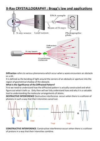
  • 1. X-Ray CRYSTALLOGRAPHY : Bragg’s law and applications Diffraction refers to various phenomena which occur when a wave encounters an obstacle or a slit. It is defined as the bending of light around the corners of an obstacle or aperture into the region of geometrical shadow of the obstacle. What is the Significance of the Diffracted Pattern? First we need to understand how the diffracted pattern is actually constructed and what figure out what it tells us. Only then will we fully understand how and why it is a valuable tool to understanding the molecular arrangements of atoms. DESTRUCTIVE INTERFERENCE Destructive interference occurs when there is a collision of photons in such a way that their intensities cancel out. CONSTRUCTIVE INTERFERENCE: Constructive interference occurs when there is a collision of protons in a way that their intensities combine.
  • 2. After we observe the reflections of the photons on the detector, we change the angle at which the x ray beam hits the crystal. The crystal should be as perfect as possible! Why does the crystal have to be perfect? Think of it this way. Atoms of the same type and that are in the same place within the structure will result in an additive diffraction, which would definitely be easier to detect! Therefore, the more perfect a crystal is, the more atoms that will be present in the same places in the structure, which would be easier to detect. Imperfect crystals usually result in fuzzy patterns that are hard to interpret. We would then have to combine all the reflections together to find the three-dimensional diffraction pattern. But why do we need to look at it at different angles???? What is Bragg’s Law and why is it Important? This equation explains why the faces of crystals appear to reflect (diffract) X-ray beams at certain angles of incidence θ. nλ = 2d sinθ d : lattice interplanar spacing of the crystal θ : x-ray incidence angle (Bragg angle) λ : wavelength of the characteristic x-rays Deriving Bragg’s Law: Bragg's Law can easily be derived by considering the conditions necessary to make the phases of the beams coincide when the incident angle equals and reflecting angle. The rays of the incident beam are always in phase and parallel up to the point at which the top beam strikes the top layer at atom z. The second beam continues to the next layer where it is scattered by atom B. The second beam must travel the extra distance AB + BC if the two beams are to continue traveling adjacent and parallel. This extra distance must be an integral (n) multiple of the wavelength (λ) for the phases of the two beams to be the same: nλ = AB +BC (2).
  • 3. Isomorphous Replacement (SIR, MIR) To solve the phase of a protein crystal the complexity of the problem (i.e. the number of variables) has to be reduced. One way of doing that is to soak the crystal in a heavy atom solution and compare the diffraction pattern of this derivative crystal to the diffraction pattern of the native crystal. The heavy atoms in the structure contribute enormously to the diffraction which makes their contribution easy to detect and the difference of the two diffraction patterns can be taken With direct methods the position of the heavy atoms (i.e. the heavy atom substructure) can be determined. Knowing these positions it is possible calculate the heavy atom contribution to each structure factor. This in turn means that for each structure factor one can determine the structure factors of the heavy atoms (FH) and the amplitudes of the native protein (|FP|) and the heavy atom derivative (|FPH|). Drawing this relationship in the space of complex numbers is called the Harker construction gives two possible solutions for the phase of each structure factor. Solving the structure this way is called single isomorphous replacement (SIR). By using at least one other heavy atom derivative this phase ambiguity can be solved which is then called multiple isomporphous replacement (MIR).
  • 4. The difference between a native structure factor (FP) and the derivative structure factor (FPH) equals the heavy atom structure factor (FH) (left figure). With direct methods we can solve the heavy atom structure factors using the amplitudes of from the native and the derivative crystal. Drawing a circle with the amplitudes of the native crystal as radius around one end of the heavy atom structure factor and another circle with the amplitude of the derivative crystal as radius we get two possible phases for the native crystal structure factor. Considerations for Selecting GC/MS or LC/MS Gas chromatography and mass spectrometry (GC/MS) are an effective combination for the analysis of volatile chemicals. Gas chromatography uses a carrier gas to move analytes through acoated, fused silica capillary. Separation occurs based on differential partition between the gas phase and the coating inside the capillary. GC/MS requires the analyte to be vaporized in order for migration through the capillary to occur. Analytes, therefore, must be volatile or amenable to chemical derivatization to render them volatile. Electron ionization (EI) is the most commonly used GC/MS ionization technique. It is very robust and reproducible. It does not suffer from ion Suppression. Electron ionization causes characteristic mass spectral fragmentation patterns. Thus, EI spectra from unknowns can be searched against libraries of spectra to achieve identification. Liquid chromatography can separate metabolites that are not volatile and have not been derivatized. As a result, LC/MS can analyze a much wider range of chemical species than GC/MS. Electrospray ionization (ESI) is most commonly used in LC/MS. Unlike EI for GC/MS, ion suppression can occur so co-eluting compounds may be underestimated or not detected at all.
  • 5. Therefore, for complex samples, greater separation is necessary for the reliable LC/MS results. The Importance of Animals in Biomedical Research Throughout history, scientists have been solving medical problems, developing new techniques and treatments, and curing diseases – all by using animals in biomedical research Why Use Animal Models?  Very limited number of studies can be done on humans  Allows for controlled experiments  Environmental variables can be controlled  Dosage/route of exposures can be controlled/varied  Experiments can be replicated  Physiology/anatomy can be matched to humans Why are animals used in Research?  organs and body systems similar to humans and other animals  susceptible to the same diseases that affect humans  short life span allows animals to be studied throughout their entire life  environment easily controllable to keep experimental variables to a minimum What animals are used in Research?  Laboratory mice are used in research more often than any other animal species;  These mice, plus other rodents such as rats and hamsters, make up more than 90% of the total number of animals used; and  Other animal species, including dogs, cats, rabbits, farm animals, fish, frogs, birds, nonhuman primates, and many others, make up the remaining 10% of animals used in research. “Natural Models” Good - Most related to the natural disease, most likely to portray all aspects of the disease Bad - Restrictive in terms of time and types of animals used, also in terms of animals developing illnesses (in aging studies). 1)Expensive 2) Long term studies 3) Restrictive at an experimental level “Semi-Natural Models”: Good – Usually good models to study disease because they are based on phenotypical presentations of the disease. Bad – Often lack appropriate controls because they are bred for a phenotype and the genetics are unknown. Lesion Models Good - to study the function of a structure and how the loss of that structure affects other cell populations Bad - because most diseases are not specific to a structure or a cell population – High mortality & variability Chemical Models
  • 6. Good - to study selective depletion of specific cell populations and examine the role of transmitters / inducers in disease, more specific than lesions and sometimes leads to full phenotype expression Bad - because it is artificially induced and is incomplete with regards to phenotype (pathology/not pathology), it does not always translate to the real population Transgenic Models Good - excellent to study the role of genes and genetic mutations in disease Bad - because diseases like all biological events are a mix of environment and genetics so a lot of times the genetic mutations do not recapitulate the disease. Also, while some of the genetics are highly conserved a mouse/fish/fly/worm do not respond the same way than humans do nor do animals of the same species!!! (BACKGROUNDS!!!) Alternative to Animals in Research Cell culture and computer-generated models Cell Culture  Preliminary screen to check the toxicity of substances  Can answer fundamental questions about biology  Cannot provide information about potential impacts on entire living organism Alternatives to Animal Use in Research: A suitable replacement for animals should:  Reliably predict biological phenomena  Be at least as good (or better!) and a consistent model for risk assessment in humans as well as in animals  Be extensively validated, tested and accepted by regulatory agencies as suitable substitutes Model organisms are found among Prokaryotes, Protists , Fungi, Plants and Animals.  Among the prokaryotes : Escherichia coli is the classical model of molecular biology, the favorite organism to study transcription, translation, recombination, DNA repair and the regulation of gene activity.  The human parasite Plasmodium, the causative agent of malaria, and the genetic model organisms such as Dictyostelium and Volvox are the most prominent in the protists  In the fungi group several models of great importance for genetics like Saccharomyces cerevisiae Schizosaccharomyces pombe and Neurospora crassa.  Yeasts, which are unicellular ascomycotan fungi, have proved to be extremely useful organisms for the study of basic eukaryotic phenomena (genetic recombination, macromolecular synthesis, metabolic regulation, cell cycle and general cell biology)  The most prominent animal model organisms are fruit flies (Drosophila melanogaster) , mouse (Mus musculus), nematode (Caenorhabditis elegans) .  The regenerative capacity of stem cells offers unprecedented opportunities for developing medical therapies for debilitating diseases and represents a source of new, healthy tissue to treat or replace diseased or injured human organs.  They are an excellent model that will help us to understand how they transform into the myriad of specialized cells that make us what we are.
  • 7. Flow Cytometry Applications: The uses of flow in research has boomed since the mid-1980s, and is now the gold standard for a variety of applications. Immunophenotyping DNA cell cycle/tumor ploidy Membrane potential Ion flux Cell viability Intracellular protein staining pH changes Cell tracking and proliferation Sorting Redox state Chromatin structure Total protein Lipids Surface charge Membrane fusion/runover Enzyme activity Oxidative metabolism Sulfhydryl groups/glutathione DNA synthesis DNA degradation Gene expression Phagocytosis Microparticle analysis What Is Flow Cytometry? Flow ~ motion, Cyto ~ cell Metry ~ measure Measuring both intrinsic and extrinsic properties of cells while in a moving fluid stream What Happens in a Flow Cytometer? • Cells in suspension flow single file • through a focused laser where they scatter light and emit fluorescence that is filtered, • measured, then converted to digitized values that are stored in a file • which can then be analyzed and interpreted within specialized software. Fluidics Interrogation Electronics Interpretation • The Fluidics System “Cells in suspension flow single file” • Cells must flow one-by-one into the Cytometer to do single cell analysis • Accomplished through a pressurized laminar flow system. • The sample is injected into a sheath fluid as it passes through a small orifice (50µm- 300µm) Interrogation : Light source needs to be focused on the same point where cells are focused. Light source 99%=Lasers Lasers : Light amplification by stimulated emission of radiation • Lasers provide a single wavelength of light (monochromatic) • They can provide milliwatts to watts of power • Low divergence
  • 8. • Provide coherent light • Gas, dye, or solid state Coherent: all emitting photons have same wavelength, phase and direction as stimulation photons END If you want Word/PDF??? Soft copy of Word/PDF = 0.013$ (US dollar) (5Rs Indian rupee) Contact :(I will share ppt via Email/WhatsApp/Telegram Email: gnccmysore@gmail.com Telegram:+919738137533(only for Chat) *Once you done payment, Please intimate me.