2. Introduction to Spectroscopical Methods
Measurements based on light and other forms of
electromagnetic radiation are widely used throughout
analytical chemistry. The interactions of radiation and
matter are the subject of the science called spectroscopy.
Spectroscopic analytical methods are based on measuring
the amount of radiation produced or absorbed by
molecular or atomic species of interest.
3. Classification
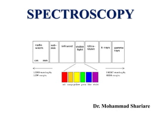
We can classify spectroscopic methods according to the
region of the electromagnetic spectrum involved in the
measurement. The regions include –ray, X-ray,
ultraviolet (UV), visible, infrared (IR), microwave and
radio frequency (RF).
Spectrochemical methods have provided the most widely
used tools for the elucidation of molecular structure as
well as the quantitative and qualitative determination of
both inorganic and organic compounds.
4. The Electromagnetic Spectrum
The electromagnetic spectrum covers an enormous range of
energies (frequencies) and thus wavelengths.
5. Electromagnetic spectrum and absorption of Radiation
The arrangement of all types of of electromagnetic radiations in order of their
increasing wavelengths or decreasing frequencies is known as complete
electromagnetic spectrum.
• Radiation have longer
wavelength are less energetic
compared to radiation with
shorter wavelength
• Microwaves are used for
telephone transmission
• Although all types of radiations
travel as waves with the same
velocity, but they differ from one
another in certain properties
• For example X-rays can pass through glass and muscle tissues, radio waves can pass
through air,
6. If some light radiation is passed through a sample of an organic compound, then
some of the wavelength are absorbed while others belonging to that light source
remain unaffected. A molecule can absorb radiation of certain frequency, when
light radiation is passed through an organic compound, then electrons of the
compound atom are excited. The wavelengths absorbed are measured with the
help of spectrometer.
Spectroscopic Techniques and Chemistry they Probe
UV spec. UV-vis region bonding electrons
Atomic Absorption UV-vis region atomic transitions (val. e-)
FT-IR IR/Microwave vibrations, rotations
Raman IR/UV vibrations
FT-NMR Radio waves nuclear spin states
X-Ray Spectroscopy X-rays inner electrons, elemental
X-ray Crystallography X-rays 3-D structure
7. Spectroscopic Techniques and Common Uses
UV-vis UV-vis region Quantitative analysis
Atomic Absorption UV-vis region Quantitative analysis
FT-IR IR/Microwave Functional Group Analysis
Raman IR/UV
Functional Group
Analysis/quant
FT-NMR Radio waves Structure determination
X-Ray Spectroscopy X-rays Elemental Analysis
X-ray Crystallography X-rays 3-D structure Anaylysis
8. Spectroscopic Measurements
Spectroscopists use the interactions of radiation
with mater to obtain information about a sample.
The sample is stimulated by applying energy in the
form of heat, electrical energy, light, or a chemical
reaction. The analyte is predominately in its
lowest-energy or ground state. The stimulus then
causes some analyte species to undergo a transition
to a higher-energy or excited state. We obtain
information about the analyte by measuring the
electromagnetic radiation emitted as it returns to
the ground state or by measuring the amount of
electromagnetic radiation absorbed as a result of
excitation.
10. …continued…
When the sample is stimulated by application of an
external electromagnetic radiation source, several
processes are possible. Some of the incident
radiation can be absorbed and promote some of the
analyte species to an excited state. In absorption
spectroscopy, the amount of light absorbed as a
function of wavelength is measured, which can
give qualitative and quantitative information about
the sample.
11. Energy Level Diagram for a Simple Molecule
E0
E1
E2
Ground State
Excitation to the next
electronic energy
level caused by
absorption of
specific wavelengths
e4
e3
e2
e1
Vibrational Energy
Levels
Relaxation from the E2
energy state to E0 may
go to different vibrational
energy states, emitting
different wavelengths.
12. The Absorption Process
The absorption law, also known as the Beer-Lambert law
or just Beer’s law, tells us quantitatively how the amount
of attenuation depends on the concentration of the
absorbing molecules and the path length over which
absorption occurs. As light traverses a medium containing
an absorbing analyte, decreases in intensity occur as the
analyte becomes excited. For an analyte solution of a
given concentration, the longer the length of the medium
through which the light passes (path length of light), the
more absorbers are in the path and the greater the
attenuation. Also for a given path length of light, the
higher the concentration of absorbers, the stronger the
attenuation.
13.
14. Measuring Transmittance and Absorbance
Ordinarily, transmittance and absorbance, cannot
be measured as shown because the solution to be
studied must be held in some sort of container
(cell or cuvette). Reflection and scattering losses
can occur at the cell walls. These losses can be
substantial. Light can also be scattered in all
directions from the surface of large molecules or
particles, such as dust, in the solvent, and this can
also cause further attenuation of the beam as it
passes through the solution.
15.
16. …continued…
To compensate for these effects, the power of the
beam transmitted through a cell containing the
analyte solution is compared with one that
traverses an identical cell containing only the
solvent or a reagent blank. An experimental
absorbance that closely approximates the true
absorbance for the solution is thus obtained; that
is
A = log P0 / P log Psolvent / Psolution
17. Beer’s Law
According to Beer’s law, absorbance A is directly
proportional to the concentration of the absorbing
species c and the pathlength b of the absorbing
medium
A = log P0 / P = abc
Here, a is a proportionality constant called the
absorptivity. Because absorbance is a unitless
quantity, the absorptivity must have units that
cancel the units of b and c. If, for example, c has
the units of grams per liter (g L-1) and b has the
units of centimeters (cm), absorptivity has the units
of liters per gram centimeter (L g-1 cm-1).
18. Limits to Beer’s Law
There are few exception to the linear relationship
between absorbance and path length at a fixed
concentration. We frequently observe deviations
from the direct proportionality between absorbance
and concentration where b is a constant. Some of
these deviations, called real deviations, are
fundamental and represent real limitations to the
law. Others occur as a consequence of the manner
in which the absorbance measurements are made
or as a result of chemical changes associated with
concentration changes. These deviations are called
instrumental deviations and chemical deviation
respectively.
19. Real Limitations to Beer’s Law
Beer’s law describes the absorption behavior of
dilute solutions only and is a limiting law. At
concentrations exceeding about 0.01 M, the average
distances between ions or molecules are diminished
to the point where each particle affects the charge
distribution, and thus the extent of absorption of its
neighbors. The occurrence of this phenomenon
causes deviations from the linear relationship
between absorbance and concentration. When ions
are in close proximity, the molar absorptivity of the
analyte can be altered because of electrostatic
interactions, which can lead to departures from
Beer’s law.
20. Chemical Deviations
Deviations from Beer’s law appear when the absorbing species
undergoes association, dissociation, or reaction with the solvent to
give products that absorb differently from the analyte.
Unfortunately, we are usually unaware that such processes are
affecting the analyte, so compensation is often impossible.
21. Instrumental Deviations
The need for monochromatic radiation and the absence of stray
radiation are practical factors that limit the applicability of Beer’s
law. Beer’s law strictly applies only when measurements are made
with monochromatic source radiation. To avoid deviation, it is
advisable to select a wavelength band near the wavelength of
maximum absorption where the analyte absorptivity changes little
with wavelength.
22. …continued…
Stray radiation, commonly called stray light, is defined as radiation
from the instrument that is outside the nominal wavelength band
chosen for the determination. This stray radiation is often the result
of scattering and reflection off the surfaces of gratings, lenses or
mirrors, filters, and windows.
Stray light always causes the apparent absorbance to be lower than
the true absorbance. The deviations due to stray light are most
significant at high absorbance values.
23. Another deviation is caused by mismatched cells. If the cells holding the
analyte and blank solutions are not of equal pathlength and equivalent in
optical characteristics, and intercept will occur in the calibration curve.
This error can be avoided either by using matched cells or by using a
linear regression procedure to calculate both the slope and intercept of the
calibration curve.
24. Absorption spectra
An absorption spectrum is a plot of absorbance versus
wavelength. Absorbance could also be plotted against
wavenumber or frequency. Most modern scanning instruments
can produce such an absorption spectrum directly.
25. All atoms and molecules are capable of absorbing energy in accordance with their
own structure variation and so the kind and amount of radiation absorbed by a
molecule depend upon: -
•The structure of the molecule.
•The number of molecules interacting with the radiation.
The study of these dependencies is called absorption spectroscopy. When
electromagnetic radiation (may be regarded as energy propagated in a wave form
i.e. visible light) is absorbed by a molecule, it undergoes transition from a state of
lower to state of higher energy. If the molecule is monoatomic, the energy
absorbed can only be used to raise the energy levels of electrons. If the molecule
consists of more than one atom, the radiation absorbed may bring about changes in
electronic, rotational, vibrational or translational energy.
Molecular Absorption
26. • Electronic energy is associated with the motion of electrons around the nucli.
• Rotational energy is associated with the overall rotation of the molecule.
• Vibrational energy is associated with the movement of atoms within the
molecules.
• Translational energy is associated with the motion of the molecule as a
whole.
Electronic transitions give absorption in the visible and ultraviolet regions of the
spectrum where as translational, rotational and vibrational changes give
absorption in the far and near infrared spectrum.
The overall energy E associated with a molecule in a given state can be written
as
E = Eelectronic + Evibrational + Erotaitonal + Etranslation
where Eelectronic is the electronic energy of the molecule, Evibrational is its
vibrational energy, Erotaitonal is its rotational energy and Etranslation is its
translation energy.
Molecular Absorption
27. Principles, application, strength and limitation of UV-Vis
spectroscopy
Principles: Radiation in the wavelength range 180 – 780 nm is passed through a
solution of a compound. The electron in the bonds within the molecule become
excited and occupy a higher quantum state by absorbing some of the energy which
is passed through the solution. The more loosely held the electrons are within the
bonds of the molecule the longer the wavelength of the radiation is absorbed.
Application:
• Quantification of drugs in formulation
• Determination of pKa values for some drugs
• Determination of partition coefficient and solubility of some drugs
• Used to determine the release rate of drugs from formulation
• The UV spectrum of drugs is used as one of the method for pharmacopoeial
identity checks.
28. Strength:
• Easy to used, cheap and robust method
• Precise method for quantitative analysis of drugs in formulation
• Routine method for determining some of the physicochemical properties
of drugs
Limitation:
• Moderately selective, selectivity of drug depend on the chromophore
present in the drug
• Not readily applicable for the mixture of drugs
Principles, application, strength and limitation of UV
spectroscopy
29. Factors governing UV/visible radiation absorption
Most drug molecules absorb radiation in the ultraviolet (UV) region of the
spectrum and colored molecule absorb radiation in the visible region. Radiation
in the UV/visible region is absorbed through excitation of the electrons involved
in the bonds between the atoms making up the molecules. Strong bond in
organic molecules need short wavelength UV radiation (<150 nm) to break,
which is vary damaging to living organism. Molecules with weaker bonds are
of more interest to analysts because they can be excited by longer wavelength
UV radiation (>200nm), which is at a longer wavelength than the region at
which air and common solvents are absorb. E.g. – ethylene
A single double bond is not useful as a chromophore for determining analytes by
UV spectroscopy since it is still in the region where air and solvent absorb.
Conjugated dienes are useful as a chromophore e.g.- benzene, toluene etc.
30. Instrumentation of UV spectrometer
The modern UV spectrometer consist of –
• Light Source
• Monochromator
• Detector
• Amplifier and
• Recording devices
31. The most suitable sources of light are:
• Tungsten filament lamp: Tungsten filament lamp is particularly rich in red radiations, i.e.
radiations with wavelength 375nm.
• Deuterium discharge lamp: The intensity of the deuterium discharge source falls above
360nm.
The primary source of light is divided into two beams of equal intensity with the help of a
rotating prism. The various wavelengths of a light source are separated with a prism and then
selected by slits for recording purposes. The selected beam is monochromatic which is then
divided into two beams of equal intensity. Light from the first dispersion is passed through a slit
and then sent to the second dispersion. After the second dispersion, light passes through the
exit slit result in increase the band width of the emergent light which is almost monochromatic.
One of the beams of selected monochromatic light is passed through the sample solution
and the other beam of equal intensity is passed through the reference solvent. The solvent as
well as solution of the sample may be contained in cells made of a material which is transparent.
Each absorbance measurement on the solution is accompanied by a simultaneous
measurement on the pure solvent.
Instrumentation of UV spectrometer
32. After the beams pass through the sample cell as well as the reference cell, the
intensities of the respective transmitted beams are then compared over the whole
wavelength range of the instrument. The spectrometer electronically subtracts the
absorption of the solvent in the reference beam from the absorption of the
solution. Hence the effects due to the absorption of light by the solvent are
minimized.
In this way, the absorbance or the transmittance characteristic of the
compound alone can be measured. The signal for the intensity of absorbance Vs
corresponding wavelength is automatically recorded on the graph. The spectrum
is usually plotted as absorbance A (log10 I0/I) against wavelength (abscissa).
The plot is often represented as mas (Extinction coefficient) against wavelength.
Instrumentation of UV spectrometer
33. Instrument calibration
The instrument used to make the measurement must be properly
calibrated with respect to its –
• Wavelength and
• Absorption scale
In addition check for stray light and spectral resolution are tested
34. Type of Electronic Transition
Molecular orbital theory states that when a molecule is excited by
the absorption of energy (UV – visible) its electron are promoted
from a bonding to an antibonding orbital. According to this theory
there are four types of electronic transition –
1. σ σ*
2. n σ*
3. π π*
4. n π*
35. Chromophore:
A chromophore is the part of a molecule responsible for its color. The color
arises when a molecule absorbs certain wavelengths of UV - visible light and
transmits or reflects others. The chromophore is a region in the molecule
where the energy difference between two different molecular orbitals falls
within the range of the UV - visible spectrum. UV - visible light that hits the
chromophore can thus be absorbed by exciting an electron from its ground
state into an excited state.
It is also defined as any isolated covalently bonded group that shows a
characteristic absorption in the ultraviolet or visible region.
e.g. -
Beta-carotene-conjugation
36. Auxochrome:
An auxochrome can be defined as any group which does not itself
act as a chromophore but whose presence brings about a shift of the
absorption band towards the red end of the spectrum (longer
wavelength). Absorption at longer wavelength is due to the
combination of a chromophore and auxochrome to give rise
another chromophore. An auxochrome group is called color
enhancing group. Some common auxochromic groups are –OH, -
OR, -NH2, -NHR.
38. Solvent effect during UV spectroscopy
For UV analysis a most suitable solvent is one which does not
itself absorb in the region under investigation. A dilute solution
of the sample is always prepared for the spectral analysis. Most
commonly used solvent is 95% ethanol. Ethanol is a best solvent
as it is cheap and is transparent down to 210nm. Other solvents
used for UV analysis are – methanol, hexane water etc.
39. Questions: Theoretically there should be sharp band in UV spectroscopy, but
practically, broad bands are absorbed. Why?
Answer:
Nature is very specific, if a molecule absorbs UV radiation it usually causes electronic
transition. In this case, the molecule absorbs radiation of a particular wavelength. So
the UV spectrum should contain a sharp band.
But at normal conditions, UV radiation causes vibrational and/or rotational transitions
along with electronic transition. So molecules usually absorbed radiation at a relatively
wide range. That is why, a broad band is observed.
Intensity
(Wavelength, nm)
A B
40. Answer:
Compounds having double or triple bonds contain electrons which are
excited relatively easily. In molecules containing a series of alternating
double bonds, the electrons are delocalized and require less energy for
excitation. So absorption occurs at higher wavelength. When there is more
conjugation in the compound it will absorb less energy radiation.
Therefore the effect of increasing number of double bonds of a compound
on its UV-Visible spectrum will increase the absorption characteristics more
easily and absorb less energy radiation.
Questions: What is the effect of increasing the number of double bonds of
compounds on its UV-Visible spectrum?