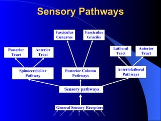
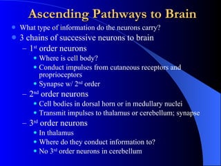
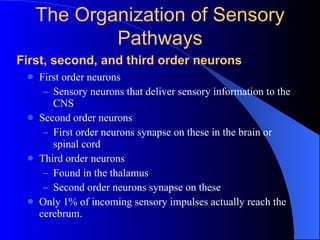
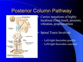
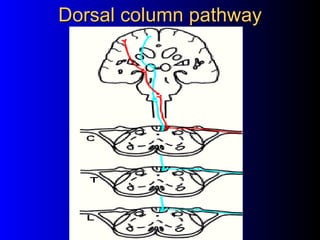
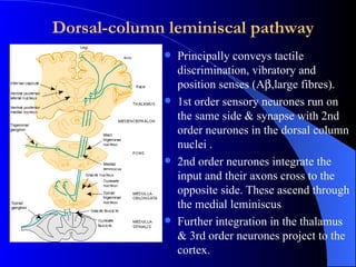
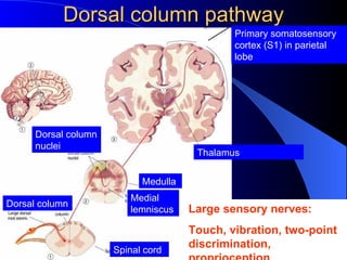
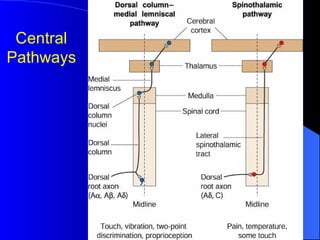
Dorsal column pathway DR SYED TOUSIF AHMED
General Sensory Receptors Sensory pathways Spinocerebellar Pathway Posterior Column Pathways Anteriolatheral Pathways Posterior Tract Anterior Tract Fasciculus Cuneatus Fasciculus Gracilis Latheral Tract Anterior Tract Sensory Pathways
Cutaneous sensory receptors Smooth skin (glaborous ) Hairy skin Free nerve endings The receptor location and its associated structure can alter the stimulus and influence the response Subcutis Dermis Epidermis Merkel disks detect steady pressure & are slowly adapting Free nerve endings around hair root can  be either rapid or slowly adapting - depends  on hair type Meissner’s corpuscles detect flutter & are rapidly adapting Pacinian corpuscles detect  vibration & are very rapidly adapting Ruffini corpuscles detect steady pressure at higher threshold & are slowly adapting Free nerve endings in the skin are modality specific and can detect  either  pain  or  touch  or   pressure  or  temperature
Tactile Sensations arise by activation encapsulated mechanoreceptors touch pressure vibration arise by activation of free nerve endings itch tickle
Touch Crude touch ability to perceive that something contacted skin exact location, shape, size, or texture cannot be detected Fine touch provides specific information about location, shape, size, and texture of stimuli
Proprioreceptors Intrinsic knowledge of limb position is known as kinaesthesia. Information is provided by sensory input from muscle spindles (Ia & II) and Golgi tendon organs. These are mechanoreceptors and provide the CNS with information on muscle length, position and tension. Muscle spindle group Ia afferent fibres are rapidly adapting (dynamic) and are sensitive to rapid changes in muscle length. Muscle spindle group II afferent fibres are slowly adapting (static) and single the fixed length of the muscle.
Joint Kinesthetic Receptors located within and around articular capsules of synovial joints perception of body movements several types free nerve endings and type II cutaneous mechanoreceptors in capsules of joint and respond to pressure small lamellated corpuscles  respond to acceleration and deceleration of joints during movement articular ligaments contain receptors similar to tendon organs adjust the contraction of adjacent muscles when excessive strain is placed on joint
Two-Point Discrimination
Processing at Receptor Level Receptor must have specificity for stimulus activity Stimulus must be applied to receptive field Transduction must occur Generator potential in 1 st  order neuron must reach threshold How is information about stimulus encoded? Tonic receptors (slow adapting) Phasic receptors (fast adapting) Adaptation
Rapidly adapting cutaneous mechanoreceptors signal the onset and offset (phasic) of a stimulus and give rise to sensations such as vibration, touch, and movement  Slowly adapting mechanoreceptors continuously signal (tonic) the intensity of the stimulus and give rise to the sensation of pressure.  Cutaneous receptors with small receptive fields are involved in fine spatial discrimination, whereas receptors with larger receptive fields are less spatially precise. Overlap of receptive fields allows lateral inhibition to occur in the ascending pathways and increase sensory acuity. Cutaneous mechanoreceptors
Somatosensory system receptor classification
First-Order Neuron  Sensory neuron delivers information to CNS Cell body is located in dorsal root ganglion Synapses with interneurons in CNS
Second-Order Neuron Usually interneurons receiving information from first order neurons  Axons cross to opposite sides of body Decussation  May be located in the spinal cord or brain stem
Third-Order Neuron Located in thalamus Carries information to cerebrum Synapses with neurons of the primary sensory cortex
Ascending Pathways to Brain What type of information do the neurons carry? 3 chains of successive neurons to brain 1 st  order neurons Where is cell body? Conduct impulses from cutaneous receptors and proprioceptors Synapse w/ 2 nd  order 2 nd  order neurons Cell bodies in dorsal horn or in medullary nuclei Transmit impulses to thalamus or cerebellum; synapse 3 rd  order neurons In thalamus Where do they conduct information to? No 3 rd  order neurons in cerebellum
First order neurons Sensory neurons that deliver sensory information to the CNS Second order neurons First order neurons synapse on these in the brain or spinal cord Third order neurons Found in the thalamus Second order neurons synapse on these Only 1% of incoming sensory impulses actually reach the cerebrum. First, second, and third order neurons The Organization of Sensory Pathways
Posterior Column Pathway Carries sensations of highly localized (fine) touch, pressure, vibration, proprioception Spinal Tracts Involved: Left/right fasciculus gracilis   Left/right fasciculus cuneatus
Peripheral nerves system
Sensory Pathways and Ascending Tracts in the Spinal Cord
dorsal  cloumn pathway
Dorsal column pathway
Dorsal-column leminiscal pathway Principally conveys tactile discrimination, vibratory and position senses (A  ,large fibres). 1st order sensory neurones run on the same side & synapse with 2nd order neurones in the dorsal column nuclei . 2nd order neurones integrate the input and their axons cross to the opposite side. These ascend through the medial leminiscus  Further integration in the thalamus & 3rd order neurones project to the cortex.
The Posterior Column Pathway and the Spinothalamic Tracts The area of sensory cortex devoted to a body region is relative to the number of sensory receptors.
Dorsal column pathway Large sensory nerves: Touch, vibration, two-point discrimination, proprioception Primary somatosensory cortex (S1) in parietal lobe Thalamus  Medulla Medial lemniscus Spinal cord Dorsal column Dorsal column nuclei
Dorsal column damage dorsal column  pathway Left spinal cord injury Loss of sense of: touch proprioception vibration in left leg
Dorsal column damage Sensory ataxia Patient staggers; cannot perceive position or movement of legs Visual clues help movement
Central Pathways
3.3 Spinocerebellar pathway Carries unconscious proprioception signals Receptors in muscles & joints 1 st  neuron: enters spinal cord through dorsal root 2 nd  neuron: ascends to cerebellum No 3 rd  neuron to cortex, hence  unconscious
Coding in the sensory system could theoretically follow: The  labeled-line principle  in which each receptor responds to a limited range of stimuli  and sends a direct line to the brain. 2 .  Across-fiber pattern  in which each receptor responds to a wider range of stimuli and contributes to the perception of each of them.
THANK YOU

Dorsal column pathway

  • 1.
    Dorsal column pathwayDR SYED TOUSIF AHMED
  • 2.
    General Sensory ReceptorsSensory pathways Spinocerebellar Pathway Posterior Column Pathways Anteriolatheral Pathways Posterior Tract Anterior Tract Fasciculus Cuneatus Fasciculus Gracilis Latheral Tract Anterior Tract Sensory Pathways
  • 3.
    Cutaneous sensory receptorsSmooth skin (glaborous ) Hairy skin Free nerve endings The receptor location and its associated structure can alter the stimulus and influence the response Subcutis Dermis Epidermis Merkel disks detect steady pressure & are slowly adapting Free nerve endings around hair root can be either rapid or slowly adapting - depends on hair type Meissner’s corpuscles detect flutter & are rapidly adapting Pacinian corpuscles detect vibration & are very rapidly adapting Ruffini corpuscles detect steady pressure at higher threshold & are slowly adapting Free nerve endings in the skin are modality specific and can detect either pain or touch or pressure or temperature
  • 4.
    Tactile Sensations ariseby activation encapsulated mechanoreceptors touch pressure vibration arise by activation of free nerve endings itch tickle
  • 5.
    Touch Crude touchability to perceive that something contacted skin exact location, shape, size, or texture cannot be detected Fine touch provides specific information about location, shape, size, and texture of stimuli
  • 6.
    Proprioreceptors Intrinsic knowledgeof limb position is known as kinaesthesia. Information is provided by sensory input from muscle spindles (Ia & II) and Golgi tendon organs. These are mechanoreceptors and provide the CNS with information on muscle length, position and tension. Muscle spindle group Ia afferent fibres are rapidly adapting (dynamic) and are sensitive to rapid changes in muscle length. Muscle spindle group II afferent fibres are slowly adapting (static) and single the fixed length of the muscle.
  • 7.
    Joint Kinesthetic Receptorslocated within and around articular capsules of synovial joints perception of body movements several types free nerve endings and type II cutaneous mechanoreceptors in capsules of joint and respond to pressure small lamellated corpuscles respond to acceleration and deceleration of joints during movement articular ligaments contain receptors similar to tendon organs adjust the contraction of adjacent muscles when excessive strain is placed on joint
  • 8.
  • 9.
    Processing at ReceptorLevel Receptor must have specificity for stimulus activity Stimulus must be applied to receptive field Transduction must occur Generator potential in 1 st order neuron must reach threshold How is information about stimulus encoded? Tonic receptors (slow adapting) Phasic receptors (fast adapting) Adaptation
  • 10.
    Rapidly adapting cutaneousmechanoreceptors signal the onset and offset (phasic) of a stimulus and give rise to sensations such as vibration, touch, and movement Slowly adapting mechanoreceptors continuously signal (tonic) the intensity of the stimulus and give rise to the sensation of pressure. Cutaneous receptors with small receptive fields are involved in fine spatial discrimination, whereas receptors with larger receptive fields are less spatially precise. Overlap of receptive fields allows lateral inhibition to occur in the ascending pathways and increase sensory acuity. Cutaneous mechanoreceptors
  • 11.
  • 12.
    First-Order Neuron Sensory neuron delivers information to CNS Cell body is located in dorsal root ganglion Synapses with interneurons in CNS
  • 13.
    Second-Order Neuron Usuallyinterneurons receiving information from first order neurons Axons cross to opposite sides of body Decussation May be located in the spinal cord or brain stem
  • 14.
    Third-Order Neuron Locatedin thalamus Carries information to cerebrum Synapses with neurons of the primary sensory cortex
  • 15.
    Ascending Pathways toBrain What type of information do the neurons carry? 3 chains of successive neurons to brain 1 st order neurons Where is cell body? Conduct impulses from cutaneous receptors and proprioceptors Synapse w/ 2 nd order 2 nd order neurons Cell bodies in dorsal horn or in medullary nuclei Transmit impulses to thalamus or cerebellum; synapse 3 rd order neurons In thalamus Where do they conduct information to? No 3 rd order neurons in cerebellum
  • 16.
    First order neuronsSensory neurons that deliver sensory information to the CNS Second order neurons First order neurons synapse on these in the brain or spinal cord Third order neurons Found in the thalamus Second order neurons synapse on these Only 1% of incoming sensory impulses actually reach the cerebrum. First, second, and third order neurons The Organization of Sensory Pathways
  • 17.
    Posterior Column PathwayCarries sensations of highly localized (fine) touch, pressure, vibration, proprioception Spinal Tracts Involved: Left/right fasciculus gracilis Left/right fasciculus cuneatus
  • 18.
  • 19.
    Sensory Pathways andAscending Tracts in the Spinal Cord
  • 20.
  • 21.
  • 22.
    Dorsal-column leminiscal pathwayPrincipally conveys tactile discrimination, vibratory and position senses (A  ,large fibres). 1st order sensory neurones run on the same side & synapse with 2nd order neurones in the dorsal column nuclei . 2nd order neurones integrate the input and their axons cross to the opposite side. These ascend through the medial leminiscus Further integration in the thalamus & 3rd order neurones project to the cortex.
  • 23.
    The Posterior ColumnPathway and the Spinothalamic Tracts The area of sensory cortex devoted to a body region is relative to the number of sensory receptors.
  • 24.
    Dorsal column pathwayLarge sensory nerves: Touch, vibration, two-point discrimination, proprioception Primary somatosensory cortex (S1) in parietal lobe Thalamus Medulla Medial lemniscus Spinal cord Dorsal column Dorsal column nuclei
  • 25.
    Dorsal column damagedorsal column pathway Left spinal cord injury Loss of sense of: touch proprioception vibration in left leg
  • 26.
    Dorsal column damageSensory ataxia Patient staggers; cannot perceive position or movement of legs Visual clues help movement
  • 27.
  • 28.
    3.3 Spinocerebellar pathwayCarries unconscious proprioception signals Receptors in muscles & joints 1 st neuron: enters spinal cord through dorsal root 2 nd neuron: ascends to cerebellum No 3 rd neuron to cortex, hence unconscious
  • 29.
    Coding in thesensory system could theoretically follow: The labeled-line principle in which each receptor responds to a limited range of stimuli and sends a direct line to the brain. 2 . Across-fiber pattern in which each receptor responds to a wider range of stimuli and contributes to the perception of each of them.
  • 30.