SlideShare a Scribd company logo
1 of 54
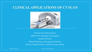
CLINICALAPPLICATIONS OF CT SCAN
Presenter: Dr. Dheeraj Kumar
MRIT, Ph.D. (Radiology and Imaging)
Assistant Professor
Medical Radiology and Imaging Technology
School of Health Sciences, CSJM University, Kanpur
September 26, 2023 Assist. Prof. Dheeraj Kumar 1
CONTENTS
• INTRODUCTION
• HISTORY
• CLINICAL APPLICATIONS
• SUMMARY
• REFERENCES
September 26, 2023 Assist. Prof. Dheeraj Kumar 2
INTRODUCTION
• Computed tomography (CT) is an essential tool in diagnostic imaging for evaluating many
clinical conditions.
• A computerized tomography (CT) scan combines a series of X-ray images taken from
different angles around your body and uses computer processing to create cross-sectional
images (slices) of the bones, blood vessels and soft tissues inside your body.
• In recent years, there have been several notable advances in CT technology that already have
had or are expected to have a significant clinical impact, including extreme multidetector
CT, iterative reconstruction algorithms, dual-energy CT, cone-beam CT, portable CT, and
phase-contrast CT.
September 26, 2023 Assist. Prof. Dheeraj Kumar 3
HISTORY
• The first commercially available CT scanner was created
by British engineer Sir Godfrey Hounsfield of EMI
(Electronic Musical Instruments) Laboratories in 1972.
• He co-invented the technology with physicist Dr. Allan
Cormack. Both researchers were later on jointly awarded
the 1979 Nobel Prize in Physiology and Medicine. By
1981, Hounsfield was knighted and became Sir Godfrey
Hounsfield.
September 26, 2023 Assist. Prof. Dheeraj Kumar 4
• However, it was the mathematical theory of Johann
Radon way back in 1917, called “Radon
transform,” that brought the technology to life.
Another mathematical advancement that Hounsfield
built on is the “Algebraic Reconstruction
Technique,” which was formulated by Polish
mathematician Stefan Kaczmarz in 1937.
• Both theories were adopted by Hounsfield to create
one of the greatest advancements in medical history.
September 26, 2023 Assist. Prof. Dheeraj Kumar 5
ADVANTEGES OF CT
MULTI SLICE SCANNING ADVANTAGE OF OVER SINGLE SLICE
September 26, 2023 Assist. Prof. Dheeraj Kumar 6
September 26, 2023 Assist. Prof. Dheeraj Kumar 7
ADVANTAGES CT SCANNER
• This has improved the diagnostic capabilities of CT scanners. Recently new scanners capable of
producing 32, 40 and even 64 images have been announced. These scanners will increase the diagnostic
capabilities of CT scanners even further, resulting in clearer images and lower doses of radiation
• Multi-slice scanners mean that it takes less time to complete a CT scan. Additionally, the amount of
radiation is reduced. The amount of radiation experienced depends on two factors. First, the design of the
scanner impacts the amount of radiation required. Secondly, how the scanner is used determines the
amount of radiation used.
• One of the key differences between single slice scanners and multi-slice scanners is the geometric
efficiency of the scan. Additionally, the amount of radiation used depends on the scan’s parameters- kV,
rotation time, mA, scan field of view, focal spot size, pitch and slice width.
September 26, 2023 Assist. Prof. Dheeraj Kumar 8
ADVANTAGES WITH MULTI SLICE CT
SCANNERS
• With each rotation, it produces higher simultaneous 0.5 mm slices and gives
isotropic volumetric data with a better resolution
• Thin slice volume data reconstructed
• Post processing advanced visualization algorithms allow the extraction of
specific body parts
• Allow to understand complex anatomy and diseases
• Open new clinical possibilities
• Uncompromised image quality at a level never seen before
September 26, 2023 Assist. Prof. Dheeraj Kumar 9
APPLICATIONS WITH MULTISLICE
CT SCANNER
• CT Head, Abdomen and Extremities
• CT Angiography (CTA)
• Coronary CT Angiography (CCTA)
• Visualization of Cardiac and Other Structure
• Cardiac Calcium Scoring
• Routine CT Scanning with Better Resolution
• Virtual Bronchoscopy
• Virtual Colonoscopy
September 26, 2023 Assist. Prof. Dheeraj Kumar 10
CT HEAD
Indications
• Bone abnormalities.
• Brain mass/tumor.
• Fluid collection, such as an abscess.
• Haemorrhage.
• Hydrocephalus.
• Ischemic process, such as a stroke.
• Trauma or fracture of the skull.
September 26, 2023 Assist. Prof. Dheeraj Kumar 11
BONE ABNORMALITIES
CT HEAD CONGENITAL
CALVARIAL DEFECTS
CT HEAD CONGENITAL
CALVARIAL SPECTRUM
CT HEAD SKULL FRACTURE
September 26, 2023 Assist. Prof. Dheeraj Kumar 12
BRAIN MASS/TUMOR
NCCT BRAINTUMOR
September 26, 2023 Assist. Prof. Dheeraj Kumar 13
FLUID COLLECTION, SUCH AS AN
ABCESS
September 26, 2023 Assist. Prof. Dheeraj Kumar 14
HAEMORRHAGE
September 26, 2023 Assist. Prof. Dheeraj Kumar 15
HYDROCEPHALUS
September 26, 2023 Assist. Prof. Dheeraj Kumar 16
ISCHEMIC PROCESS, SUCH AS A
STROKE
September 26, 2023 Assist. Prof. Dheeraj Kumar 17
TRAUMA OR FRACTURE OF THE
SKULL
September 26, 2023 Assist. Prof. Dheeraj Kumar 18
CT ABDOMEN
Indications
• Abdominal pain.
• Difficulties in breathing
• Abdominal sepsis.
• Bowel obstruction.
• Postoperative complications.
• Trauma.
• Vascular compromise, e.g. aortic aneurysm.
September 26, 2023 Assist. Prof. Dheeraj Kumar 19
DIFFICULTIES IN BREATHING
Consolidation
September 26, 2023 Assist. Prof. Dheeraj Kumar 20
ABDOMINAL SEPSIS
Abscess
sepsis
September 26, 2023 Assist. Prof. Dheeraj Kumar 21
BOWEL OBSTRUCTION
September 26, 2023 Assist. Prof. Dheeraj Kumar 22
POSTOPERATIVE COMPLICATIONS
September 26, 2023 Assist. Prof. Dheeraj Kumar 23
ABDOMINAL TRAUMA
September 26, 2023 Assist. Prof. Dheeraj Kumar 24
VASCULAR COMPROMISE, E.G.
AORTIC ANEURYSM
September 26, 2023 Assist. Prof. Dheeraj Kumar 25
CT EXTREMITY
Reasons for an Extremity CT Scan
• Evaluate pain, swelling, or trauma.
• Identify and localize a known mass.
• Examine complex fractures.
• Diagnose arthritis.
• Scan for a collection of pus (abscess)
• Monitor scar tissue and healing after surgery.
September 26, 2023 Assist. Prof. Dheeraj Kumar 26
CT ANGIOGRAPHY (CTA)
With ultra fast scanning, arteries serving the brain, lungs, kidneys, arms and legs can
be evaluated non-invasively.
 Cerebral aneurysm
 Carotid stenosis
 Pulmonary embolism
 Renal artery stenosis
 Aortic aneurysm / dissection
 Mesenteric ischemia
 Hepatic artery anatomy (For Surgery)
September 26, 2023 Assist. Prof. Dheeraj Kumar 27
CT Angiography- Technique
• Bolus tracking
• Amount and rate of contrast media
• Exposure factors
• Pitch/ Collimation
September 26, 2023 Assist. Prof. Dheeraj Kumar 28
September 26, 2023 Assist. Prof. Dheeraj Kumar 29
September 26, 2023 Assist. Prof. Dheeraj Kumar 30
September 26, 2023 Assist. Prof. Dheeraj Kumar 31
September 26, 2023 Assist. Prof. Dheeraj Kumar 32
September 26, 2023 Assist. Prof. Dheeraj Kumar 33
CT PERFUSION
• Computed tomography (CT) perfusion
imaging shows which areas of the specific
organ are adequately supplied or perfused with
blood and provides detailed information on
delivery of blood or blood flow to the brain
• CT perfusion scanning is a non-invasive
medical test that helps physicians diagnose and
treat medical conditions
September 26, 2023 Assist. Prof. Dheeraj Kumar 34
• Xenon gas previously used
Patients not very tolerant
Scans taken over 5-10 minutes at 1
minute intervals
• Faster scanning means ionic
contrast can now be used
Continuous scanning of the brain
during contrast injection
Scan time < 1 Minute
September 26, 2023 Assist. Prof. Dheeraj Kumar 35
CORONARYANGIOGRAPHY
•Only 30% conventional angiographies intervention for
therapeutic purpose
•Rest 65-70% - Only for diagnostic purpose
(AHA- Heart and stroke statistics update, 2001)
September 26, 2023 Assist. Prof. Dheeraj Kumar 36
CT CORONARYANGIOGRAPHY
CLINICALAPPLICATIONS
• ASYMPTOMATIC PATIENT
• High risk
• High calcium score
• SYMPATOMATIC PATIENT
• No history of CAD
• Atypical chest pain
• Inconclusive stress test
• FOLOOW UP OF POST BTPASS AND POST STENT PATIENTS
• TO RULE OUT CONGENITAL ANOMALIES
September 26, 2023 Assist. Prof. Dheeraj Kumar 37
CALCIUM SCORE
• Cardiac computed tomography (CT) for Calcium Scoring uses special x-ray
equipment to produce pictures of the coronary arteries to determine if they
are blocked or narrowed by the build-up of plaque – an indicator for
atherosclerosis or coronary artery disease (CAD).
• The information obtained can help evaluate whether you are at increased risk
for heart attack.
September 26, 2023 Assist. Prof. Dheeraj Kumar 38
September 26, 2023 Assist. Prof. Dheeraj Kumar 39
RISK FACTORS OF CAD
The major risk factors for CAD are:
• high blood cholesterol levels
• family history of heart attacks
• diabetes
• high blood pressure
• cigarette smoking
• overweight or obese
• physical inactivity
September 26, 2023 Assist. Prof. Dheeraj Kumar 40
CALCIUM SCORE- INTERPRETATION
The result of the test is usually given as a number called an Agatston score. The
score reflects the total area of calcium deposits and the density of the calcium.
• A score of zero means no calcium is seen in the heart. It suggests a low
chance of developing a heart attack in the future.
• When calcium is present, the higher the score, the higher your risk of heart
disease.
• A score of 100 to 300 means moderate plaque deposits. It's associated with a
relatively high risk of a heart attack or other heart disease over the next three
to five years.
• A score greater than 300 is a sign of very high to severe disease and heart
attack risk.
September 26, 2023 Assist. Prof. Dheeraj Kumar 41
September 26, 2023 Assist. Prof. Dheeraj Kumar 42
PLAQUE CHARACTERIZATION
(Schroder, JACC 2001)
PLAQUES CT DENSITY
Soft < 50 HU
Fibrotic 50-130 HU
Calcified > 130 HU
September 26, 2023 Assist. Prof. Dheeraj Kumar 43
CT SOFTWARES AVAILABLE
Curved MPR
Dynamic CT MPI
September 26, 2023 Assist. Prof. Dheeraj Kumar 44
September 26, 2023 Assist. Prof. Dheeraj Kumar 45
VIRTUAL COLONOSCOPY
• Emerging noninvasive imaging technology for detecting colon polyps and
cancer
• Trends towards using this as screening gold standards as it permits complete
visualization of the entire colon, hence proving the opportunity to identify
precancerous polyps and cancer
• Accepted application include incomplete colonoscopy
September 26, 2023 Assist. Prof. Dheeraj Kumar 46
ADVANTAGES OF CT COLONOSCOPY
• more comfortable
• No sedation is required
• Evidence that CTC is better able to
detect polyps than fecal occult blood
testing, Ba enema and sigmoidoscopy
• Take less time than either conventional
colonoscopy or lower GI Series
• Secondary benefits of the revealing
diseases or abnormalities outside the
colon
September 26, 2023 Assist. Prof. Dheeraj Kumar 47
VIRTUAL BRONCHOSCOPY
• Virtual bronchoscopy (VB) is a novel
computed tomography (CT)-based
imaging technique that allows a non-
invasive intraluminal evaluation of the
tracheobronchial tree.
• Several studies have shown that VB can
accurately show the lumen and the
diameter of the trachea, the left and
right main stem bronchi, and the
bronchial tree down to the fourth order
of bronchial orifices and branches
September 26, 2023 Assist. Prof. Dheeraj Kumar 48
Applications
Normal Anatomic Features And Variants
Tracheobronchial Stenosis
Bronchogenic Carcinoma
Endoluminal Lesion
Foreign Body Aspiration
Trauma
Stent Planning And Follow-up
Burn Injury
Tracheoesophageal Fistula
September 26, 2023 Assist. Prof. Dheeraj Kumar 49
NORMAL ANATOMIC FEATURES and
TRACHEOBRONCHIAL STENOSIS
• 3D CT can depict the airway down to the 6th and 7th order of
subdivision
• The 3d map can be used to guide bronchoscopy or to direct
transbronchail needle biopsy
• The stenosis to lumen ratios determined with VB and Conventional
bronchoscopy were found to be within 10 % of each other
• Especially valuable for evaluation of suspected tracheobronchial
stenosis in children
• Less invasive and safer than fiberoptic bronchoscopy
• The advantage of depicting the adjustment structures such as
vascular rings, which can be a cause of stridor in children.
September 26, 2023 Assist. Prof. Dheeraj Kumar 50
BRONCHOGENIC CARCINOMA
CT is the primary imaging technique for the detection, staging and follow-up of the
primary malignant tumors of the lung
CT with VB
Sensitivity- 100% for Obstructive lesions
16% for Mucosal lesions
90% for Endoluminal lesions
• Specificity for malignant tumors -100%
• Advantage of VB over fiberoptic bronchoscopy, can image beyond the site of
obstruction
• Visualization of the smaller airways, which are not accessible with fiberoptic
bronchoscopy
September 26, 2023 Assist. Prof. Dheeraj Kumar 51
CT Scan Procedure May Be More Comfortable For The Patient
Carry Fewer Risks Of Complications
Sometimes Replace More Invasive Procedure
New Technology Providing Its Worth In Routine Scanning
It More Specialized Of Medical Image
CT Doses-
Higher For Some Exams But Could Be Lower For Other
Thin Slice Doses Lower Than On 4 Slice
Are Being Addressed By Dose Reduction Features
September 26, 2023 Assist. Prof. Dheeraj Kumar 52
REFERENCES
• De wever W, vandecaveye V, lanciotti S, verschakelen JA. Multidetector ct-generated
virtual bronchoscopy: an illustrated review of the potential clinical indications. European
respiratory journal. 2004 may 1;23(5):776-82.
• Himi t, kataura a, sakata m, odawara y, satoh ji, sawaishi m. Three-dimensional imaging
of the temporal bone using a helical CT scan and its application in patients with cochlear
implantation. Orl. 1996;58(6):298-300.
• Ganz sd. Computer-aided design/computer-aided manufacturing applications using CT
and cone beam CT scanning technology. Dental clinics of north america. 2008 oct
1;52(4):777-808.
• De chiffre l, carmignato s, kruth jp, schmitt r, weckenmann a. Industrial applications of
computed tomography. CIRP annals. 2014 jan 1;63(2):655-77.
September 26, 2023 Assist. Prof. Dheeraj Kumar 53
September 26, 2023 Assist. Prof. Dheeraj Kumar 54

More Related Content

What's hot

Computed Tomography Equipments.pptx
Computed Tomography Equipments.pptxComputed Tomography Equipments.pptx
Computed Tomography Equipments.pptxDr. Dheeraj Kumar
 
ULTRASOUND COMPONENTS AND THEIR USES.pptx
ULTRASOUND COMPONENTS AND THEIR USES.pptxULTRASOUND COMPONENTS AND THEIR USES.pptx
ULTRASOUND COMPONENTS AND THEIR USES.pptxDr. Dheeraj Kumar
 
History of Ultrasound Imaging.pptx
History of Ultrasound Imaging.pptxHistory of Ultrasound Imaging.pptx
History of Ultrasound Imaging.pptxDr. Dheeraj Kumar
 
Physical Principle of MRI.pptx
Physical Principle of MRI.pptxPhysical Principle of MRI.pptx
Physical Principle of MRI.pptxDr. Dheeraj Kumar
 
Guidelines for X-Ray Equipments.pptx
Guidelines for X-Ray Equipments.pptxGuidelines for X-Ray Equipments.pptx
Guidelines for X-Ray Equipments.pptxDr. Dheeraj Kumar
 
Discograpgy (intradiscal procedure)
Discograpgy (intradiscal procedure)Discograpgy (intradiscal procedure)
Discograpgy (intradiscal procedure)SKSHAHWAZ
 
Emergency in Radiology Department.pptx
Emergency in Radiology Department.pptxEmergency in Radiology Department.pptx
Emergency in Radiology Department.pptxDr. Dheeraj Kumar
 
Clinical Applications and Procedure of MRI.pptx
Clinical Applications and Procedure of MRI.pptxClinical Applications and Procedure of MRI.pptx
Clinical Applications and Procedure of MRI.pptxDr. Dheeraj Kumar
 
Range of Secondary Electrons and Electron Build-Up: Impact on Scatter in Homo...
Range of Secondary Electrons and Electron Build-Up: Impact on Scatter in Homo...Range of Secondary Electrons and Electron Build-Up: Impact on Scatter in Homo...
Range of Secondary Electrons and Electron Build-Up: Impact on Scatter in Homo...Dr. Dheeraj Kumar
 
Ultrasound Transducer Constriction And It’s Physics.pptx
Ultrasound Transducer Constriction And It’s Physics.pptxUltrasound Transducer Constriction And It’s Physics.pptx
Ultrasound Transducer Constriction And It’s Physics.pptxDr. Dheeraj Kumar
 
Positron emission Tomography (PET)
Positron emission Tomography (PET) Positron emission Tomography (PET)
Positron emission Tomography (PET) chetanath
 
DUAL ENERGY CT-1.pptx
DUAL ENERGY CT-1.pptxDUAL ENERGY CT-1.pptx
DUAL ENERGY CT-1.pptxJyotiDalal22
 
ct image quality
ct image qualityct image quality
ct image qualitydypradio
 
Ptc )percutaneous transhepatic cholangiography
Ptc )percutaneous transhepatic cholangiographyPtc )percutaneous transhepatic cholangiography
Ptc )percutaneous transhepatic cholangiographyYashawant Yadav
 

What's hot (20)

Computed Tomography Equipments.pptx
Computed Tomography Equipments.pptxComputed Tomography Equipments.pptx
Computed Tomography Equipments.pptx
 
ULTRASOUND COMPONENTS AND THEIR USES.pptx
ULTRASOUND COMPONENTS AND THEIR USES.pptxULTRASOUND COMPONENTS AND THEIR USES.pptx
ULTRASOUND COMPONENTS AND THEIR USES.pptx
 
History of Ultrasound Imaging.pptx
History of Ultrasound Imaging.pptxHistory of Ultrasound Imaging.pptx
History of Ultrasound Imaging.pptx
 
Angiography.pptx
Angiography.pptxAngiography.pptx
Angiography.pptx
 
Physical Principle of MRI.pptx
Physical Principle of MRI.pptxPhysical Principle of MRI.pptx
Physical Principle of MRI.pptx
 
Catheter and Guidewire.pptx
Catheter and Guidewire.pptxCatheter and Guidewire.pptx
Catheter and Guidewire.pptx
 
Guidelines for X-Ray Equipments.pptx
Guidelines for X-Ray Equipments.pptxGuidelines for X-Ray Equipments.pptx
Guidelines for X-Ray Equipments.pptx
 
Discograpgy (intradiscal procedure)
Discograpgy (intradiscal procedure)Discograpgy (intradiscal procedure)
Discograpgy (intradiscal procedure)
 
Emergency in Radiology Department.pptx
Emergency in Radiology Department.pptxEmergency in Radiology Department.pptx
Emergency in Radiology Department.pptx
 
Arthrograms(3)
Arthrograms(3)Arthrograms(3)
Arthrograms(3)
 
Clinical Applications and Procedure of MRI.pptx
Clinical Applications and Procedure of MRI.pptxClinical Applications and Procedure of MRI.pptx
Clinical Applications and Procedure of MRI.pptx
 
Range of Secondary Electrons and Electron Build-Up: Impact on Scatter in Homo...
Range of Secondary Electrons and Electron Build-Up: Impact on Scatter in Homo...Range of Secondary Electrons and Electron Build-Up: Impact on Scatter in Homo...
Range of Secondary Electrons and Electron Build-Up: Impact on Scatter in Homo...
 
Ultrasound Transducer Constriction And It’s Physics.pptx
Ultrasound Transducer Constriction And It’s Physics.pptxUltrasound Transducer Constriction And It’s Physics.pptx
Ultrasound Transducer Constriction And It’s Physics.pptx
 
Positron emission Tomography (PET)
Positron emission Tomography (PET) Positron emission Tomography (PET)
Positron emission Tomography (PET)
 
Ct scan final (2)
Ct scan final (2)Ct scan final (2)
Ct scan final (2)
 
Ct head protocols
Ct head protocolsCt head protocols
Ct head protocols
 
DUAL ENERGY CT-1.pptx
DUAL ENERGY CT-1.pptxDUAL ENERGY CT-1.pptx
DUAL ENERGY CT-1.pptx
 
ct image quality
ct image qualityct image quality
ct image quality
 
Ptc )percutaneous transhepatic cholangiography
Ptc )percutaneous transhepatic cholangiographyPtc )percutaneous transhepatic cholangiography
Ptc )percutaneous transhepatic cholangiography
 
X-Ray tube.pptx
X-Ray tube.pptxX-Ray tube.pptx
X-Ray tube.pptx
 

Similar to CLINICAL APPLICATIONS OF CT SCAN.pptx

Intracoronary optical coherence tomography
Intracoronary optical coherence tomographyIntracoronary optical coherence tomography
Intracoronary optical coherence tomographyBHAWANI SHANKAR
 
Examination of carotid pre-bifurcation expansion to predict boundary layer s...
 Examination of carotid pre-bifurcation expansion to predict boundary layer s... Examination of carotid pre-bifurcation expansion to predict boundary layer s...
Examination of carotid pre-bifurcation expansion to predict boundary layer s...Minh Anh Nguyen
 
HDMICS WSS Koutsiaris 2007
HDMICS WSS Koutsiaris 2007HDMICS WSS Koutsiaris 2007
HDMICS WSS Koutsiaris 2007Koutsiaris Aris
 
surgical mgt ICH of hemorraghic stroke. indications for surgery.
surgical mgt ICH of hemorraghic stroke. indications for surgery.surgical mgt ICH of hemorraghic stroke. indications for surgery.
surgical mgt ICH of hemorraghic stroke. indications for surgery.blessingmichael5
 
INTRA-ARTERIAL BLOOD PRESSURE MONITORING
INTRA-ARTERIAL BLOOD PRESSURE MONITORING INTRA-ARTERIAL BLOOD PRESSURE MONITORING
INTRA-ARTERIAL BLOOD PRESSURE MONITORING vikramnaidu2311
 
VAC Study material VAC
VAC Study material VAC VAC Study material VAC
VAC Study material VAC Vamsi kumar
 
Ilumienation of lightening - 4final.pptx
Ilumienation of lightening - 4final.pptxIlumienation of lightening - 4final.pptx
Ilumienation of lightening - 4final.pptxShreyaKedia10
 
Angio ct of heart and blood vessels
Angio ct of heart and blood vesselsAngio ct of heart and blood vessels
Angio ct of heart and blood vesselsSkrillTunes
 
Chest x-ray ppt
Chest x-ray pptChest x-ray ppt
Chest x-ray pptMhmwdFthy
 
bbbbbbasic chest x-ray interpretation.ppt
bbbbbbasic chest x-ray interpretation.pptbbbbbbasic chest x-ray interpretation.ppt
bbbbbbasic chest x-ray interpretation.pptketut9
 
Uro instruments- cystoscopy &amp; fibreoptics
Uro instruments- cystoscopy &amp; fibreopticsUro instruments- cystoscopy &amp; fibreoptics
Uro instruments- cystoscopy &amp; fibreopticsGovtRoyapettahHospit
 
Ct scan centres in mumbai - iScan
Ct scan centres in mumbai - iScanCt scan centres in mumbai - iScan
Ct scan centres in mumbai - iScaniscanimaging
 
Carotid stenosis
Carotid stenosisCarotid stenosis
Carotid stenosiskiwaasa
 

Similar to CLINICAL APPLICATIONS OF CT SCAN.pptx (20)

Radiographic Exposure.pptx
Radiographic Exposure.pptxRadiographic Exposure.pptx
Radiographic Exposure.pptx
 
Intracoronary optical coherence tomography
Intracoronary optical coherence tomographyIntracoronary optical coherence tomography
Intracoronary optical coherence tomography
 
Examination of carotid pre-bifurcation expansion to predict boundary layer s...
 Examination of carotid pre-bifurcation expansion to predict boundary layer s... Examination of carotid pre-bifurcation expansion to predict boundary layer s...
Examination of carotid pre-bifurcation expansion to predict boundary layer s...
 
MEDICAL IMAGING NOTES
MEDICAL IMAGING NOTESMEDICAL IMAGING NOTES
MEDICAL IMAGING NOTES
 
HDMICS WSS Koutsiaris 2007
HDMICS WSS Koutsiaris 2007HDMICS WSS Koutsiaris 2007
HDMICS WSS Koutsiaris 2007
 
surgical mgt ICH of hemorraghic stroke. indications for surgery.
surgical mgt ICH of hemorraghic stroke. indications for surgery.surgical mgt ICH of hemorraghic stroke. indications for surgery.
surgical mgt ICH of hemorraghic stroke. indications for surgery.
 
Critical Limb Ischemia
Critical Limb IschemiaCritical Limb Ischemia
Critical Limb Ischemia
 
Basics of CT Scan
Basics of CT ScanBasics of CT Scan
Basics of CT Scan
 
INTRA-ARTERIAL BLOOD PRESSURE MONITORING
INTRA-ARTERIAL BLOOD PRESSURE MONITORING INTRA-ARTERIAL BLOOD PRESSURE MONITORING
INTRA-ARTERIAL BLOOD PRESSURE MONITORING
 
MRI BRAIN
MRI BRAINMRI BRAIN
MRI BRAIN
 
VAC Study material VAC
VAC Study material VAC VAC Study material VAC
VAC Study material VAC
 
Cardiac ct
Cardiac ctCardiac ct
Cardiac ct
 
Ilumienation of lightening - 4final.pptx
Ilumienation of lightening - 4final.pptxIlumienation of lightening - 4final.pptx
Ilumienation of lightening - 4final.pptx
 
Angio ct of heart and blood vessels
Angio ct of heart and blood vesselsAngio ct of heart and blood vessels
Angio ct of heart and blood vessels
 
Chest x-ray ppt
Chest x-ray pptChest x-ray ppt
Chest x-ray ppt
 
bbbbbbasic chest x-ray interpretation.ppt
bbbbbbasic chest x-ray interpretation.pptbbbbbbasic chest x-ray interpretation.ppt
bbbbbbasic chest x-ray interpretation.ppt
 
Uro instruments- cystoscopy &amp; fibreoptics
Uro instruments- cystoscopy &amp; fibreopticsUro instruments- cystoscopy &amp; fibreoptics
Uro instruments- cystoscopy &amp; fibreoptics
 
Dr.tatchanaamurrti
Dr.tatchanaamurrtiDr.tatchanaamurrti
Dr.tatchanaamurrti
 
Ct scan centres in mumbai - iScan
Ct scan centres in mumbai - iScanCt scan centres in mumbai - iScan
Ct scan centres in mumbai - iScan
 
Carotid stenosis
Carotid stenosisCarotid stenosis
Carotid stenosis
 

More from Dr. Dheeraj Kumar

Measurement of Radiation and Dosimetric Procedure.pptx
Measurement of Radiation and Dosimetric Procedure.pptxMeasurement of Radiation and Dosimetric Procedure.pptx
Measurement of Radiation and Dosimetric Procedure.pptxDr. Dheeraj Kumar
 
Radiation Dosimetry Parameters and Isodose Curves.pptx
Radiation Dosimetry Parameters and Isodose Curves.pptxRadiation Dosimetry Parameters and Isodose Curves.pptx
Radiation Dosimetry Parameters and Isodose Curves.pptxDr. Dheeraj Kumar
 
Culture and Health Disorders Social change.pptx
Culture and Health Disorders Social change.pptxCulture and Health Disorders Social change.pptx
Culture and Health Disorders Social change.pptxDr. Dheeraj Kumar
 
Units of Radiation Measurements, Quality Specification, Half-Value Thickness,...
Units of Radiation Measurements, Quality Specification, Half-Value Thickness,...Units of Radiation Measurements, Quality Specification, Half-Value Thickness,...
Units of Radiation Measurements, Quality Specification, Half-Value Thickness,...Dr. Dheeraj Kumar
 
X-Ray Beam Restrictors/Beam Modifying devices.pptx
X-Ray Beam Restrictors/Beam Modifying devices.pptxX-Ray Beam Restrictors/Beam Modifying devices.pptx
X-Ray Beam Restrictors/Beam Modifying devices.pptxDr. Dheeraj Kumar
 
Transmission of X-ray through body tissues linear energy transfer..pptx
Transmission of X-ray through body tissues linear energy transfer..pptxTransmission of X-ray through body tissues linear energy transfer..pptx
Transmission of X-ray through body tissues linear energy transfer..pptxDr. Dheeraj Kumar
 
X-ray Production A Journey Through History and the X-ray Tube.pptx
X-ray Production A Journey Through History and the X-ray Tube.pptxX-ray Production A Journey Through History and the X-ray Tube.pptx
X-ray Production A Journey Through History and the X-ray Tube.pptxDr. Dheeraj Kumar
 
Population of India and Family welfare programme.pptx
Population of India and Family welfare programme.pptxPopulation of India and Family welfare programme.pptx
Population of India and Family welfare programme.pptxDr. Dheeraj Kumar
 
Artificial Radionuclide Generators in Medicine Applications in Radiotherapy.pptx
Artificial Radionuclide Generators in Medicine Applications in Radiotherapy.pptxArtificial Radionuclide Generators in Medicine Applications in Radiotherapy.pptx
Artificial Radionuclide Generators in Medicine Applications in Radiotherapy.pptxDr. Dheeraj Kumar
 
Effects of variation of tube voltage current, filtration..pptx
Effects of variation of tube voltage current, filtration..pptxEffects of variation of tube voltage current, filtration..pptx
Effects of variation of tube voltage current, filtration..pptxDr. Dheeraj Kumar
 
Radioactivity spectrum of diagnostic imaging and therapy X ray..pptx
Radioactivity spectrum of diagnostic imaging and therapy X ray..pptxRadioactivity spectrum of diagnostic imaging and therapy X ray..pptx
Radioactivity spectrum of diagnostic imaging and therapy X ray..pptxDr. Dheeraj Kumar
 
Atomic structure as applied to generation of X-rays.pptx
Atomic structure as applied to generation of X-rays.pptxAtomic structure as applied to generation of X-rays.pptx
Atomic structure as applied to generation of X-rays.pptxDr. Dheeraj Kumar
 
FILM PROCESSING AND DEVELOPMENT.pptx
FILM PROCESSING AND DEVELOPMENT.pptxFILM PROCESSING AND DEVELOPMENT.pptx
FILM PROCESSING AND DEVELOPMENT.pptxDr. Dheeraj Kumar
 
Physical Principle of Ultrasound.pptx
Physical Principle of Ultrasound.pptxPhysical Principle of Ultrasound.pptx
Physical Principle of Ultrasound.pptxDr. Dheeraj Kumar
 
Radiographic Latent Image .pptx
Radiographic Latent Image .pptxRadiographic Latent Image .pptx
Radiographic Latent Image .pptxDr. Dheeraj Kumar
 

More from Dr. Dheeraj Kumar (17)

Measurement of Radiation and Dosimetric Procedure.pptx
Measurement of Radiation and Dosimetric Procedure.pptxMeasurement of Radiation and Dosimetric Procedure.pptx
Measurement of Radiation and Dosimetric Procedure.pptx
 
Radiation Dosimetry Parameters and Isodose Curves.pptx
Radiation Dosimetry Parameters and Isodose Curves.pptxRadiation Dosimetry Parameters and Isodose Curves.pptx
Radiation Dosimetry Parameters and Isodose Curves.pptx
 
Culture and Health Disorders Social change.pptx
Culture and Health Disorders Social change.pptxCulture and Health Disorders Social change.pptx
Culture and Health Disorders Social change.pptx
 
Units of Radiation Measurements, Quality Specification, Half-Value Thickness,...
Units of Radiation Measurements, Quality Specification, Half-Value Thickness,...Units of Radiation Measurements, Quality Specification, Half-Value Thickness,...
Units of Radiation Measurements, Quality Specification, Half-Value Thickness,...
 
X-Ray Beam Restrictors/Beam Modifying devices.pptx
X-Ray Beam Restrictors/Beam Modifying devices.pptxX-Ray Beam Restrictors/Beam Modifying devices.pptx
X-Ray Beam Restrictors/Beam Modifying devices.pptx
 
Transmission of X-ray through body tissues linear energy transfer..pptx
Transmission of X-ray through body tissues linear energy transfer..pptxTransmission of X-ray through body tissues linear energy transfer..pptx
Transmission of X-ray through body tissues linear energy transfer..pptx
 
X-ray Production A Journey Through History and the X-ray Tube.pptx
X-ray Production A Journey Through History and the X-ray Tube.pptxX-ray Production A Journey Through History and the X-ray Tube.pptx
X-ray Production A Journey Through History and the X-ray Tube.pptx
 
Population of India and Family welfare programme.pptx
Population of India and Family welfare programme.pptxPopulation of India and Family welfare programme.pptx
Population of India and Family welfare programme.pptx
 
Artificial Radionuclide Generators in Medicine Applications in Radiotherapy.pptx
Artificial Radionuclide Generators in Medicine Applications in Radiotherapy.pptxArtificial Radionuclide Generators in Medicine Applications in Radiotherapy.pptx
Artificial Radionuclide Generators in Medicine Applications in Radiotherapy.pptx
 
Effects of variation of tube voltage current, filtration..pptx
Effects of variation of tube voltage current, filtration..pptxEffects of variation of tube voltage current, filtration..pptx
Effects of variation of tube voltage current, filtration..pptx
 
Radioactivity spectrum of diagnostic imaging and therapy X ray..pptx
Radioactivity spectrum of diagnostic imaging and therapy X ray..pptxRadioactivity spectrum of diagnostic imaging and therapy X ray..pptx
Radioactivity spectrum of diagnostic imaging and therapy X ray..pptx
 
Atomic structure as applied to generation of X-rays.pptx
Atomic structure as applied to generation of X-rays.pptxAtomic structure as applied to generation of X-rays.pptx
Atomic structure as applied to generation of X-rays.pptx
 
Radiation and Spectra.pptx
Radiation and Spectra.pptxRadiation and Spectra.pptx
Radiation and Spectra.pptx
 
FILM PROCESSING AND DEVELOPMENT.pptx
FILM PROCESSING AND DEVELOPMENT.pptxFILM PROCESSING AND DEVELOPMENT.pptx
FILM PROCESSING AND DEVELOPMENT.pptx
 
Physical Principle of Ultrasound.pptx
Physical Principle of Ultrasound.pptxPhysical Principle of Ultrasound.pptx
Physical Principle of Ultrasound.pptx
 
Radiographic Latent Image .pptx
Radiographic Latent Image .pptxRadiographic Latent Image .pptx
Radiographic Latent Image .pptx
 
Radiographic Grid.pptx
Radiographic Grid.pptxRadiographic Grid.pptx
Radiographic Grid.pptx
 

Recently uploaded

Call Girls Service Mohali {7435815124} ❤️VVIP PALAK Call Girl in Mohali Punjab
Call Girls Service Mohali {7435815124} ❤️VVIP PALAK Call Girl in Mohali PunjabCall Girls Service Mohali {7435815124} ❤️VVIP PALAK Call Girl in Mohali Punjab
Call Girls Service Mohali {7435815124} ❤️VVIP PALAK Call Girl in Mohali PunjabSheetaleventcompany
 
Premium Call Girls Bangalore {7304373326} ❤️VVIP POOJA Call Girls in Bangalor...
Premium Call Girls Bangalore {7304373326} ❤️VVIP POOJA Call Girls in Bangalor...Premium Call Girls Bangalore {7304373326} ❤️VVIP POOJA Call Girls in Bangalor...
Premium Call Girls Bangalore {7304373326} ❤️VVIP POOJA Call Girls in Bangalor...Sheetaleventcompany
 
Erode Call Girls 👙 6297143586 👙 Genuine WhatsApp Number for Real Meet
Erode Call Girls 👙 6297143586 👙 Genuine WhatsApp Number for Real MeetErode Call Girls 👙 6297143586 👙 Genuine WhatsApp Number for Real Meet
Erode Call Girls 👙 6297143586 👙 Genuine WhatsApp Number for Real MeetCall Girls Service
 
bhopal Call Girls 👙 6297143586 👙 Genuine WhatsApp Number for Real Meet
bhopal Call Girls 👙 6297143586 👙 Genuine WhatsApp Number for Real Meetbhopal Call Girls 👙 6297143586 👙 Genuine WhatsApp Number for Real Meet
bhopal Call Girls 👙 6297143586 👙 Genuine WhatsApp Number for Real MeetCall Girls Service
 
Call Girl in Bangalore 9632137771 {LowPrice} ❤️ (Navya) Bangalore Call Girls ...
Call Girl in Bangalore 9632137771 {LowPrice} ❤️ (Navya) Bangalore Call Girls ...Call Girl in Bangalore 9632137771 {LowPrice} ❤️ (Navya) Bangalore Call Girls ...
Call Girl in Bangalore 9632137771 {LowPrice} ❤️ (Navya) Bangalore Call Girls ...mahaiklolahd
 
Hubli Call Girls 👙 6297143586 👙 Genuine WhatsApp Number for Real Meet
Hubli Call Girls 👙 6297143586 👙 Genuine WhatsApp Number for Real MeetHubli Call Girls 👙 6297143586 👙 Genuine WhatsApp Number for Real Meet
Hubli Call Girls 👙 6297143586 👙 Genuine WhatsApp Number for Real MeetCall Girls Service
 
Bhagalpur Call Girls 👙 6297143586 👙 Genuine WhatsApp Number for Real Meet
Bhagalpur Call Girls 👙 6297143586 👙 Genuine WhatsApp Number for Real MeetBhagalpur Call Girls 👙 6297143586 👙 Genuine WhatsApp Number for Real Meet
Bhagalpur Call Girls 👙 6297143586 👙 Genuine WhatsApp Number for Real MeetCall Girls Service
 
kozhikode Call Girls 👙 6297143586 👙 Genuine WhatsApp Number for Real Meet
kozhikode Call Girls 👙 6297143586 👙 Genuine WhatsApp Number for Real Meetkozhikode Call Girls 👙 6297143586 👙 Genuine WhatsApp Number for Real Meet
kozhikode Call Girls 👙 6297143586 👙 Genuine WhatsApp Number for Real MeetCall Girls Service
 
Dehradun Call Girls 8854095900 Call Girl in Dehradun Uttrakhand
Dehradun Call Girls 8854095900 Call Girl in Dehradun  UttrakhandDehradun Call Girls 8854095900 Call Girl in Dehradun  Uttrakhand
Dehradun Call Girls 8854095900 Call Girl in Dehradun Uttrakhandindiancallgirl4rent
 
Thoothukudi Call Girls 👙 6297143586 👙 Genuine WhatsApp Number for Real Meet
Thoothukudi Call Girls 👙 6297143586 👙 Genuine WhatsApp Number for Real MeetThoothukudi Call Girls 👙 6297143586 👙 Genuine WhatsApp Number for Real Meet
Thoothukudi Call Girls 👙 6297143586 👙 Genuine WhatsApp Number for Real MeetCall Girls Service
 
Patna Call Girls 👙 6297143586 👙 Genuine WhatsApp Number for Real Meet
Patna Call Girls 👙 6297143586 👙 Genuine WhatsApp Number for Real MeetPatna Call Girls 👙 6297143586 👙 Genuine WhatsApp Number for Real Meet
Patna Call Girls 👙 6297143586 👙 Genuine WhatsApp Number for Real MeetCall Girls Service
 
Bareilly Call Girls 👙 6297143586 👙 Genuine WhatsApp Number for Real Meet
Bareilly Call Girls 👙 6297143586 👙 Genuine WhatsApp Number for Real MeetBareilly Call Girls 👙 6297143586 👙 Genuine WhatsApp Number for Real Meet
Bareilly Call Girls 👙 6297143586 👙 Genuine WhatsApp Number for Real MeetCall Girls Service
 
Best Lahore Escorts 😮‍💨03250114445 || VIP escorts in Lahore
Best Lahore Escorts 😮‍💨03250114445 || VIP escorts in LahoreBest Lahore Escorts 😮‍💨03250114445 || VIP escorts in Lahore
Best Lahore Escorts 😮‍💨03250114445 || VIP escorts in LahoreDeny Daniel
 
Ernakulam Call Girls 👙 6297143586 👙 Genuine WhatsApp Number for Real Meet
Ernakulam Call Girls 👙 6297143586 👙 Genuine WhatsApp Number for Real MeetErnakulam Call Girls 👙 6297143586 👙 Genuine WhatsApp Number for Real Meet
Ernakulam Call Girls 👙 6297143586 👙 Genuine WhatsApp Number for Real MeetCall Girls Chandigarh
 
surat Call Girls 👙 6297143586 👙 Genuine WhatsApp Number for Real Meet
surat Call Girls 👙 6297143586 👙 Genuine WhatsApp Number for Real Meetsurat Call Girls 👙 6297143586 👙 Genuine WhatsApp Number for Real Meet
surat Call Girls 👙 6297143586 👙 Genuine WhatsApp Number for Real MeetCall Girls Chandigarh
 
Thrissur Call Girls 👙 6297143586 👙 Genuine WhatsApp Number for Real Meet
Thrissur Call Girls 👙 6297143586 👙 Genuine WhatsApp Number for Real MeetThrissur Call Girls 👙 6297143586 👙 Genuine WhatsApp Number for Real Meet
Thrissur Call Girls 👙 6297143586 👙 Genuine WhatsApp Number for Real MeetCall Girls Service
 
neemuch Call Girls 👙 6297143586 👙 Genuine WhatsApp Number for Real Meet
neemuch Call Girls 👙 6297143586 👙 Genuine WhatsApp Number for Real Meetneemuch Call Girls 👙 6297143586 👙 Genuine WhatsApp Number for Real Meet
neemuch Call Girls 👙 6297143586 👙 Genuine WhatsApp Number for Real MeetCall Girls Service
 
palanpur Call Girls 👙 6297143586 👙 Genuine WhatsApp Number for Real Meet
palanpur Call Girls 👙 6297143586 👙 Genuine WhatsApp Number for Real Meetpalanpur Call Girls 👙 6297143586 👙 Genuine WhatsApp Number for Real Meet
palanpur Call Girls 👙 6297143586 👙 Genuine WhatsApp Number for Real MeetCall Girls Service
 
kochi Call Girls 👙 6297143586 👙 Genuine WhatsApp Number for Real Meet
kochi Call Girls 👙 6297143586 👙 Genuine WhatsApp Number for Real Meetkochi Call Girls 👙 6297143586 👙 Genuine WhatsApp Number for Real Meet
kochi Call Girls 👙 6297143586 👙 Genuine WhatsApp Number for Real MeetCall Girls Service
 
nagpur Call Girls 👙 6297143586 👙 Genuine WhatsApp Number for Real Meet
nagpur Call Girls 👙 6297143586 👙 Genuine WhatsApp Number for Real Meetnagpur Call Girls 👙 6297143586 👙 Genuine WhatsApp Number for Real Meet
nagpur Call Girls 👙 6297143586 👙 Genuine WhatsApp Number for Real MeetCall Girls Service
 

Recently uploaded (20)

Call Girls Service Mohali {7435815124} ❤️VVIP PALAK Call Girl in Mohali Punjab
Call Girls Service Mohali {7435815124} ❤️VVIP PALAK Call Girl in Mohali PunjabCall Girls Service Mohali {7435815124} ❤️VVIP PALAK Call Girl in Mohali Punjab
Call Girls Service Mohali {7435815124} ❤️VVIP PALAK Call Girl in Mohali Punjab
 
Premium Call Girls Bangalore {7304373326} ❤️VVIP POOJA Call Girls in Bangalor...
Premium Call Girls Bangalore {7304373326} ❤️VVIP POOJA Call Girls in Bangalor...Premium Call Girls Bangalore {7304373326} ❤️VVIP POOJA Call Girls in Bangalor...
Premium Call Girls Bangalore {7304373326} ❤️VVIP POOJA Call Girls in Bangalor...
 
Erode Call Girls 👙 6297143586 👙 Genuine WhatsApp Number for Real Meet
Erode Call Girls 👙 6297143586 👙 Genuine WhatsApp Number for Real MeetErode Call Girls 👙 6297143586 👙 Genuine WhatsApp Number for Real Meet
Erode Call Girls 👙 6297143586 👙 Genuine WhatsApp Number for Real Meet
 
bhopal Call Girls 👙 6297143586 👙 Genuine WhatsApp Number for Real Meet
bhopal Call Girls 👙 6297143586 👙 Genuine WhatsApp Number for Real Meetbhopal Call Girls 👙 6297143586 👙 Genuine WhatsApp Number for Real Meet
bhopal Call Girls 👙 6297143586 👙 Genuine WhatsApp Number for Real Meet
 
Call Girl in Bangalore 9632137771 {LowPrice} ❤️ (Navya) Bangalore Call Girls ...
Call Girl in Bangalore 9632137771 {LowPrice} ❤️ (Navya) Bangalore Call Girls ...Call Girl in Bangalore 9632137771 {LowPrice} ❤️ (Navya) Bangalore Call Girls ...
Call Girl in Bangalore 9632137771 {LowPrice} ❤️ (Navya) Bangalore Call Girls ...
 
Hubli Call Girls 👙 6297143586 👙 Genuine WhatsApp Number for Real Meet
Hubli Call Girls 👙 6297143586 👙 Genuine WhatsApp Number for Real MeetHubli Call Girls 👙 6297143586 👙 Genuine WhatsApp Number for Real Meet
Hubli Call Girls 👙 6297143586 👙 Genuine WhatsApp Number for Real Meet
 
Bhagalpur Call Girls 👙 6297143586 👙 Genuine WhatsApp Number for Real Meet
Bhagalpur Call Girls 👙 6297143586 👙 Genuine WhatsApp Number for Real MeetBhagalpur Call Girls 👙 6297143586 👙 Genuine WhatsApp Number for Real Meet
Bhagalpur Call Girls 👙 6297143586 👙 Genuine WhatsApp Number for Real Meet
 
kozhikode Call Girls 👙 6297143586 👙 Genuine WhatsApp Number for Real Meet
kozhikode Call Girls 👙 6297143586 👙 Genuine WhatsApp Number for Real Meetkozhikode Call Girls 👙 6297143586 👙 Genuine WhatsApp Number for Real Meet
kozhikode Call Girls 👙 6297143586 👙 Genuine WhatsApp Number for Real Meet
 
Dehradun Call Girls 8854095900 Call Girl in Dehradun Uttrakhand
Dehradun Call Girls 8854095900 Call Girl in Dehradun  UttrakhandDehradun Call Girls 8854095900 Call Girl in Dehradun  Uttrakhand
Dehradun Call Girls 8854095900 Call Girl in Dehradun Uttrakhand
 
Thoothukudi Call Girls 👙 6297143586 👙 Genuine WhatsApp Number for Real Meet
Thoothukudi Call Girls 👙 6297143586 👙 Genuine WhatsApp Number for Real MeetThoothukudi Call Girls 👙 6297143586 👙 Genuine WhatsApp Number for Real Meet
Thoothukudi Call Girls 👙 6297143586 👙 Genuine WhatsApp Number for Real Meet
 
Patna Call Girls 👙 6297143586 👙 Genuine WhatsApp Number for Real Meet
Patna Call Girls 👙 6297143586 👙 Genuine WhatsApp Number for Real MeetPatna Call Girls 👙 6297143586 👙 Genuine WhatsApp Number for Real Meet
Patna Call Girls 👙 6297143586 👙 Genuine WhatsApp Number for Real Meet
 
Bareilly Call Girls 👙 6297143586 👙 Genuine WhatsApp Number for Real Meet
Bareilly Call Girls 👙 6297143586 👙 Genuine WhatsApp Number for Real MeetBareilly Call Girls 👙 6297143586 👙 Genuine WhatsApp Number for Real Meet
Bareilly Call Girls 👙 6297143586 👙 Genuine WhatsApp Number for Real Meet
 
Best Lahore Escorts 😮‍💨03250114445 || VIP escorts in Lahore
Best Lahore Escorts 😮‍💨03250114445 || VIP escorts in LahoreBest Lahore Escorts 😮‍💨03250114445 || VIP escorts in Lahore
Best Lahore Escorts 😮‍💨03250114445 || VIP escorts in Lahore
 
Ernakulam Call Girls 👙 6297143586 👙 Genuine WhatsApp Number for Real Meet
Ernakulam Call Girls 👙 6297143586 👙 Genuine WhatsApp Number for Real MeetErnakulam Call Girls 👙 6297143586 👙 Genuine WhatsApp Number for Real Meet
Ernakulam Call Girls 👙 6297143586 👙 Genuine WhatsApp Number for Real Meet
 
surat Call Girls 👙 6297143586 👙 Genuine WhatsApp Number for Real Meet
surat Call Girls 👙 6297143586 👙 Genuine WhatsApp Number for Real Meetsurat Call Girls 👙 6297143586 👙 Genuine WhatsApp Number for Real Meet
surat Call Girls 👙 6297143586 👙 Genuine WhatsApp Number for Real Meet
 
Thrissur Call Girls 👙 6297143586 👙 Genuine WhatsApp Number for Real Meet
Thrissur Call Girls 👙 6297143586 👙 Genuine WhatsApp Number for Real MeetThrissur Call Girls 👙 6297143586 👙 Genuine WhatsApp Number for Real Meet
Thrissur Call Girls 👙 6297143586 👙 Genuine WhatsApp Number for Real Meet
 
neemuch Call Girls 👙 6297143586 👙 Genuine WhatsApp Number for Real Meet
neemuch Call Girls 👙 6297143586 👙 Genuine WhatsApp Number for Real Meetneemuch Call Girls 👙 6297143586 👙 Genuine WhatsApp Number for Real Meet
neemuch Call Girls 👙 6297143586 👙 Genuine WhatsApp Number for Real Meet
 
palanpur Call Girls 👙 6297143586 👙 Genuine WhatsApp Number for Real Meet
palanpur Call Girls 👙 6297143586 👙 Genuine WhatsApp Number for Real Meetpalanpur Call Girls 👙 6297143586 👙 Genuine WhatsApp Number for Real Meet
palanpur Call Girls 👙 6297143586 👙 Genuine WhatsApp Number for Real Meet
 
kochi Call Girls 👙 6297143586 👙 Genuine WhatsApp Number for Real Meet
kochi Call Girls 👙 6297143586 👙 Genuine WhatsApp Number for Real Meetkochi Call Girls 👙 6297143586 👙 Genuine WhatsApp Number for Real Meet
kochi Call Girls 👙 6297143586 👙 Genuine WhatsApp Number for Real Meet
 
nagpur Call Girls 👙 6297143586 👙 Genuine WhatsApp Number for Real Meet
nagpur Call Girls 👙 6297143586 👙 Genuine WhatsApp Number for Real Meetnagpur Call Girls 👙 6297143586 👙 Genuine WhatsApp Number for Real Meet
nagpur Call Girls 👙 6297143586 👙 Genuine WhatsApp Number for Real Meet
 

CLINICAL APPLICATIONS OF CT SCAN.pptx

  • 1. CLINICALAPPLICATIONS OF CT SCAN Presenter: Dr. Dheeraj Kumar MRIT, Ph.D. (Radiology and Imaging) Assistant Professor Medical Radiology and Imaging Technology School of Health Sciences, CSJM University, Kanpur September 26, 2023 Assist. Prof. Dheeraj Kumar 1
  • 2. CONTENTS • INTRODUCTION • HISTORY • CLINICAL APPLICATIONS • SUMMARY • REFERENCES September 26, 2023 Assist. Prof. Dheeraj Kumar 2
  • 3. INTRODUCTION • Computed tomography (CT) is an essential tool in diagnostic imaging for evaluating many clinical conditions. • A computerized tomography (CT) scan combines a series of X-ray images taken from different angles around your body and uses computer processing to create cross-sectional images (slices) of the bones, blood vessels and soft tissues inside your body. • In recent years, there have been several notable advances in CT technology that already have had or are expected to have a significant clinical impact, including extreme multidetector CT, iterative reconstruction algorithms, dual-energy CT, cone-beam CT, portable CT, and phase-contrast CT. September 26, 2023 Assist. Prof. Dheeraj Kumar 3
  • 4. HISTORY • The first commercially available CT scanner was created by British engineer Sir Godfrey Hounsfield of EMI (Electronic Musical Instruments) Laboratories in 1972. • He co-invented the technology with physicist Dr. Allan Cormack. Both researchers were later on jointly awarded the 1979 Nobel Prize in Physiology and Medicine. By 1981, Hounsfield was knighted and became Sir Godfrey Hounsfield. September 26, 2023 Assist. Prof. Dheeraj Kumar 4
  • 5. • However, it was the mathematical theory of Johann Radon way back in 1917, called “Radon transform,” that brought the technology to life. Another mathematical advancement that Hounsfield built on is the “Algebraic Reconstruction Technique,” which was formulated by Polish mathematician Stefan Kaczmarz in 1937. • Both theories were adopted by Hounsfield to create one of the greatest advancements in medical history. September 26, 2023 Assist. Prof. Dheeraj Kumar 5
  • 6. ADVANTEGES OF CT MULTI SLICE SCANNING ADVANTAGE OF OVER SINGLE SLICE September 26, 2023 Assist. Prof. Dheeraj Kumar 6
  • 7. September 26, 2023 Assist. Prof. Dheeraj Kumar 7
  • 8. ADVANTAGES CT SCANNER • This has improved the diagnostic capabilities of CT scanners. Recently new scanners capable of producing 32, 40 and even 64 images have been announced. These scanners will increase the diagnostic capabilities of CT scanners even further, resulting in clearer images and lower doses of radiation • Multi-slice scanners mean that it takes less time to complete a CT scan. Additionally, the amount of radiation is reduced. The amount of radiation experienced depends on two factors. First, the design of the scanner impacts the amount of radiation required. Secondly, how the scanner is used determines the amount of radiation used. • One of the key differences between single slice scanners and multi-slice scanners is the geometric efficiency of the scan. Additionally, the amount of radiation used depends on the scan’s parameters- kV, rotation time, mA, scan field of view, focal spot size, pitch and slice width. September 26, 2023 Assist. Prof. Dheeraj Kumar 8
  • 9. ADVANTAGES WITH MULTI SLICE CT SCANNERS • With each rotation, it produces higher simultaneous 0.5 mm slices and gives isotropic volumetric data with a better resolution • Thin slice volume data reconstructed • Post processing advanced visualization algorithms allow the extraction of specific body parts • Allow to understand complex anatomy and diseases • Open new clinical possibilities • Uncompromised image quality at a level never seen before September 26, 2023 Assist. Prof. Dheeraj Kumar 9
  • 10. APPLICATIONS WITH MULTISLICE CT SCANNER • CT Head, Abdomen and Extremities • CT Angiography (CTA) • Coronary CT Angiography (CCTA) • Visualization of Cardiac and Other Structure • Cardiac Calcium Scoring • Routine CT Scanning with Better Resolution • Virtual Bronchoscopy • Virtual Colonoscopy September 26, 2023 Assist. Prof. Dheeraj Kumar 10
  • 11. CT HEAD Indications • Bone abnormalities. • Brain mass/tumor. • Fluid collection, such as an abscess. • Haemorrhage. • Hydrocephalus. • Ischemic process, such as a stroke. • Trauma or fracture of the skull. September 26, 2023 Assist. Prof. Dheeraj Kumar 11
  • 12. BONE ABNORMALITIES CT HEAD CONGENITAL CALVARIAL DEFECTS CT HEAD CONGENITAL CALVARIAL SPECTRUM CT HEAD SKULL FRACTURE September 26, 2023 Assist. Prof. Dheeraj Kumar 12
  • 13. BRAIN MASS/TUMOR NCCT BRAINTUMOR September 26, 2023 Assist. Prof. Dheeraj Kumar 13
  • 14. FLUID COLLECTION, SUCH AS AN ABCESS September 26, 2023 Assist. Prof. Dheeraj Kumar 14
  • 15. HAEMORRHAGE September 26, 2023 Assist. Prof. Dheeraj Kumar 15
  • 16. HYDROCEPHALUS September 26, 2023 Assist. Prof. Dheeraj Kumar 16
  • 17. ISCHEMIC PROCESS, SUCH AS A STROKE September 26, 2023 Assist. Prof. Dheeraj Kumar 17
  • 18. TRAUMA OR FRACTURE OF THE SKULL September 26, 2023 Assist. Prof. Dheeraj Kumar 18
  • 19. CT ABDOMEN Indications • Abdominal pain. • Difficulties in breathing • Abdominal sepsis. • Bowel obstruction. • Postoperative complications. • Trauma. • Vascular compromise, e.g. aortic aneurysm. September 26, 2023 Assist. Prof. Dheeraj Kumar 19
  • 20. DIFFICULTIES IN BREATHING Consolidation September 26, 2023 Assist. Prof. Dheeraj Kumar 20
  • 21. ABDOMINAL SEPSIS Abscess sepsis September 26, 2023 Assist. Prof. Dheeraj Kumar 21
  • 22. BOWEL OBSTRUCTION September 26, 2023 Assist. Prof. Dheeraj Kumar 22
  • 23. POSTOPERATIVE COMPLICATIONS September 26, 2023 Assist. Prof. Dheeraj Kumar 23
  • 24. ABDOMINAL TRAUMA September 26, 2023 Assist. Prof. Dheeraj Kumar 24
  • 25. VASCULAR COMPROMISE, E.G. AORTIC ANEURYSM September 26, 2023 Assist. Prof. Dheeraj Kumar 25
  • 26. CT EXTREMITY Reasons for an Extremity CT Scan • Evaluate pain, swelling, or trauma. • Identify and localize a known mass. • Examine complex fractures. • Diagnose arthritis. • Scan for a collection of pus (abscess) • Monitor scar tissue and healing after surgery. September 26, 2023 Assist. Prof. Dheeraj Kumar 26
  • 27. CT ANGIOGRAPHY (CTA) With ultra fast scanning, arteries serving the brain, lungs, kidneys, arms and legs can be evaluated non-invasively.  Cerebral aneurysm  Carotid stenosis  Pulmonary embolism  Renal artery stenosis  Aortic aneurysm / dissection  Mesenteric ischemia  Hepatic artery anatomy (For Surgery) September 26, 2023 Assist. Prof. Dheeraj Kumar 27
  • 28. CT Angiography- Technique • Bolus tracking • Amount and rate of contrast media • Exposure factors • Pitch/ Collimation September 26, 2023 Assist. Prof. Dheeraj Kumar 28
  • 29. September 26, 2023 Assist. Prof. Dheeraj Kumar 29
  • 30. September 26, 2023 Assist. Prof. Dheeraj Kumar 30
  • 31. September 26, 2023 Assist. Prof. Dheeraj Kumar 31
  • 32. September 26, 2023 Assist. Prof. Dheeraj Kumar 32
  • 33. September 26, 2023 Assist. Prof. Dheeraj Kumar 33
  • 34. CT PERFUSION • Computed tomography (CT) perfusion imaging shows which areas of the specific organ are adequately supplied or perfused with blood and provides detailed information on delivery of blood or blood flow to the brain • CT perfusion scanning is a non-invasive medical test that helps physicians diagnose and treat medical conditions September 26, 2023 Assist. Prof. Dheeraj Kumar 34
  • 35. • Xenon gas previously used Patients not very tolerant Scans taken over 5-10 minutes at 1 minute intervals • Faster scanning means ionic contrast can now be used Continuous scanning of the brain during contrast injection Scan time < 1 Minute September 26, 2023 Assist. Prof. Dheeraj Kumar 35
  • 36. CORONARYANGIOGRAPHY •Only 30% conventional angiographies intervention for therapeutic purpose •Rest 65-70% - Only for diagnostic purpose (AHA- Heart and stroke statistics update, 2001) September 26, 2023 Assist. Prof. Dheeraj Kumar 36
  • 37. CT CORONARYANGIOGRAPHY CLINICALAPPLICATIONS • ASYMPTOMATIC PATIENT • High risk • High calcium score • SYMPATOMATIC PATIENT • No history of CAD • Atypical chest pain • Inconclusive stress test • FOLOOW UP OF POST BTPASS AND POST STENT PATIENTS • TO RULE OUT CONGENITAL ANOMALIES September 26, 2023 Assist. Prof. Dheeraj Kumar 37
  • 38. CALCIUM SCORE • Cardiac computed tomography (CT) for Calcium Scoring uses special x-ray equipment to produce pictures of the coronary arteries to determine if they are blocked or narrowed by the build-up of plaque – an indicator for atherosclerosis or coronary artery disease (CAD). • The information obtained can help evaluate whether you are at increased risk for heart attack. September 26, 2023 Assist. Prof. Dheeraj Kumar 38
  • 39. September 26, 2023 Assist. Prof. Dheeraj Kumar 39
  • 40. RISK FACTORS OF CAD The major risk factors for CAD are: • high blood cholesterol levels • family history of heart attacks • diabetes • high blood pressure • cigarette smoking • overweight or obese • physical inactivity September 26, 2023 Assist. Prof. Dheeraj Kumar 40
  • 41. CALCIUM SCORE- INTERPRETATION The result of the test is usually given as a number called an Agatston score. The score reflects the total area of calcium deposits and the density of the calcium. • A score of zero means no calcium is seen in the heart. It suggests a low chance of developing a heart attack in the future. • When calcium is present, the higher the score, the higher your risk of heart disease. • A score of 100 to 300 means moderate plaque deposits. It's associated with a relatively high risk of a heart attack or other heart disease over the next three to five years. • A score greater than 300 is a sign of very high to severe disease and heart attack risk. September 26, 2023 Assist. Prof. Dheeraj Kumar 41
  • 42. September 26, 2023 Assist. Prof. Dheeraj Kumar 42
  • 43. PLAQUE CHARACTERIZATION (Schroder, JACC 2001) PLAQUES CT DENSITY Soft < 50 HU Fibrotic 50-130 HU Calcified > 130 HU September 26, 2023 Assist. Prof. Dheeraj Kumar 43
  • 44. CT SOFTWARES AVAILABLE Curved MPR Dynamic CT MPI September 26, 2023 Assist. Prof. Dheeraj Kumar 44
  • 45. September 26, 2023 Assist. Prof. Dheeraj Kumar 45
  • 46. VIRTUAL COLONOSCOPY • Emerging noninvasive imaging technology for detecting colon polyps and cancer • Trends towards using this as screening gold standards as it permits complete visualization of the entire colon, hence proving the opportunity to identify precancerous polyps and cancer • Accepted application include incomplete colonoscopy September 26, 2023 Assist. Prof. Dheeraj Kumar 46
  • 47. ADVANTAGES OF CT COLONOSCOPY • more comfortable • No sedation is required • Evidence that CTC is better able to detect polyps than fecal occult blood testing, Ba enema and sigmoidoscopy • Take less time than either conventional colonoscopy or lower GI Series • Secondary benefits of the revealing diseases or abnormalities outside the colon September 26, 2023 Assist. Prof. Dheeraj Kumar 47
  • 48. VIRTUAL BRONCHOSCOPY • Virtual bronchoscopy (VB) is a novel computed tomography (CT)-based imaging technique that allows a non- invasive intraluminal evaluation of the tracheobronchial tree. • Several studies have shown that VB can accurately show the lumen and the diameter of the trachea, the left and right main stem bronchi, and the bronchial tree down to the fourth order of bronchial orifices and branches September 26, 2023 Assist. Prof. Dheeraj Kumar 48
  • 49. Applications Normal Anatomic Features And Variants Tracheobronchial Stenosis Bronchogenic Carcinoma Endoluminal Lesion Foreign Body Aspiration Trauma Stent Planning And Follow-up Burn Injury Tracheoesophageal Fistula September 26, 2023 Assist. Prof. Dheeraj Kumar 49
  • 50. NORMAL ANATOMIC FEATURES and TRACHEOBRONCHIAL STENOSIS • 3D CT can depict the airway down to the 6th and 7th order of subdivision • The 3d map can be used to guide bronchoscopy or to direct transbronchail needle biopsy • The stenosis to lumen ratios determined with VB and Conventional bronchoscopy were found to be within 10 % of each other • Especially valuable for evaluation of suspected tracheobronchial stenosis in children • Less invasive and safer than fiberoptic bronchoscopy • The advantage of depicting the adjustment structures such as vascular rings, which can be a cause of stridor in children. September 26, 2023 Assist. Prof. Dheeraj Kumar 50
  • 51. BRONCHOGENIC CARCINOMA CT is the primary imaging technique for the detection, staging and follow-up of the primary malignant tumors of the lung CT with VB Sensitivity- 100% for Obstructive lesions 16% for Mucosal lesions 90% for Endoluminal lesions • Specificity for malignant tumors -100% • Advantage of VB over fiberoptic bronchoscopy, can image beyond the site of obstruction • Visualization of the smaller airways, which are not accessible with fiberoptic bronchoscopy September 26, 2023 Assist. Prof. Dheeraj Kumar 51
  • 52. CT Scan Procedure May Be More Comfortable For The Patient Carry Fewer Risks Of Complications Sometimes Replace More Invasive Procedure New Technology Providing Its Worth In Routine Scanning It More Specialized Of Medical Image CT Doses- Higher For Some Exams But Could Be Lower For Other Thin Slice Doses Lower Than On 4 Slice Are Being Addressed By Dose Reduction Features September 26, 2023 Assist. Prof. Dheeraj Kumar 52
  • 53. REFERENCES • De wever W, vandecaveye V, lanciotti S, verschakelen JA. Multidetector ct-generated virtual bronchoscopy: an illustrated review of the potential clinical indications. European respiratory journal. 2004 may 1;23(5):776-82. • Himi t, kataura a, sakata m, odawara y, satoh ji, sawaishi m. Three-dimensional imaging of the temporal bone using a helical CT scan and its application in patients with cochlear implantation. Orl. 1996;58(6):298-300. • Ganz sd. Computer-aided design/computer-aided manufacturing applications using CT and cone beam CT scanning technology. Dental clinics of north america. 2008 oct 1;52(4):777-808. • De chiffre l, carmignato s, kruth jp, schmitt r, weckenmann a. Industrial applications of computed tomography. CIRP annals. 2014 jan 1;63(2):655-77. September 26, 2023 Assist. Prof. Dheeraj Kumar 53
  • 54. September 26, 2023 Assist. Prof. Dheeraj Kumar 54