Physical Principle of Ultrasound.pptx
•Download as PPTX, PDF•
0 likes•89 views

Welcome to the presentation on the Physical Principles of Ultrasound. Today, we will discuss the fundamental principles underlying medical ultrasound imaging, a crucial tool in radiology. Sound waves with frequencies higher than the upper audible limit of human hearing are called ultrasound.

Recommended
More Related Content
Similar to Physical Principle of Ultrasound.pptx
Similar to Physical Principle of Ultrasound.pptx (20)
More from Dr. Dheeraj Kumar
More from Dr. Dheeraj Kumar (20)
Recently uploaded
Recently uploaded (20)
Physical Principle of Ultrasound.pptx
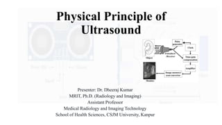
- 1. Physical Principle of Ultrasound Presenter: Dr. Dheeraj Kumar MRIT, Ph.D. (Radiology and Imaging) Assistant Professor Medical Radiology and Imaging Technology School of Health Sciences, CSJM University, Kanpur
- 2. Introduction • Welcome to the presentation on the Physical Principles of Ultrasound. Today, we will discuss the fundamental principles underlying medical ultrasound imaging, a crucial tool in radiology. Sound waves with frequencies higher than the upper audible limit of human hearing are called ultrasound. • The limit varies from person to person but is approximately 20,000 Hertz. The physical properties of ultrasound are similar to the normal audible sound. Monday, 16 October 2023 Physical Principle of Ultrasound By- Dr. Dheeraj Kumar 2
- 3. • Significance: Understanding these principles is essential for radiology professionals as it forms the basis for image formation and interpretation. • Agenda: We will explore ultrasound waves, their generation, the piezoelectric effect, transmission through tissues, reception, and imaging modes. Monday, 16 October 2023 Physical Principle of Ultrasound By- Dr. Dheeraj Kumar 3
- 4. Ultrasound Waves • Ultrasound Waves: Ultrasound is a high-frequency sound wave beyond the range of human hearing (>20,000 Hz). In medical imaging, frequencies typically range from 2 to 18 MHz. Monday, 16 October 2023 Physical Principle of Ultrasound By- Dr. Dheeraj Kumar 4
- 5. • Properties: Ultrasound waves have properties such as frequency (number of cycles per second), wavelength (distance between two wave cycles), and speed (typically 1540 m/s in soft tissues). • Inaudible Sound: These waves are inaudible to the human ear, making them safe for diagnostic purposes. Monday, 16 October 2023 Physical Principle of Ultrasound By- Dr. Dheeraj Kumar 5
- 6. Ultrasound Frequency Definition: Ultrasound frequency refers to the number of sound waves or cycles that pass through a point in one second. It is typically measured in Hertz (Hz) and falls within the range of sound frequencies beyond the upper limit of human hearing, typically above 20,000 Hz. • Example: Medical ultrasound imaging commonly uses frequencies in the range of 2 to 18 megahertz (MHz). For instance, a medical ultrasound machine operating at 5 MHz emits 5 million sound waves per second. Monday, 16 October 2023 Physical Principle of Ultrasound By- Dr. Dheeraj Kumar 6
- 7. Amplitude Definition: Amplitude in ultrasound refers to the maximum displacement of particles in a medium from their equilibrium position as a sound wave passes through. It indicates the strength or intensity of the ultrasound wave. A higher amplitude corresponds to a more powerful wave, while a lower amplitude indicates a weaker wave. • Example: In medical ultrasound, the amplitude of the sound wave determines the brightness of the images produced. Higher amplitude waves may be used to visualize structures with greater detail, such as when assessing the heart's valves and chambers, while lower amplitude waves are suitable for imaging soft tissues like the liver or kidneys. Monday, 16 October 2023 Physical Principle of Ultrasound By- Dr. Dheeraj Kumar 7
- 8. Wavelength Definition: Wavelength in ultrasound refers to the distance between two successive points that are in phase, meaning they are at the same point in their respective cycles. It is inversely related to frequency, meaning higher frequencies have shorter wavelengths and vice versa. • Example: If an ultrasound system emits waves at a frequency of 10 MHz, the corresponding wavelength in soft tissue will be approximately 0.15 millimeters. Shorter wavelengths are advantageous for high-resolution imaging because they can detect smaller structures, while longer wavelengths are better for deeper penetration, as they suffer less attenuation. Monday, 16 October 2023 Physical Principle of Ultrasound By- Dr. Dheeraj Kumar 8
- 9. Relationship The relationship between velocity (v), frequency (f), and wavelength (λ) in medical ultrasound can be expressed using the following formula: v = f x λ Where: • v represents the velocity of sound in the medium (typically soft tissue in medical ultrasound, approximately 1540 m/s). • f is the frequency of the ultrasound wave. • λ is the wavelength of the ultrasound wave. Monday, 16 October 2023 Physical Principle of Ultrasound By- Dr. Dheeraj Kumar 9
- 10. Tissue Type Ultrasound Velocity (m/s) Soft Tissue (Muscle) 1540 m/s Adipose (Fat) Tissue 1450 m/s Blood 1570 m/s Bone (Cortical) 4080 m/s Bone (Trabecular) 3080 m/s Brain 1540 m/s Liver 1550 m/s Kidney 1560 m/s Lung 480 m/s Water 1480 m/s Monday, 16 October 2023 Physical Principle of Ultrasound By- Dr. Dheeraj Kumar 10
- 11. Generation of Ultrasound • Generation: Ultrasound waves are generated using a transducer. A transducer is a critical component in an ultrasound machine, and it operates based on the piezoelectric effect. • Transducer: The transducer contains piezoelectric crystals that convert electrical energy into mechanical vibrations. • Vibration: When an electrical current is applied, the crystals vibrate, generating ultrasound waves that travel into the patient's body. Monday, 16 October 2023 Physical Principle of Ultrasound By- Dr. Dheeraj Kumar 11
- 12. (A) An ultrasound wave is depicted as a sine wave, propagating through tissue. Its wavelength depends on frequency, inversely related to it. Amplitude measures the energy or pressure change in the wave, influencing loudness (amplitude) and pitch (frequency). (B) Frequency determines pitch, while amplitude represents loudness in sound waves. (C) Pulse length is inversely related to transducer frequency, with higher frequencies producing shorter pulses. These pulses are emitted at a specific rate known as pulse repetition frequency. Monday, 16 October 2023 Physical Principle of Ultrasound By- Dr. Dheeraj Kumar 12
- 13. Piezoelectric Effect • Piezoelectric Effect: This effect is central to ultrasound technology. It refers to the ability of certain materials (e.g., quartz, PZT) to generate an electric charge when subjected to mechanical stress. • Two-Way Process: The piezoelectric effect is a two-way process. It generates mechanical vibrations when an electric current is applied (for ultrasound transmission) and generates electrical signals when mechanical vibrations are received (for ultrasound reception). Monday, 16 October 2023 Physical Principle of Ultrasound By- Dr. Dheeraj Kumar 13
- 14. Ultrasound Transmission • Transmission Through Tissues: Ultrasound waves travel through the body, encountering different tissues. They can be transmitted, reflected, refracted, and scattered. • Refraction: When ultrasound waves change direction as they pass through tissues with varying acoustic properties. • Reflection: When waves bounce back at tissue boundaries (e.g., bone-soft tissue interface), leading to echo formation. • Scattering: Random redirection of waves due to small structures within tissues. Monday, 16 October 2023 Physical Principle of Ultrasound By- Dr. Dheeraj Kumar 14
- 15. Ultrasound Reception • Receiving Ultrasound Echoes: The same transducer used for transmission also receives echoes. Here's how it works: • When ultrasound waves encounter tissues, some are reflected back. • These returning waves cause mechanical vibrations in the transducer's piezoelectric crystals. • These mechanical vibrations are then converted into electrical signals. • Echo Formation: The time it takes for the ultrasound waves to return and the amplitude of the returning waves are used to form an image. Monday, 16 October 2023 Physical Principle of Ultrasound By- Dr. Dheeraj Kumar 15
- 16. Ultrasound Imaging Modes • Imaging Modes: Ultrasound can produce various imaging modes, each with a specific clinical application. • B-Mode (Brightness Mode): This is the most common mode, providing a 2D cross-sectional image of tissues. It relies on the intensity of returning echoes to create brightness variations. • Doppler Mode: Used to assess blood flow by detecting frequency shifts in echoes caused by moving blood cells. Monday, 16 October 2023 Physical Principle of Ultrasound By- Dr. Dheeraj Kumar 16
- 17. Monday, 16 October 2023 Physical Principle of Ultrasound By- Dr. Dheeraj Kumar 17
- 18. • M-Mode (Motion Mode): This mode displays motion over time, suitable for cardiac imaging. • Color Doppler: Combines color and Doppler modes to visualize blood flow direction and velocity. • 3D and 4D Imaging: Advanced techniques that offer three-dimensional and real-time images of structures, enhancing diagnostic capabilities. Monday, 16 October 2023 Physical Principle of Ultrasound By- Dr. Dheeraj Kumar 18
- 19. Advantages of Ultrasound Understanding the physical principles of ultrasound helps us appreciate its numerous advantages in medical imaging. • Non-Invasive: Ultrasound is non-invasive, avoiding the use of ionizing radiation. • Real-Time Imaging: It provides real-time imaging, making it suitable for dynamic processes. • High Spatial Resolution: Ultrasound has excellent spatial resolution, enabling fine detail visualization. • Safe for All Ages: It is safe for patients of all ages, including pregnant women and infants. Monday, 16 October 2023 Physical Principle of Ultrasound By- Dr. Dheeraj Kumar 19
- 20. Limitations Despite its advantages, ultrasound has some limitations: • Depth Limitations: Limited depth penetration in obese patients or when imaging deep structures. • Sound Attenuation: Ultrasound waves weaken as they travel through tissues, potentially leading to reduced image quality at greater depths. • Artifacts: Ultrasound images can have artifacts like shadowing, reverberation, and refraction artifacts. • Operator-Dependent: Image quality can vary based on the operator's skill. Monday, 16 October 2023 Physical Principle of Ultrasound By- Dr. Dheeraj Kumar 20
- 21. Conclusion In conclusion, understanding the physical principles of ultrasound is vital for radiology professionals. It forms the foundation of medical ultrasound imaging and is central to providing safe, high-quality patient care. Monday, 16 October 2023 Physical Principle of Ultrasound By- Dr. Dheeraj Kumar 21
- 22. References 1. Kremkau, F. W. (2015). Sonography: Introduction to Normal Structure and Function. Saunders. 2. Hagen-Ansert, S. (2019). Textbook of Diagnostic Sonography: 2-Volume Set. Mosby. 3. Rumack, C. M., Wilson, S. R., & Charboneau, J. W. (2017). Diagnostic Ultrasound. Elsevier. 4. Oelze, M. L. (2008). Introduction to Infrasound: Piezoelectric Sensors. CRC Press. 5. Sauerbrey, G. (1959). Use of quartz vibration in microbalance studies. Journal of Physics and Chemistry of Solids, 4(2), 206-212. 6. Stuart, D. A., Yuen, J. M., & Lyandres, O. (2008). In vivo nanoimaging of membrane-associated biomolecules. Accounts of Chemical Research, 41(12), 1722-1731. Monday, 16 October 2023 Physical Principle of Ultrasound By- Dr. Dheeraj Kumar 22
- 23. Thank You Monday, 16 October 2023 Physical Principle of Ultrasound By- Dr. Dheeraj Kumar 23