Downloaded 1,491 times

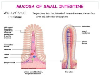
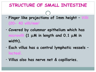
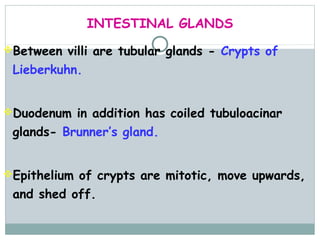
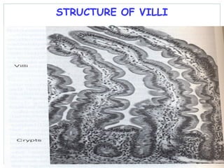
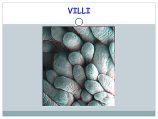
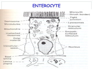
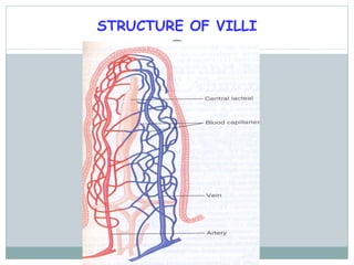
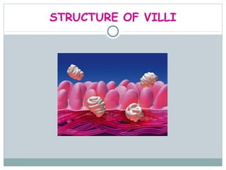
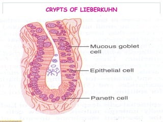
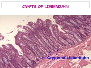
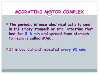
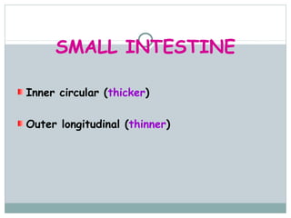
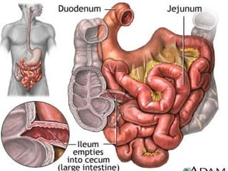
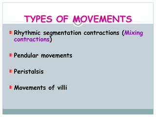
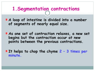
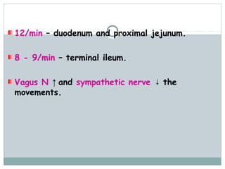
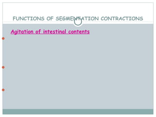
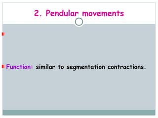
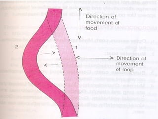
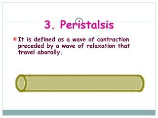
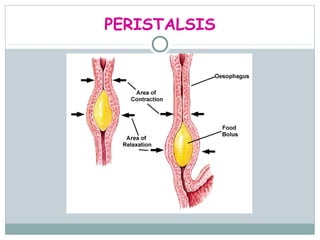
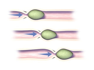

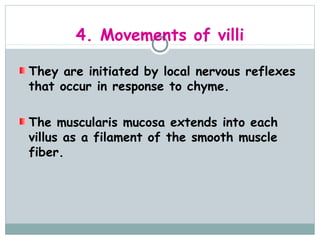
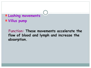
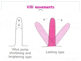


The small intestine is responsible for the digestion and absorption of nutrients. It is made up of the duodenum, jejunum, and ileum. The small intestine contains finger-like villi covered in microvilli that increase surface area for absorption. Glands including crypts of Lieberkuhn and Brunner's glands secrete enzymes and mucus. Movement patterns in the small intestine include segmentation contractions that mix contents, peristalsis that propels contents aborally, and villi movements that enhance absorption. Disorders can impair digestion or absorption.


![GI PHYSIOLOGY new].pptx](https://cdn.slidesharecdn.com/ss_thumbnails/giphysiologynew-230405152805-b9462356-thumbnail.jpg?width=640&height=640&fit=bounds)


![CASE_PRESENTATION_ON_subdural_hematoma(SDH)[1 FINAL PPT]-1.pptx](https://cdn.slidesharecdn.com/ss_thumbnails/casepresentationonsubduralhematomasdh1finalppt-1-260129172522-d405d375-thumbnail.jpg?width=640&height=640&fit=bounds)


