Git 8-csbrp
•Download as PPT, PDF•
8 likes•3,585 views

for Undergraduate Medical Students (MBBS)

Recommended
More Related Content
What's hot
What's hot (20)
Viewers also liked
Viewers also liked (20)
Similar to Git 8-csbrp
Similar to Git 8-csbrp (20)
More from Prasad CSBR
More from Prasad CSBR (20)
Recently uploaded
Recently uploaded (20)
Git 8-csbrp
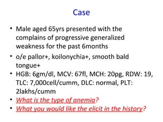
- 1. Case • Male aged 65yrs presented with the complains of progressive generalized weakness for the past 6months • o/e pallor+, koilonychia+, smooth bald tongue+ • HGB: 6gm/dl, MCV: 67fl, MCH: 20pg, RDW: 19, TLC: 7,000cell/cumm, DLC: normal, PLT: 2lakhs/cumm • What is the type of anemia? • What you would like the elicit in the history?
- 2. Case…. continued • Male aged 65yrs presented with the complains of progressive generalized weakness for the past 6months • o/e pallor+, koilonychia+, smooth bald tongue+ • HGB: 6gm/dl, MCV: 67fl, MCH: 20pg, RDW: 19, TLC: 7,000cell/cumm, DLC: normal, PLT: 2lakhs/cumm • No h/o hemorrhoids, peptic ulcer, hematuria • Stool for ova/parasites: Negative • Stool for occult blood: Positive • What is the next step?
- 3. Case…. continued • Colonoscopy: Large mass in the caecum • Diagnosis: Carcinoma of colon
- 4. Terms • Loss of heterozygosity – LOH • Constipation / obstipation
- 7. AdenocarcinomaAdenocarcinoma Small intestine: •Uncommon site for neoplasms •Tumors that may occur: – Adenocarcinoma – Carcinoid – Lymphoma – Sarcoma Large intestine: •Common site for adenocarcinoma
- 8. Colorectal carcinoma -Colorectal carcinoma - AdenocarcinomaAdenocarcinoma
- 9. Colorectal carcinomaColorectal carcinoma • Mostly Adenocarcinomas • 60-70yrs • < 20% of cases occur before 50yrs • M>F • Most common in developed countries – 30x less common in India / Africa
- 10. Colorectal carcinomaColorectal carcinoma Risk factors: •Low Dietary fiber •Diet rich in fat and refined carbohydrates •Deficiencies of vitamins A, C, and E
- 11. Colorectal carcinomaColorectal carcinoma Risk factors: •High fat intake enhances the hepatic synthesis of cholesterol and bile acids, which can be converted into carcinogens by intestinal bacteria
- 13. Colorectal carcinomaColorectal carcinoma Risk factors: •Dietary modification •Pharmacologic chemoprevention
- 14. Colorectal carcinomaColorectal carcinoma Risk factors: Dietary modification – Low fat & High fiber diet
- 15. Colorectal carcinomaColorectal carcinoma Risk factors: Pharmacologic chemoprevention – Aspirin or other NSAIDs have a protective effect
- 17. PathogenesisPathogenesis • Heterogeneous genetic abnormalities • Genetic and Epigenetic abnormalities Two distinct genetic pathways: 1.APC/β-catenin pathway – associated with WNT and the classic adenoma- carcinoma sequence 1.The microsatellite instability pathway
- 18. APC/β-Catenin Pathway • APC (WNT signaling pathway) - control cell fate, adhesion, and cell polarity during embryonic development • APC gene (5q21) down-regulate growth- promoting signals – ‘Tumor suppressor’ • An important function of the APC protein is to down-regulate β-catenin
- 19. APC/β-Catenin Pathway WNT signals through a family of cell surface receptors called frizzled (FRZ)
- 20. • 80% of sporadic colon tumors • Mutation of APC (LOH) • Additional mutations: – Activating mutations in KRAS – Mutations in SMAD2 and SMAD4 – p53 mutation – Methylation of promoter region – Increase expression of telomerase PathogenesisPathogenesis 1- Adenoma-carcinoma sequence1- Adenoma-carcinoma sequence
- 21. PathogenesisPathogenesis 1- Adenoma-carcinoma sequence1- Adenoma-carcinoma sequence
- 22. PathogenesisPathogenesis 2- Microsatellite instability2- Microsatellite instability • DNA mismatch repair deficiency • Accumulation of mutations in microsatellite repeats (Microsatellite instability) • Most of them are silent • Mutation resulting in MSI involving: – type II TGF-β receptor (uncontrolled cell growth) – the pro-apoptotic protein BAX (survival of genetically abnormal clones) • Mutations in the oncogene BRAF • Hypermethylation of MLH1 • KRAS and p53 are not typically mutated
- 23. PathogenesisPathogenesis 2- Microsatellite instability2- Microsatellite instability Thus, the combination of the following is the signature of this pathway of carcinogenesis: – MSI – BRAF mutation, and – Methylation of specific targets, such as MLH1
- 24. PathogenesisPathogenesis 2- Microsatellite instability2- Microsatellite instability
- 25. PathogenesisPathogenesis 3- Hypermethylation of3- Hypermethylation of promoter & absence of MSIpromoter & absence of MSI • Increased CpG island methylation in the absence of microsatellite instability • Many of these tumors harbor KRAS mutations, but p53 and BRAF mutations are uncommon
- 26. Morphology and molecular alterations • Sessile serrated adenomas: Mismatch repair deficiency and MSI • Tumors with prominent mucinous differentiation and peritumoral lymphocytic infiltrates: – MSI – Frequently located in the right colon
- 27. Morphology – Carcinoma of Colon • Can occur in any part of the colon In the proximal colon: – Polypoid, exophytic masses – Rarely cause obstruction – Symptoms occur LATELY In the distal colon: – Annular lesions – “Napkin-ring” constriction – Luminal narrowing > Obstruction – Symptoms occur EARLY
- 28. Carcinoma colonCarcinoma colon Exophytic type Napkin ring type
- 29. Napkin rings
- 30. Carcinoma colonCarcinoma colon Exophytic type Napkin ring type
- 31. Colorectal carcinoma – infiltration in to the muscle coat
- 32. Morphology – Microscopic features • Most tumors are composed of glands lined by tall columnar cells with features of malignancy • Invasive tumors elicits desmoplastic response • Poorly differentiated tumors form few glands • Some produce abundant mucin - poor prognosis • Some poorly differentiated tumors composed of signet-ring cells
- 33. Morphology – Microscopic features
- 34. Morphology – Microscopic features
- 35. Clinical FeaturesClinical Features • Right sided tumors: – Go undetected for long periods – Present with fatigue and weakness due to iron deficiency anemia • Left-sided tumors: – Occult bleeding – Changes in bowel habits – Cramping left lower quadrant discomfort
- 36. Clinical FeaturesClinical Features Clinical MAXIMClinical MAXIM:: •The underlying cause of iron deficiency anemia in an older man or postmenopausal woman is GI cancer until proven otherwise
- 37. The two most important Prognostic Factors 1. Depth of invasion • Invasion into the muscularis propria confers significantly reduced survival 1. Lymph node metastases
- 38. Metastases • Metastases may involve regional lymph nodes, lungs and bones • but LIVER is the most common site of metastatic lesions (as a result of portal drainage of the colon) • Mets in the liver are often umbilicated
- 44. HNPCCHNPCC
- 45. E N D
- 47. Dr.Denis Birkitt Fiber and Colonic carcinoma America is a constipated nation.... If you pass small stools, you have to have large hospitals – Denis Burkitt
- 49. Sister Joseph Lilly nodule
Editor's Notes
- obstipation [ˌɒbstɪˈpeɪʃən]n(Medicine) Pathol a severe form of constipation, usually resulting from obstruction of the intestinal tract [from Latin obstīpātiō, from ob- (intensive) + stīpāre to press together]
- Most of them adenocarcinomas. Colorectal cancer means adenocarcinoma, otherwise specified.
- Epidemiology. Each year in the United States there are more than 130,000 new cases and 55,000 deaths from colorectal adenocarcinoma. This represents nearly 15% of all cancer-related deaths, and is second only to lung cancer. Colorectal cancer incidence peaks at 60 to 70 years of age, and fewer than 20% of cases occur before age 50. Males are affected slightly more often than females. Colorectal carcinoma is most prevalent in the United States, Canada, Australia, New Zealand, Denmark, Sweden, and other developed countries. The incidence of this cancer is as much as 30-fold lower in India, South America, and Africa. In Japan, where incidence was previously very low, rates have now risen to intermediate levels (similar to those in the United Kingdom), presumably as a result of changes in lifestyle and diet.
- The dietary factors most closely associated with increased colorectal cancer rates are low intake of unabsorbable vegetable fiber and high intake of refined carbohydrates and fat. Although these associations are clear, the mechanistic relationship between diet and risk remains poorly understood. However, it is theorized that reduced fiber content leads to decreased stool bulk and altered composition of the intestinal microbiota. This change may increase synthesis of potentially toxic oxidative by-products of bacterial metabolism, which would be expected to remain in contact with the colonic mucosa for longer periods of time as a result of reduced stool bulk. Deficiencies of vitamins A, C, and E, which act as free-radical scavengers, may compound damage caused by oxidants. High fat intake enhances the hepatic synthesis of cholesterol and bile acids, which can be converted into carcinogens by intestinal bacteria.
- In addition to dietary modification, pharmacologic chemoprevention has become an area of great interest. Several epidemiologic studies suggest that aspirin or other NSAIDs have a protective effect. This is consistent with studies showing that some NSAIDs cause polyp regression in FAP patients in whom the rectum was left in place after colectomy. It is suspected that this effect is mediated by inhibition of the enzyme cyclooxygenase-2 (COX-2), which is highly expressed in 90% of colorectal carcinomas and 40% to 90% of adenomas. COX-2 is necessary for production of prostaglandin E2, which promotes epithelial proliferation, particularly after injury.[114] Of further interest, COX-2 expression is regulated by TLR4, which recognizes lipopolysaccharide and is also overexpressed in adenomas and carcinomas.
- An important function of the APC protein is to down-regulate β-catenin. In the absence of WNT signaling APC causes degradation of β-catenin, preventing its accumulation in the cytoplasm.[85] It does so by forming a macromolecular complex with β-catenin, axin, and GSK3β, which leads to the phosphorylation and eventually ubiquitination of β-catenin and destruction by the proteasome. Signaling by WNT blocks the APC-AXIN-GSK3β destruction complex, allowing β-catenin to translocate from the cytoplasm to the nucleus. In the cell nucleus, β-catenin forms a complex with TCF, a transcription factor that up-regulates cellular proliferation by increasing the transcription of c-MYC, cyclin D1, and other genes. Since inactivation of the APC gene disrupts the destruction complex, β-catenin survives and translocates to the nucleus, where it can activate transcription in cooperation with TCF.[85] Thus, cells with loss of APC behave as if they are under continuous WNT signaling. The importance of the APC/β-catenin signaling pathway in tumorigenesis is attested to by the fact that colon tumors that have normal APC genes harbor mutations in β-catenin that prevent its destruction by APC, allowing the mutant protein to accumulate in the nucleus. Dysregulation of the APC/β-catenin pathway is not restricted to colon cancers; mutations in the β-catenin gene are present in more than 50% of hepatoblastomas and in approximately 20% of hepatocellular carcinomas.[86] As mentioned in Chapter 3 , β-catenin binds to the cytoplasmic tail of E-cadherin, a cell surface protein that maintains intercellular adhesiveness. Loss of cell-cell contact, such as in a wound or injury to the epithelium, disrupts the interaction between E-cadherin and β-catenin, and allows β-catenin to travel to the nucleus and stimulate proliferation; this is an appropriate response to injury that can help repair the wound. Re-establishment of these E-cadherin contacts as the wound heals leads to β-catenin again being sequestered at the membrane and reduction in the proliferative signal; these cells are said to be “contact-inhibited.” Loss of contact inhibition, by mutation of the E-cadherin/β-catenin axis, or by other methods, is a key characteristic of carcinomas. Furthermore, loss of cadherins can favor the malignant phenotype by allowing easy disaggregation of cells, which can then invade locally or metastasize. Reduced cell surface expression of E-cadherin has been noted in many types of cancers, including those that arise in the esophagus, colon, breast, ovary, and prostate.[87] Germline mutations of the E-cadherin gene can predispose to familial gastric carcinoma, and mutation of the gene and decreased E-cadherin expression are present in a variable proportion of gastric cancers of the diffuse type. The molecular basis of reduced E-cadherin expression is varied. In a small proportion of cases, there are mutations in the E-cadherin gene (located on 16q); in other cancers, E-cadherin expression is reduced as a secondary effect of mutations in β-catenin genes. Additionally, E-cadherin may be down-regulated by transcription repressors, such as SNAIL, which have been implicated in epithelial-to-mesenchymal transition and metastasis[88] (discussed below).
- FIGURE 7-33 A, The role of APC in regulating the stability and function of β-catenin. APC and β-catenin are components of the WNT signaling pathway. In resting cells (not exposed to WNT), β-catenin forms a macromolecular complex containing the APC protein. This complex leads to the destruction of β-catenin, and intracellular levels of β-catenin are low. B, When cells are stimulated by WNT molecules, the destruction complex is deactivated, β-catenin degradation does not occur, and cytoplasmic levels increase. β-catenin translocates to the nucleus, where it binds to TCF, a transcription factor that activates genes involved in cell cycle progression. C, When APC is mutated or absent, the destruction of β-catenin cannot occur. β-catenin translocates to the nucleus and coactivates genes that promote entry into the cell cycle, and cells behave as if they are under constant stimulation by the WNT pathway.
- Figure 1. Wnt doesn&apos;t bind to the receptor. Axin, GSK and APC form a &quot;destruction complex,&quot; and β-Cat is destroyed. Figure 2. Wnt binds to (activates) the receptor. Axin is removed from the &quot;destruction complex.&quot; β-Cat moves into the nucleus, binds to a transcription factor on DNA, and activates transcription of a protein. &quot;P&quot; represents phosphate.
- The classic adenoma-carcinoma sequence, which accounts for as much as 80% of sporadic colon tumors, typically includes mutation of APC early in the neoplastic process ( Fig. 17-49 ). Both copies of the APC gene must be functionally inactivated, either by mutation or epigenetic events, for adenomas to develop. APC is a key negative regulator of β-catenin, a component of the WNT signaling pathway (see Chapter 7 ). The APC protein normally binds to and promotes degradation of β-catenin. With loss of APC function, β-catenin accumulates and translocates to the nucleus, where it activates the transcription of genes, such as those encoding MYC and cyclin D1, which promote proliferation. This is followed by additional mutations, including activating mutations in KRAS, which also promote growth and prevent apoptosis. The conclusion that mutation of KRAS is a late event is supported by the observation that mutations are present in fewer than 10% of adenomas less than 1 cm in diameter but are found in 50% of adenomas greater than 1 cm in diameter and 50% of invasive adenocarcinomas. Neoplastic progression is also associated with mutations in other tumor suppressor genes such as those encoding SMAD2 and SMAD4, which are effectors of TGF-β signaling. Because TGF-β signaling normally inhibits the cell cycle, loss of these genes may allow unrestrained cell growth. The tumor suppressor p53 is mutated in 70% to 80% of colon cancers, but is uncommonly affected in adenomas, suggesting that p53 mutations also occur at late stages of tumor progression. “Loss of function” of p53 and other tumor suppressor genes is often caused by chromosomal deletions, pointing out that chromosomal instability is a hallmark of the APC/β-catenin pathway. Alternatively, tumor suppressor genes may be silenced by methylation of a CpG-rich zone, or CpG island, a 5′ region of some genes that frequently includes the promoter and transcriptional start site. Expression of telomerase also increases as lesions become more advanced.
- FIGURE 17-49 Morphologic and molecular changes in the adenoma-carcinoma sequence. It is postulated that loss of one normal copy of the tumor suppressor gene APC occurs early. Individuals may be born with one mutant allele, making them extremely prone to develop colon cancer, or inactivation of APC may occur later in life. This is the “first hit” according to Knudson&apos;s hypothesis ( Chapter 7 ). The loss of the intact copy of APC follows (“second hit”). Other mutations include those on KRAS, losses at 18q21 involving SMAD2 and SMAD4, and inactivation of the tumor suppressor gene p53, lead to the emergence of carcinoma, in which additional mutations occur. Although there seems to be a temporal sequence of changes, the accumulation of mutations, rather than their occurrence in a specific order, is most critical.
- FIGURE 17-50 Morphologic and molecular changes in the mismatch repair pathway of colon carcinogenesis. Defects in mismatch repair genes result in microsatellite instability and permit accumulation of mutations in numerous genes. If these mutations affect genes involved in cell survival and proliferation, cancer may develop.
- Right sided tumors go undetected for long periods.
- FIGURE 17-51 Colorectal carcinoma. A, Circumferential, ulcerated rectal cancer. Note the anal mucosa at the bottom of the image. B, Cancer of the sigmoid colon that has invaded through the muscularis propria and is present within subserosal adipose tissue (left). Areas of chalky necrosis are present within the colon wall (arrow).
- The general microscopic characteristics of right- and left-sided colonic adenocarcinomas are similar. Most tumors are composed of tall columnar cells that resemble dysplastic epithelium found in adenomas ( Fig. 17-52A ). The invasive component of these tumors elicits a strong stromal desmoplastic response, which is responsible for their characteristic firm consistency. Some poorly differentiated tumors form few glands ( Fig. 17-52B ). Others may produce abundant mucin that accumulates within the intestinal wall, and these are associated with poor prognosis. Tumors may also be composed of signet-ring cells that are similar to those in gastric cancer.
- FIGURE 17-52 Histologic appearance of colorectal carcinoma. A, Well-differentiated adenocarcinoma. Note the elongated, hyperchromatic nuclei. Necrotic debris, present in the gland lumen, is typical. B, Poorly differentiated adenocarcinoma forms a few glands but is largely composed of infiltrating nests of tumor cells. C, Mucinous adenocarcinoma with signet-ring cells and extracellular mucin pools.
- FIGURE 17-52 Histologic appearance of colorectal carcinoma. A, Well-differentiated adenocarcinoma. Note the elongated, hyperchromatic nuclei. Necrotic debris, present in the gland lumen, is typical. B, Poorly differentiated adenocarcinoma forms a few glands but is largely composed of infiltrating nests of tumor cells. C, Mucinous adenocarcinoma with signet-ring cells and extracellular mucin pools.
- Note: carcinomas of the anal region that metastasize often circumvent the liver. Umbilication is due to central necrosis.
- Note the numerous mass lesions that are of variable size. Some of the larger ones demonstrate central necrosis. The masses are metastases to the liver. The obstruction from such masses generally elevates alkaline phosphatase, but not all bile ducts are obstructed, so hyperbilirubinemia is typically not present. Also, the transaminases are usually not greatly elevated.
- FIGURE 17-53 Metastatic colorectal carcinoma. A, Lymph node metastasis. Note the glandular structures within the subcapsular sinus. B, Solitary subpleural nodule of colorectal carcinoma metastatic to the lung. C, Liver containing two large and many smaller metastases. Note the central necrosis within metastases
- America is a constipated nation.... If you pass small stools, you have to have large hospitals – Denis Burkitt