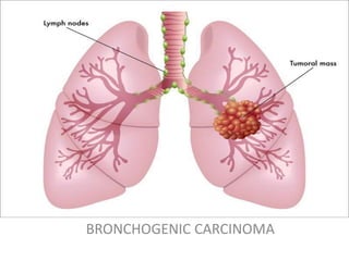
Bronchogenic Carcinoma
•Download as PPTX, PDF•
19 likes•7,046 views

Bronchogenic Carcinoma

Recommended
More Related Content
What's hot
What's hot (20)
Similar to Bronchogenic Carcinoma
Similar to Bronchogenic Carcinoma (20)
More from Dr. Varughese George
More from Dr. Varughese George (20)
Recently uploaded
Recently uploaded (20)
Bronchogenic Carcinoma
- 2. Learning Objectives At the end of this presentation, you should know • Different histological types of bronchogenic carcinoma • Gross appearance of each type • Microscopic appearance of each type. • Differences between each type.
- 3. BRONCHOGENIC CARCINOMA There are four main histologic types of primary bronchogenic carcinoma : - 1. Squamous cell carcinoma 2. Adenocarcinoma 3. Bronchioalveolar carcinoma 4. Small cell carcinoma 5. Large cell carcinoma
- 4. SQUAMOUS CELL CARCINOMA GROSS : • The tumour is often hilar or central arising from a large bronchus • Variable size and invades adjacent lung parenchyma • Cut surface of tumour shows extensive necrosis and cavitation C/S grey-white fleshy tumour in bronchus at its bifurcation and occluding lumen partly (arrow) The tumour is seen extending directly into adjacent lung parenchyma and hilar nodes
- 5. SQUAMOUS CELL CARCINOMA MICROSCOPY : • Varying grades of differentiation from well-differentiated with keratinisation to poorly differentiated • Intercellular bridges or keratinisation often seen in well differentiated • Edge of tumour often shows squamous metaplasia, epithelial dysplasia and carcinoma insitu Islands of invading malignant squamous cells are seen. A few well-developed cell nests with keratinisation are evident.
- 6. ADENOCARCINOMA GROSS : • Classically peripheral tumors which does not form a cavitary lesion • Single or multiple solid firm yellow- white nodule or mass which may invade pleura Peripheral tumor
- 7. ADENOCARCINOMA MICROSCOPY : • Invasive malignant epithelial tumor with glandular differentiation or mucin production by tumor cells • Various patterns- acinar, lepidic, papillary, micropapillary, and solid with mucin formation • Tumors show desmoplastic reaction, lymphovascular or pleural invasion with areas of necrosis Gland-forming adenocarcinoma
- 8. BRONCHIOALVEOLAR CARCINOMA • Variant of adenocarcinoma • Growth along existing alveolar walls ( lepidic pattern) • No association with smoking
- 9. Small cell carcinoma GROSS : • The tumour is frequently hilar or central in location • The tumour appears as a nodule measuring 1-5 cm in diameter with ulcerated surface • Cut surface - tumour is yellowish- white with areas of necrosis and haemorrhages
- 10. SMALL CELL CARCINOMA MICROSCOPY : • Tumour cells -uniform, small, larger than lymphocytes • Dense round or oval nuclei having diffuse chromatin, inconspicuous nucleoli and scanty cytoplasm • Tumour cells arranged in cords, aggregates and ribbons, or around small blood vessels forming pseudorosettes The tumour cells are arranged in sheets,cords, aggregates and at places form pseudorosettes. The individual tumour cells are small, uniform, lymphocyte-like with scanty cytoplasm.
- 11. LARGE CELL CARCINOMA GROSS : • The tumor is peripheral with lobulated apperance and bulging borders • Cut surface shows gray white "fish flesh" areas with areas of hemorrhage and necrosis
- 12. LARGE CELL CARCINOMA MICROSCOPY : • Tumour cells - large, polygonal and anaplastic cells growing in sheets or solid nests • No clear adenocarcinoma, squamous or neuroendocrine morphology • Moderately abundant cytoplasm, well defined cell borders, vesicular nuclei, prominent nucleoli • Foci of central necrosis and hemorrhage may be present The tumor cells are pleomorphic and show no evidence of squamous or glandular differentiation.