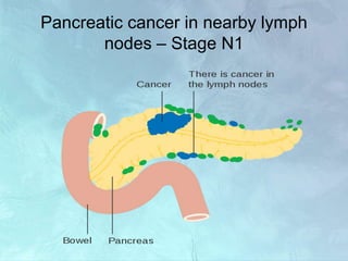
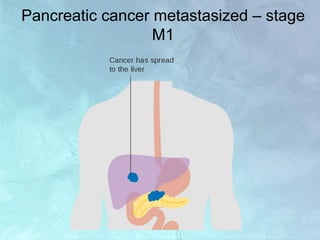
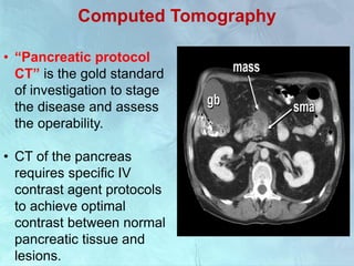
Case scenario
A 63-year-old Caucasian male was admitted to the hospital
from the emergency room with symptoms of epigastric pain
that radiated toward the back, abdominal distention,
vomiting, and jaundice
Laboratory tests:
• Bilirubin and liver enzymes; elevated
• CBC values WNL
• Hepatitis B, & C testing, negative
• CEA: 34.2 ng/mL; CA 19-9 > 12000 U/mL
• CT reveals 3.5 cm × 3.7 cm mass in the head of the
pancreas and multiple liver nodules; also, indicates an
obstruction of the bile duct.
• Diagnosis: stage IV pancreatic cancer with liver metastasis
Care of patient with Ca
Pancreas
Presenter:
Saumya P.Srivastava
Msc.nsg 2nd year
CON,AIIMS
Moderator:
Dr.L.Gopichandran
Lecturer
CON,AIIMS
Objectives
At the end of this session, you will be able to:
• To introduce the topic
• Explain epidemiology of pancreatic cancer
• Describe anatomy and physiology of pancreas
• Enumerate Etiology and risk factors associated with
pancreatic cancer.
• Explain Staging of pancreatic cancer
• Enlist Types of pancreatic cancer
• Explain pathogenesis of pancreatic cancer
• Describe clinical presentation of pancreatic cancer
• Enlist investigations
• Explain management(medical,surgical and nursing)
Introduction
• Pancreatic cancers can arise from the exocrine and
endocrine portions of the pancreas,93% of them
develop from the exocrine portion.
• Fourth leading cause of cancer deaths, being
responsible for 7% of all cancer-related deaths in
men and 8% in women.
• Approximately 75% of all pancreatic carcinomas
occur within the head or neck of the pancreas, 15-
20% occur in the body of the pancreas, and 5-10%
occur in the tail.
• It is a biologically aggressive tumor from the onset .
• Remains clinically queisent for a long time and
hence present in advanced state.
Location of pancreatic cancer
Epidemiology
• Pancreatic cancer is the ninth most common cancer in
women and the tenth most common cancer in men.
• The 5-year survival rate for people with pancreatic cancer
is 9%.
• The overall incidence rate of pancreatic cancer increased
by about 1% per year from 2006 to 2015.
• Incidence rates are 25% higher in black people than in
white people.
• Most pancreatic cancers (93%) are exocrine
adenocarcinoma
Cont..
• pancreatic cancer remains one of the most lethal
malignant neoplasms that caused 432,242 new
deaths in 2018 (GLOBOCAN 2018 estimates).
• Annual incidence 10 new cases per 100000
population.
• The American Cancer Society estimates that in the
United States in 2019, about 45,750 people (23,800
men and 21,950 women) will die of pancreatic
cancer.
Age and pancreatic cancer
Median age is 69 years in whites and
65 years in blacks
Anatomy Of Pancreas
• Pancreas is a long
retroperitoneal organ 15 to 20
cm in length.
• Weighs about 80 gms ,lies
against L1 & L2 Vertebra.
• It is arbitarily divided into
HEAD,NECK BODY & TAIL
• Head lies within the concavity of
duodenum against second
lumbar vertebra and body
overlies the first lumbar vertebra
Cont..
• The pancreas is made up of 2 types of glands:
 Exocrine
 Endocrine
Arterial and venous supply of
pancreas
Ducts of pancreas
Functions of the pancreas
The pancreas has digestive and hormonal functions:
• The enzymes secreted by the exocrine gland in the
pancreas help break down carbohydrates, fats,
proteins, and acids in the duodenum.
• The hormones secreted by the endocrine gland in
the pancreas are insulin and glucagon, which
regulate the level of glucose in the blood, and
somatostatin, which prevents the release of insulin
and glucagon.
Etiology and risk factors
• Older Age
• Male sex
• Heredity - cancer family syndromes
• Cigarette smoking
• Obesity
• Diet – high intake of red meat.
• Occupational
• Exposure to radiations
• Gastric surgeries
• Diabetes mellitus/pernicious anaemia/ chronic
Pancreatitis
Etiology-hereditary factors
• Hereditary pancreatitis
• Multiple endocrine neoplasia (MEN)
• Hereditary nonpolyposis rectal cancer (HNPCC)
• Familial adenomatous polyposis (FAP) and
Gardner syndrome
• Familial atypical multiple mole melanoma
(FAMMM) syndrome
• Hippel-Lindau syndrome (VHL), and germline
mutations in the BRCA1 and BRCA2 genes
Etiology – Diabetes – Is it a cause or
effect
• Several studies have shown an increased incidence
of pancreatic cancer in diabetics
• Diabetes is considered as an early symptom of
pancreatic cancer rather than being a cause
• The diabetes of Pancreatic cancer is due to islet cell
dysfunction (Islet Amyloid polypeptide) and not due
to the destruction of the gland
Types of pancreatic cancer
Exocrine tumors
• Pancreatic adenocarcinoma(85%)
Rare types:
• Adenosquamous
carcinomas
• Squamous cell carcinoma
• Signet ring cell carcinomas
• Undifferentiated carcinomas
• Undifferentiated carcinomas with
giant cells
• Pancretoblastoma
• Cystadenocarcinomas
• Colloid carcinomas
Benign and precancerous growths in the
pancreas
• Serous cystic neoplasms (SCNs)
• Mucinous cystic neoplasms (MCNs)
• Intraductal papillary mucinous neoplasms
(IPMNs)(most common of all)
• Solid pseudopapillary neoplasms (SPNs)
Pancreatic Neuroendocrine Tumors
(PNETs)
• About 7 percent of pancreatic tumors are
neuroendocrine tumors (pancreatic NETs or PNETs),
also called islet cell tumors. They often grow slower
than exocrine tumors.
• Pancreatic neuroendocrine tumors are either
functional (produce hormones) or nonfunctional (do
not produce hormones). Most PNETs are
nonfunctional
Pancreatic Neuroendocrine Tumors
(PNETs)
Common PNET types are:
• Gastrinoma (gastrin)
• Glucaganoma (glucagon)
• Insulinoma (insulin)
• Somatostatinoma (somatostatin)
• VIPoma (vasoactive intestinal peptide)
• Nonfunctional Islet Cell Tumor (no hormones)
Pathological (WHO) classification
• Primary ( 93% )
• Metastatic ( 7 % )
Duct cell origin – 90%
• Duct cell
adenocarcinoma – 75 %
• Mucinous carcinoma
• Cystadenocarcinoma
Acinar cell origin – 1%
• Acinar cell carcinoma
• Cystadenocarcinoma
( acinar cell )
Pathological (WHO) classification
Uncertain
histogenesis ( 9% )
• Pancreatoblastoma
• Papillary and cystic
neoplasm
• Mixed tumours
Connective tissue origin ( 1 % )
• Malignant fibrous histocytoma
• Osteogenic sarcoma
• Leiomyosarcoma
• Hemangio pericytoma
Pathogenesis
Staging of pancreatic cancer
Stage T1 pancreatic cancer
Stage T2 pancreatic cancer
Stage T3 pancreatic cancer
Stage T4 pancreatic cancer
Pancreatic cancer in nearby lymph
nodes – Stage N1
Pancreatic cancer metastasized – stage
M1
Clinical presentation
• Initial symptoms of the disease are often quite
nonspecific.
• Nonspecific symptoms such as anorexia, malaise,
nausea, fatigue, and midepigastric or back pain.
• Midepigastric pain , with radiation of the pain to the
midback or lower-back region.
• Weight loss
• Gastric outlet obstruction
• Delayed gastric emptying
• The onset of symptoms are insidious and
progressive
• Abdominal pain is usually post prandial
and in epigastrium
Tumors in the Head of pancreas
• Weight loss –
averaging about
40%
• Obstructive jaundice
• Deep seated
abdominal pain
• Non tender palpable
gall bladder
• Cholangitis occurs in
10 % of patients
Carcinoma of body and tail of pancreas
• Weight loss
• Deep seated pain
• Jaundice- < 10 % of
patient
• New onset of diabetes
mellitus-25% of
Patient
• Migratory
thrombophlebitis-
occurs in about10%
patient
Research Input
Diabetes and Pancreatic Cancer: Both Cause and
Effect Harvey A Risch
JNCI: Journal of the National Cancer Institute, Volume 111, Issue
1, January 2019
“About 25% of patients with pancreatic cancer have diabetes
mellitus at diagnosis, and roughly another 40% have pre-
diabetes,” that “patients with long-term (five or more years) type
II diabetes have a 50% increased risk of pancreatic cancer” and
“pancreatic cancer can cause diabetes, and sometimes diabetes
is an early sign of the tumor.”
Carcinoma of ampulla of vater
• Pain occurs less
frequently – usually its
colicky
• Jaundice is often
intermittent
• Chills and fever – due
to associated
cholangitis
Physical examination
• Pain radiating to the midback or lower-back region.
• Mild to moderate midepigastric tenderness.
• Palpable gallbladder (ie, Courvoisier sign)
• skin excoriations from unrelenting pruritus.
• Ascites
• Palpable abdominal mass
• Hepatomegaly from liver metastases
• Splenomegaly from portal vein obstruction
• Sister Mary Joseph nodule
• Blumer's shelf
• A metastatic node may be palpable behind the
medial end of the left clavicle (Virchow's node).
Differential diagnosis
• Acute Pancreatitis
• Cholangitis
• Cholecystitis
• Choledochal Cysts
• Chronic Pancreatitis
• Gallstones (Cholelithiasis)
• Gastric Cancer
• Peptic Ulcer Disease
Investigations
 Lab investigations
• CBC
• LFT
• RFT
• Coagulation profile
• CXR
• ECG
• Echo
 Imaging modalities:
• Computed tomography (CT)
• Transcutaneous ultrasonography (TUS)
• Endoscopic ultrasonography (EUS)
• Magnetic resonance imaging (MRI)
• Endoscopic retrograde
cholangiopancreatography (ERCP)
• Positron emission tomography (PET)
 Tumor Markers
• Carbohydrate antigen 19-9
• Carcinoembryonic antigen
Laboratory Findings
• Mild normochromic anemia
• Thrombocytosis
• In patients with obstructive jaundice:
bilirubin (conjugated and total)
alkaline phosphatase
gamma-glutamyl transpeptidase
aspartate aminotransferase
alanine aminotransferase
• Raised Serum amylase and/or lipase levels
• low serum albumin or cholesterol level in
patients with advanced pancreatic cancer.
Computed Tomography
• “Pancreatic protocol
CT” is the gold standard
of investigation to stage
the disease and assess
the operability.
• CT of the pancreas
requires specific IV
contrast agent protocols
to achieve optimal
contrast between normal
pancreatic tissue and
lesions.
Cont..
Includes:
• dual-phase CT acquisition
• IV contrast medium administration at a flow rate of
3–5 mL/s
• Helical or multislice
Advantages
• Available easily
• Surgeons are familiar with CT
• Excellent in giving details of operability
Disadvantages
• May miss liver mets less than 1 cm
• Miss peritoneal mets
Transcutaneous Ultrasonography
• As an initial screening test in evaluating patients
who present with possible obstructive jaundice.
• TUS has less utility in pancreatic carcinoma than
CT scanning.
• TUS can detect only 60-70% of pancreatic
carcinomas, more than 40% of the lesions smaller
than 3 cm are missed.
Endoscopic Ultrasonography
• It has proven to be the most sensitive and specific
diagnostic test for pancreatic cancer.
• Diagnostic advantage is EUS-guided fine-needle
aspiration.
• EUS as complementary to CT
Endoscopic Retrograde
Cholangiopancreatography
• Highly sensitive
• Usually reserved as a therapeutic procedure for
biliary obstruction.
• Brush cytology and forceps biopsy for histologically
diagnosing pancreatic cancer.
• Therapeutic palliation of obstructive jaundice through
stent placement
MRI
• The role of MRI in pancreatic cancer has been less well
studied.
• MRCP can be used as a noninvasive method for imaging
the biliary tree and pancreatic duct.
PET Scan
• Useful in detecting occult metastatic disease.
• False-positive PET scans have been reported in
pancreatitis.
• PET-CT scanning is more sensitive than conventional
imaging for the detection of pancreatic cancer.
Needle Aspiration
• EUS-guided fine-
needle aspiration
has proven to be the
most effective
means for making a
definitive cytologic
diagnosis of
pancreatic
carcinoma.
Treatment and management
• Resection is the only chance for a cure, and
resectable patients show undergo surgery without
delay followed by adjuvant therapy
• Borderline resectable patients may benefit from
neoadjuvant therapy and then surgery
• Unresectable patients may benefit from
chemotherapy or chemoradiation
• Metastatic disease may benefit from
chemotherapy or other palliative treatments
Treatment options
• Chemotherapy
• Adjuvant therapy
• Whipple procedure
• Distal pancreatectomy
• Total pancreatectomy
• Palliative therapy
• Dietary management
Chemotherapy
following drugs are approved by the US Food and
Drug Administration (FDA) for pancreatic cancer:
• Capecitabine (Xeloda)
• Erlotinib (Tarceva), a type of Targeted therapy (see
below)
• Fluorouracil (5-FU)
• Gemcitabine (Gemzar)
• Irinotecan (Camptosar)
• Leucovorin (Wellcovorin)
• Nab-paclitaxel (Abraxane)
• Nanoliposomal irinotecan (Onivyde)
• Oxaliplatin (Eloxatin)
Chemotherapy regimens
Adjuvant chemotherapy
• Gemcitabine 1000 mg/m2 IV over 30 min weekly for
3 wk; every 4 wk for six cycles.
• mFOLFIRINOX: Oxaliplatin 85 mg/m², leucovorin
400 mg/m², irinotecan 150 mg/m², 5-FU 2.4 g/m²
over 46 hours) every 14 days for 12 cycles.
Adjuvant chemotherapy and chemoradiation
• Concurrent chemoradiation starting 1-2 wk after
gemcitabine: 5-fluorouracil (FU) 250 mg/m2/day
continuous IV infusion via pump during radiation
Cont..
Treatment recommendations for locally advanced,
unresectable disease
• Gemcitabine 1000 mg/m2 IV over 30 min weekly for
3 wk; every 28 d.
• 5-FU 500 mg/m2/day IV bolus on days 1-3 and 29-
31 with concurrent radiotherapy, 40 Gy
Treatment recommendations for metastatic
disease
• Gemcitabine 1000 mg/m2 IV over 30 min weekly for
7 wk, followed by 1 wk off, then weekly for 3 wk;
every 28 d
• Gemcitabine 1000 mg/m2 IV weekly for 3 wk; every
28 d; plus capecitabine 1660 mg/m2/day weekly for
3 wk; every 28 d
Adjuvant therapy
• Adjuvant therapy with gemcitabine is accepted
as standard therapy for surgically resected
pancreatic cancer.
• Superior survival demonstrated with modified
FOLFIRINOX (mFOLFIRINOX)
Neoadjuvant therapy
Neoadjuvant therapy for patients with resectable or
borderline resectable tumor:
• FOLFIRINOX, with or without subsequent
chemoradiation
• Gemcitabine + albumin-bound paclitaxel, with or
without subsequent chemoradiation
• Gemcitabine + cisplatin (≥2–6 cycles) followed
by chemoradiation (only for
known BRCA1/2 mutations)
Complications related to
FOLFIRINOX
• Gastrointestinal toxicity
• Myelosuppression
• Infection
• Neuropathy
• Fatigue
Cont..
• In patients with unresectable locally advanced
pancreatic cancer, local ablation has been explored
as a treatment option.
• Radiofrequency ablation (RFA)
• Irreversible electroporation
• Stereotactic body radiation therapy (SBRT)
• High-intensity focused ultrasound (HIFU)
• Iodine-125
• Iodine-125–cryosurgery
• Photodynamic therapy
• Microwave ablation
Pancreaticoduodenectomy (Whipple
Procedure)
• It is a complex operation to remove the head of
the pancreas, the first part of the small intestine
(duodenum),antrum of stomach with surgical
drainage of the distal pancreatic duct and biliary
system, usually accomplished through
anastomosis to the jejunum
• It is the most often used surgery to treat pancreatic
cancer that's confined to the head of the pancreas.
Distal Pancreatectomy
• Distal pancreatectomy is a procedure in which the tail
and/or portion of the body of the pancreas are
removed, but not the head.
• Distal pancreatectomy may be an effective procedure
for tumors located in the body and tail of the pancreas.
Total Pancreatectomy
• The indication for the use of total pancreatectomy is
in cases in which the tumor involves the neck of the
pancreas.
• This can either be a situation in which the tumor
originates from the neck or is growing into the neck
Pancreatic transplantation
• Suitable donors between 20 and 50 yrs.
• Pancreatic blood flow to be maintained- warm
ischemia
• Gland should be perfused with a cold preservation
fluid hypertonic citrate solution
• Pancreas removed avoiding damage to the gland–
injection of collagenase enzyme into the pancreatic
duct under pressure.
• Pancreas transported to processing centre-within
four hours cold ischemia.
Complications related to surgery
• Pancreatic anastomotic leak
• Malabsorption
• Pancreatic stump leak
• Hemorrhage
• Endocrine insufficiency
• Insulin-dependent diabetes
• Delayed gastric emptying
• Intraabdominal abscess
• Biliary fistula
• Cholangitis
• Pancreatitis
Palliative Therapy
PAIN
 Narcotic analgesics
with tricyclic
antidepressants
 Antiemetics
 Endoscopic
decompression with
stents.
JAUNDICE
 Endoscopic placement
of plastic or metal
stents.
 Biliary decompression
 choledochojejunostomy
Palliative Therapy
DUODENAL OBSTRUCTION
 gastrojejunostomy or an
endoscopic procedure.
 Endoscopic stenting of
duodenal obstruction
Dietary management
• Pancreatic enzyme replacement supplements.
• Eat little and often, aiming for a small meal or snack
every 2-3 hours.
• Eat more protein rich foods (meat, fish, eggs,
cheese, beans, lentils and pulses).
• Avoid Foods that contain lots of cheese,high fibre
foods, spicy foods.
• Choose carbohydrates with a lower glycaemic
index (GI).
Recent updates
• MR Based, Screening Program for Individuals at Risk
for Pancreas Cancer .
• Neoadjuvant Therapy for Pancreatic
Adenocarcinoma.
• Role of biopsy
• Preop biliary drainage
• Modified FOLFIRINOX (mFOLFIRINOX) also
provides significantly longer survival than gemcitabine
in patients with pancreatic ductal adenocarcinomas
who have undergone R0 or R1 resection.
Research Input
• Early Detection of Pancreatic Cancer: The Role of
Depression and Anxiety as a Precursor for Disease
Kenner, Barbara, J., PhD Pancreas: April 2018 -
Volume 47 - Issue 4 - p 363–367
Background: The depression-before-diagnosis relationship
is particularly notable in pancreatic cancer, which has very
few symptoms and frequently is a systemic disease at the
time of detection.
A review of the literature substantiates
that depression and anxiety may be potential precursors to
a pancreatic cancer diagnosis. Studies have started to look
more closely at this link, with an eye to developing a
testable marker for pancreatic cancer.
Cont..
• Conclusion: General practitioners and mental health
professionals need to be educated that these symptoms are
critically important when reported by an individual who has
not had depression or anxiety previously, is older than 50
years, and may be at high risk for pancreatic cancer (eg,
familial, new onset of diabetes).
• In those situations, referral to a mental health practitioner
who can further assess the patient may be as important as
providing a prescription that can help alleviate the
depression or anxiety.
Post-op management
• Hospital stays vary from 12 to 23 days, depending
on the seriousness of the intervention.
• The nasogastric tube (NGT) is required between
the POD 4 and 7.
• Assess for mild signs of infection, such as a
leukocytosis and fever during the first
postoperative week.
• Exogenous pancreatic enzymes.
• Regular blood glucose measurement is important
(approximately every 3 to 6 months).
Nursing management
• Acute pain related to obstruction of pancreatic, biliary ducts
possibly evidenced by reports of pain.
• Risk for deficient fluid volume related to excessive losses:
vomiting, gastric suctioning.
• Imbalanced Nutrition: Less Than Body Requirements related
to vomiting, decreased oral intake as evidenced by aversion
to eating,weight loss.
• Risk for Infection related to Inadequate primary defenses:
stasis of body fluids, altered peristalsis, change in pH of
secretion.
• Deficient Knowledge related to lack of exposure/recall,
unfamiliarity with information resources
Acute pain related to obstruction of
pancreatic, biliary ducts possibly evidenced
by reports of pain.
• Investigate verbal reports of pain, noting specific
location and intensity (0–10 scale).
• Note factors that aggravate and relieve pain.
• Maintain bedrest during acute attack. Provide quiet,
restful environment.
• Promote position of comfort on one side with knees
flexed, sitting up and leaning forward.
• Provide alternative comfort measures (back rub),
encourage relaxation techniques (guided imagery,
visualization), quiet diversional activities (TV, radio).
Cont..
• Administer medication as indicated:Narcotic
analgesics, sedatives, antispasmodics.
• Withhold food and fluid as indicated.
• Maintain gastric suction when used
Risk for deficient fluid volume related to
excessive losses: vomiting, gastric
suctioning
• Monitor BP and measure CVP if available.
• Measure I&O including vomiting, gastric aspirate,
diarrhea. Calculate 24-hr fluid balance.
• Note decrease in urine output (less than 400 mL per
24 hr).
• Note poor skin turgor, dry skin and mucous
membranes, reports of thirst.
• Investigate changes in sensorium (confusion, slowed
responses).
• Administer fluid replacement as indicated (saline
solutions, albumin, blood, blood products, dextran).
Cont..
• Monitor laboratory studies (Hb and Hct, Protein,
albumin, electrolytes, BUN, creatinine, urine
osmolality and sodium, potassium, coagulation
studies).
Imbalanced Nutrition: Less Than Body
Requirements related to vomiting, decreased oral
intake as evidenced by aversion to eating,weight
loss
• Assess abdomen, noting presence and character of
bowel sounds, abdominal distension, and reports of
nausea.
• Provide frequent oral care.
• Assist patient in selecting food and fluids that meet
nutritional needs and restrictions when diet is resumed.
• Note signs of increased thirst and urination or changes in
mentation and visual acuity.
• Maintain NPO status and gastric suctioning in acute
phase.
• Administer medications :Vitamins: A,D,E,K, Replacement
enzymes.
Risk for Infection related to Inadequate primary
defenses: stasis of body fluids, altered peristalsis,
change in pH of secretion.
• Use strict aseptic technique when changing surgical
dressings or working with IV lines, indwelling
catheters and tubes, drains.
• Stress importance of good handwashing.
• Observe rate and characteristics of respirations,
breath sounds. Note occurrence of cough and
sputum production.
• Fever and respiratory distress in conjunction with
jaundice.
• Obtain culture specimens (blood, wound, urine,
sputum, or pancreatic aspirate).
• Administer antibiotic therapy as indicated
Deficient Knowledge related to lack of
exposure/recall, unfamiliarity with information
resources
• Review specific cause of current episode and
prognosis.
• Explore availability of treatment programs and
rehabilitation of chemical dependency if indicated.
• Stress the importance of follow-up care, and review
symptoms that need to be reported immediately to
physician.
• Review importance of initially continuing bland, low-
fat diet with frequent small feedings.
• Instruct in use of pancreatic enzyme replacements
and bile salt therapy as indicated.
Take home message
• Pancreatic cancers can arise from the exocrine
and endocrine portions of the pancreas,93% of
them develop from the exocrine portion.
• Surgical resection if possible ,is the only curative
treatment but it can play a role only in very small
percentage of cases.
• Survival can be further increased by- early
detection.
References
• Bailey & love’s- short practise of Surgery
• CANCER PRINCIPLES- de vita
• Recent advances- wolters kluwer
• Itano, J. K., & Taoka, K. N. (Eds.). (2005). Core
curriculum for oncology nursing (4th ed.). St.
Louis: Elsevier/Saunders.
• Newton, S., Hickey, M., & Marrs, J. (Eds.).
(2009). Mosby’s oncology nursing advisor: A
comprehensive guide to clinical practice.
• https://www.cancer.gov/types/pancreatic
Thankyou

Ca pancreas [autosaved]

  • 1.
    Case scenario A 63-year-oldCaucasian male was admitted to the hospital from the emergency room with symptoms of epigastric pain that radiated toward the back, abdominal distention, vomiting, and jaundice Laboratory tests: • Bilirubin and liver enzymes; elevated • CBC values WNL • Hepatitis B, & C testing, negative • CEA: 34.2 ng/mL; CA 19-9 > 12000 U/mL • CT reveals 3.5 cm × 3.7 cm mass in the head of the pancreas and multiple liver nodules; also, indicates an obstruction of the bile duct. • Diagnosis: stage IV pancreatic cancer with liver metastasis
  • 2.
    Care of patientwith Ca Pancreas Presenter: Saumya P.Srivastava Msc.nsg 2nd year CON,AIIMS Moderator: Dr.L.Gopichandran Lecturer CON,AIIMS
  • 3.
    Objectives At the endof this session, you will be able to: • To introduce the topic • Explain epidemiology of pancreatic cancer • Describe anatomy and physiology of pancreas • Enumerate Etiology and risk factors associated with pancreatic cancer. • Explain Staging of pancreatic cancer • Enlist Types of pancreatic cancer • Explain pathogenesis of pancreatic cancer • Describe clinical presentation of pancreatic cancer • Enlist investigations • Explain management(medical,surgical and nursing)
  • 4.
    Introduction • Pancreatic cancerscan arise from the exocrine and endocrine portions of the pancreas,93% of them develop from the exocrine portion. • Fourth leading cause of cancer deaths, being responsible for 7% of all cancer-related deaths in men and 8% in women. • Approximately 75% of all pancreatic carcinomas occur within the head or neck of the pancreas, 15- 20% occur in the body of the pancreas, and 5-10% occur in the tail. • It is a biologically aggressive tumor from the onset . • Remains clinically queisent for a long time and hence present in advanced state.
  • 5.
  • 7.
    Epidemiology • Pancreatic canceris the ninth most common cancer in women and the tenth most common cancer in men. • The 5-year survival rate for people with pancreatic cancer is 9%. • The overall incidence rate of pancreatic cancer increased by about 1% per year from 2006 to 2015. • Incidence rates are 25% higher in black people than in white people. • Most pancreatic cancers (93%) are exocrine adenocarcinoma
  • 8.
    Cont.. • pancreatic cancerremains one of the most lethal malignant neoplasms that caused 432,242 new deaths in 2018 (GLOBOCAN 2018 estimates). • Annual incidence 10 new cases per 100000 population. • The American Cancer Society estimates that in the United States in 2019, about 45,750 people (23,800 men and 21,950 women) will die of pancreatic cancer.
  • 9.
    Age and pancreaticcancer Median age is 69 years in whites and 65 years in blacks
  • 10.
    Anatomy Of Pancreas •Pancreas is a long retroperitoneal organ 15 to 20 cm in length. • Weighs about 80 gms ,lies against L1 & L2 Vertebra. • It is arbitarily divided into HEAD,NECK BODY & TAIL • Head lies within the concavity of duodenum against second lumbar vertebra and body overlies the first lumbar vertebra
  • 11.
    Cont.. • The pancreasis made up of 2 types of glands:  Exocrine  Endocrine
  • 12.
    Arterial and venoussupply of pancreas
  • 13.
  • 14.
    Functions of thepancreas The pancreas has digestive and hormonal functions: • The enzymes secreted by the exocrine gland in the pancreas help break down carbohydrates, fats, proteins, and acids in the duodenum. • The hormones secreted by the endocrine gland in the pancreas are insulin and glucagon, which regulate the level of glucose in the blood, and somatostatin, which prevents the release of insulin and glucagon.
  • 15.
    Etiology and riskfactors • Older Age • Male sex • Heredity - cancer family syndromes • Cigarette smoking • Obesity • Diet – high intake of red meat. • Occupational • Exposure to radiations • Gastric surgeries • Diabetes mellitus/pernicious anaemia/ chronic Pancreatitis
  • 16.
    Etiology-hereditary factors • Hereditarypancreatitis • Multiple endocrine neoplasia (MEN) • Hereditary nonpolyposis rectal cancer (HNPCC) • Familial adenomatous polyposis (FAP) and Gardner syndrome • Familial atypical multiple mole melanoma (FAMMM) syndrome • Hippel-Lindau syndrome (VHL), and germline mutations in the BRCA1 and BRCA2 genes
  • 17.
    Etiology – Diabetes– Is it a cause or effect • Several studies have shown an increased incidence of pancreatic cancer in diabetics • Diabetes is considered as an early symptom of pancreatic cancer rather than being a cause • The diabetes of Pancreatic cancer is due to islet cell dysfunction (Islet Amyloid polypeptide) and not due to the destruction of the gland
  • 18.
    Types of pancreaticcancer Exocrine tumors • Pancreatic adenocarcinoma(85%) Rare types: • Adenosquamous carcinomas • Squamous cell carcinoma • Signet ring cell carcinomas • Undifferentiated carcinomas • Undifferentiated carcinomas with giant cells • Pancretoblastoma • Cystadenocarcinomas • Colloid carcinomas
  • 19.
    Benign and precancerousgrowths in the pancreas • Serous cystic neoplasms (SCNs) • Mucinous cystic neoplasms (MCNs) • Intraductal papillary mucinous neoplasms (IPMNs)(most common of all) • Solid pseudopapillary neoplasms (SPNs)
  • 20.
    Pancreatic Neuroendocrine Tumors (PNETs) •About 7 percent of pancreatic tumors are neuroendocrine tumors (pancreatic NETs or PNETs), also called islet cell tumors. They often grow slower than exocrine tumors. • Pancreatic neuroendocrine tumors are either functional (produce hormones) or nonfunctional (do not produce hormones). Most PNETs are nonfunctional
  • 21.
    Pancreatic Neuroendocrine Tumors (PNETs) CommonPNET types are: • Gastrinoma (gastrin) • Glucaganoma (glucagon) • Insulinoma (insulin) • Somatostatinoma (somatostatin) • VIPoma (vasoactive intestinal peptide) • Nonfunctional Islet Cell Tumor (no hormones)
  • 22.
    Pathological (WHO) classification •Primary ( 93% ) • Metastatic ( 7 % ) Duct cell origin – 90% • Duct cell adenocarcinoma – 75 % • Mucinous carcinoma • Cystadenocarcinoma Acinar cell origin – 1% • Acinar cell carcinoma • Cystadenocarcinoma ( acinar cell )
  • 23.
    Pathological (WHO) classification Uncertain histogenesis( 9% ) • Pancreatoblastoma • Papillary and cystic neoplasm • Mixed tumours Connective tissue origin ( 1 % ) • Malignant fibrous histocytoma • Osteogenic sarcoma • Leiomyosarcoma • Hemangio pericytoma
  • 24.
  • 25.
  • 26.
  • 27.
  • 28.
  • 29.
  • 30.
    Pancreatic cancer innearby lymph nodes – Stage N1
  • 31.
  • 33.
    Clinical presentation • Initialsymptoms of the disease are often quite nonspecific. • Nonspecific symptoms such as anorexia, malaise, nausea, fatigue, and midepigastric or back pain. • Midepigastric pain , with radiation of the pain to the midback or lower-back region. • Weight loss • Gastric outlet obstruction • Delayed gastric emptying • The onset of symptoms are insidious and progressive • Abdominal pain is usually post prandial and in epigastrium
  • 34.
    Tumors in theHead of pancreas • Weight loss – averaging about 40% • Obstructive jaundice • Deep seated abdominal pain • Non tender palpable gall bladder • Cholangitis occurs in 10 % of patients
  • 35.
    Carcinoma of bodyand tail of pancreas • Weight loss • Deep seated pain • Jaundice- < 10 % of patient • New onset of diabetes mellitus-25% of Patient • Migratory thrombophlebitis- occurs in about10% patient
  • 36.
    Research Input Diabetes andPancreatic Cancer: Both Cause and Effect Harvey A Risch JNCI: Journal of the National Cancer Institute, Volume 111, Issue 1, January 2019 “About 25% of patients with pancreatic cancer have diabetes mellitus at diagnosis, and roughly another 40% have pre- diabetes,” that “patients with long-term (five or more years) type II diabetes have a 50% increased risk of pancreatic cancer” and “pancreatic cancer can cause diabetes, and sometimes diabetes is an early sign of the tumor.”
  • 37.
    Carcinoma of ampullaof vater • Pain occurs less frequently – usually its colicky • Jaundice is often intermittent • Chills and fever – due to associated cholangitis
  • 39.
    Physical examination • Painradiating to the midback or lower-back region. • Mild to moderate midepigastric tenderness. • Palpable gallbladder (ie, Courvoisier sign) • skin excoriations from unrelenting pruritus. • Ascites • Palpable abdominal mass • Hepatomegaly from liver metastases • Splenomegaly from portal vein obstruction • Sister Mary Joseph nodule • Blumer's shelf • A metastatic node may be palpable behind the medial end of the left clavicle (Virchow's node).
  • 40.
    Differential diagnosis • AcutePancreatitis • Cholangitis • Cholecystitis • Choledochal Cysts • Chronic Pancreatitis • Gallstones (Cholelithiasis) • Gastric Cancer • Peptic Ulcer Disease
  • 41.
    Investigations  Lab investigations •CBC • LFT • RFT • Coagulation profile • CXR • ECG • Echo  Imaging modalities: • Computed tomography (CT) • Transcutaneous ultrasonography (TUS) • Endoscopic ultrasonography (EUS) • Magnetic resonance imaging (MRI) • Endoscopic retrograde cholangiopancreatography (ERCP) • Positron emission tomography (PET)  Tumor Markers • Carbohydrate antigen 19-9 • Carcinoembryonic antigen
  • 42.
    Laboratory Findings • Mildnormochromic anemia • Thrombocytosis • In patients with obstructive jaundice: bilirubin (conjugated and total) alkaline phosphatase gamma-glutamyl transpeptidase aspartate aminotransferase alanine aminotransferase • Raised Serum amylase and/or lipase levels • low serum albumin or cholesterol level in patients with advanced pancreatic cancer.
  • 43.
    Computed Tomography • “Pancreaticprotocol CT” is the gold standard of investigation to stage the disease and assess the operability. • CT of the pancreas requires specific IV contrast agent protocols to achieve optimal contrast between normal pancreatic tissue and lesions.
  • 44.
    Cont.. Includes: • dual-phase CTacquisition • IV contrast medium administration at a flow rate of 3–5 mL/s • Helical or multislice Advantages • Available easily • Surgeons are familiar with CT • Excellent in giving details of operability Disadvantages • May miss liver mets less than 1 cm • Miss peritoneal mets
  • 45.
    Transcutaneous Ultrasonography • Asan initial screening test in evaluating patients who present with possible obstructive jaundice. • TUS has less utility in pancreatic carcinoma than CT scanning. • TUS can detect only 60-70% of pancreatic carcinomas, more than 40% of the lesions smaller than 3 cm are missed.
  • 46.
    Endoscopic Ultrasonography • Ithas proven to be the most sensitive and specific diagnostic test for pancreatic cancer. • Diagnostic advantage is EUS-guided fine-needle aspiration. • EUS as complementary to CT
  • 47.
    Endoscopic Retrograde Cholangiopancreatography • Highlysensitive • Usually reserved as a therapeutic procedure for biliary obstruction. • Brush cytology and forceps biopsy for histologically diagnosing pancreatic cancer. • Therapeutic palliation of obstructive jaundice through stent placement
  • 48.
    MRI • The roleof MRI in pancreatic cancer has been less well studied. • MRCP can be used as a noninvasive method for imaging the biliary tree and pancreatic duct.
  • 49.
    PET Scan • Usefulin detecting occult metastatic disease. • False-positive PET scans have been reported in pancreatitis. • PET-CT scanning is more sensitive than conventional imaging for the detection of pancreatic cancer.
  • 50.
    Needle Aspiration • EUS-guidedfine- needle aspiration has proven to be the most effective means for making a definitive cytologic diagnosis of pancreatic carcinoma.
  • 51.
    Treatment and management •Resection is the only chance for a cure, and resectable patients show undergo surgery without delay followed by adjuvant therapy • Borderline resectable patients may benefit from neoadjuvant therapy and then surgery • Unresectable patients may benefit from chemotherapy or chemoradiation • Metastatic disease may benefit from chemotherapy or other palliative treatments
  • 53.
    Treatment options • Chemotherapy •Adjuvant therapy • Whipple procedure • Distal pancreatectomy • Total pancreatectomy • Palliative therapy • Dietary management
  • 54.
    Chemotherapy following drugs areapproved by the US Food and Drug Administration (FDA) for pancreatic cancer: • Capecitabine (Xeloda) • Erlotinib (Tarceva), a type of Targeted therapy (see below) • Fluorouracil (5-FU) • Gemcitabine (Gemzar) • Irinotecan (Camptosar) • Leucovorin (Wellcovorin) • Nab-paclitaxel (Abraxane) • Nanoliposomal irinotecan (Onivyde) • Oxaliplatin (Eloxatin)
  • 55.
    Chemotherapy regimens Adjuvant chemotherapy •Gemcitabine 1000 mg/m2 IV over 30 min weekly for 3 wk; every 4 wk for six cycles. • mFOLFIRINOX: Oxaliplatin 85 mg/m², leucovorin 400 mg/m², irinotecan 150 mg/m², 5-FU 2.4 g/m² over 46 hours) every 14 days for 12 cycles. Adjuvant chemotherapy and chemoradiation • Concurrent chemoradiation starting 1-2 wk after gemcitabine: 5-fluorouracil (FU) 250 mg/m2/day continuous IV infusion via pump during radiation
  • 56.
    Cont.. Treatment recommendations forlocally advanced, unresectable disease • Gemcitabine 1000 mg/m2 IV over 30 min weekly for 3 wk; every 28 d. • 5-FU 500 mg/m2/day IV bolus on days 1-3 and 29- 31 with concurrent radiotherapy, 40 Gy Treatment recommendations for metastatic disease • Gemcitabine 1000 mg/m2 IV over 30 min weekly for 7 wk, followed by 1 wk off, then weekly for 3 wk; every 28 d • Gemcitabine 1000 mg/m2 IV weekly for 3 wk; every 28 d; plus capecitabine 1660 mg/m2/day weekly for 3 wk; every 28 d
  • 57.
    Adjuvant therapy • Adjuvanttherapy with gemcitabine is accepted as standard therapy for surgically resected pancreatic cancer. • Superior survival demonstrated with modified FOLFIRINOX (mFOLFIRINOX)
  • 58.
    Neoadjuvant therapy Neoadjuvant therapyfor patients with resectable or borderline resectable tumor: • FOLFIRINOX, with or without subsequent chemoradiation • Gemcitabine + albumin-bound paclitaxel, with or without subsequent chemoradiation • Gemcitabine + cisplatin (≥2–6 cycles) followed by chemoradiation (only for known BRCA1/2 mutations)
  • 59.
    Complications related to FOLFIRINOX •Gastrointestinal toxicity • Myelosuppression • Infection • Neuropathy • Fatigue
  • 60.
    Cont.. • In patientswith unresectable locally advanced pancreatic cancer, local ablation has been explored as a treatment option. • Radiofrequency ablation (RFA) • Irreversible electroporation • Stereotactic body radiation therapy (SBRT) • High-intensity focused ultrasound (HIFU) • Iodine-125 • Iodine-125–cryosurgery • Photodynamic therapy • Microwave ablation
  • 61.
    Pancreaticoduodenectomy (Whipple Procedure) • Itis a complex operation to remove the head of the pancreas, the first part of the small intestine (duodenum),antrum of stomach with surgical drainage of the distal pancreatic duct and biliary system, usually accomplished through anastomosis to the jejunum • It is the most often used surgery to treat pancreatic cancer that's confined to the head of the pancreas.
  • 62.
    Distal Pancreatectomy • Distalpancreatectomy is a procedure in which the tail and/or portion of the body of the pancreas are removed, but not the head. • Distal pancreatectomy may be an effective procedure for tumors located in the body and tail of the pancreas.
  • 63.
    Total Pancreatectomy • Theindication for the use of total pancreatectomy is in cases in which the tumor involves the neck of the pancreas. • This can either be a situation in which the tumor originates from the neck or is growing into the neck
  • 64.
    Pancreatic transplantation • Suitabledonors between 20 and 50 yrs. • Pancreatic blood flow to be maintained- warm ischemia • Gland should be perfused with a cold preservation fluid hypertonic citrate solution • Pancreas removed avoiding damage to the gland– injection of collagenase enzyme into the pancreatic duct under pressure. • Pancreas transported to processing centre-within four hours cold ischemia.
  • 65.
    Complications related tosurgery • Pancreatic anastomotic leak • Malabsorption • Pancreatic stump leak • Hemorrhage • Endocrine insufficiency • Insulin-dependent diabetes • Delayed gastric emptying • Intraabdominal abscess • Biliary fistula • Cholangitis • Pancreatitis
  • 66.
    Palliative Therapy PAIN  Narcoticanalgesics  Narcotic analgesics with tricyclic antidepressants  Antiemetics  Endoscopic decompression with stents. JAUNDICE  Endoscopic placement of plastic or metal stents.  Biliary decompression  choledochojejunostomy
  • 67.
    Palliative Therapy DUODENAL OBSTRUCTION gastrojejunostomy or an endoscopic procedure.  Endoscopic stenting of duodenal obstruction
  • 68.
    Dietary management • Pancreaticenzyme replacement supplements. • Eat little and often, aiming for a small meal or snack every 2-3 hours. • Eat more protein rich foods (meat, fish, eggs, cheese, beans, lentils and pulses). • Avoid Foods that contain lots of cheese,high fibre foods, spicy foods. • Choose carbohydrates with a lower glycaemic index (GI).
  • 69.
    Recent updates • MRBased, Screening Program for Individuals at Risk for Pancreas Cancer . • Neoadjuvant Therapy for Pancreatic Adenocarcinoma. • Role of biopsy • Preop biliary drainage • Modified FOLFIRINOX (mFOLFIRINOX) also provides significantly longer survival than gemcitabine in patients with pancreatic ductal adenocarcinomas who have undergone R0 or R1 resection.
  • 70.
    Research Input • EarlyDetection of Pancreatic Cancer: The Role of Depression and Anxiety as a Precursor for Disease Kenner, Barbara, J., PhD Pancreas: April 2018 - Volume 47 - Issue 4 - p 363–367 Background: The depression-before-diagnosis relationship is particularly notable in pancreatic cancer, which has very few symptoms and frequently is a systemic disease at the time of detection. A review of the literature substantiates that depression and anxiety may be potential precursors to a pancreatic cancer diagnosis. Studies have started to look more closely at this link, with an eye to developing a testable marker for pancreatic cancer.
  • 71.
    Cont.. • Conclusion: Generalpractitioners and mental health professionals need to be educated that these symptoms are critically important when reported by an individual who has not had depression or anxiety previously, is older than 50 years, and may be at high risk for pancreatic cancer (eg, familial, new onset of diabetes). • In those situations, referral to a mental health practitioner who can further assess the patient may be as important as providing a prescription that can help alleviate the depression or anxiety.
  • 72.
    Post-op management • Hospitalstays vary from 12 to 23 days, depending on the seriousness of the intervention. • The nasogastric tube (NGT) is required between the POD 4 and 7. • Assess for mild signs of infection, such as a leukocytosis and fever during the first postoperative week. • Exogenous pancreatic enzymes. • Regular blood glucose measurement is important (approximately every 3 to 6 months).
  • 73.
    Nursing management • Acutepain related to obstruction of pancreatic, biliary ducts possibly evidenced by reports of pain. • Risk for deficient fluid volume related to excessive losses: vomiting, gastric suctioning. • Imbalanced Nutrition: Less Than Body Requirements related to vomiting, decreased oral intake as evidenced by aversion to eating,weight loss. • Risk for Infection related to Inadequate primary defenses: stasis of body fluids, altered peristalsis, change in pH of secretion. • Deficient Knowledge related to lack of exposure/recall, unfamiliarity with information resources
  • 74.
    Acute pain relatedto obstruction of pancreatic, biliary ducts possibly evidenced by reports of pain. • Investigate verbal reports of pain, noting specific location and intensity (0–10 scale). • Note factors that aggravate and relieve pain. • Maintain bedrest during acute attack. Provide quiet, restful environment. • Promote position of comfort on one side with knees flexed, sitting up and leaning forward. • Provide alternative comfort measures (back rub), encourage relaxation techniques (guided imagery, visualization), quiet diversional activities (TV, radio).
  • 75.
    Cont.. • Administer medicationas indicated:Narcotic analgesics, sedatives, antispasmodics. • Withhold food and fluid as indicated. • Maintain gastric suction when used
  • 76.
    Risk for deficientfluid volume related to excessive losses: vomiting, gastric suctioning • Monitor BP and measure CVP if available. • Measure I&O including vomiting, gastric aspirate, diarrhea. Calculate 24-hr fluid balance. • Note decrease in urine output (less than 400 mL per 24 hr). • Note poor skin turgor, dry skin and mucous membranes, reports of thirst. • Investigate changes in sensorium (confusion, slowed responses). • Administer fluid replacement as indicated (saline solutions, albumin, blood, blood products, dextran).
  • 77.
    Cont.. • Monitor laboratorystudies (Hb and Hct, Protein, albumin, electrolytes, BUN, creatinine, urine osmolality and sodium, potassium, coagulation studies).
  • 78.
    Imbalanced Nutrition: LessThan Body Requirements related to vomiting, decreased oral intake as evidenced by aversion to eating,weight loss • Assess abdomen, noting presence and character of bowel sounds, abdominal distension, and reports of nausea. • Provide frequent oral care. • Assist patient in selecting food and fluids that meet nutritional needs and restrictions when diet is resumed. • Note signs of increased thirst and urination or changes in mentation and visual acuity. • Maintain NPO status and gastric suctioning in acute phase. • Administer medications :Vitamins: A,D,E,K, Replacement enzymes.
  • 79.
    Risk for Infectionrelated to Inadequate primary defenses: stasis of body fluids, altered peristalsis, change in pH of secretion. • Use strict aseptic technique when changing surgical dressings or working with IV lines, indwelling catheters and tubes, drains. • Stress importance of good handwashing. • Observe rate and characteristics of respirations, breath sounds. Note occurrence of cough and sputum production. • Fever and respiratory distress in conjunction with jaundice. • Obtain culture specimens (blood, wound, urine, sputum, or pancreatic aspirate). • Administer antibiotic therapy as indicated
  • 80.
    Deficient Knowledge relatedto lack of exposure/recall, unfamiliarity with information resources • Review specific cause of current episode and prognosis. • Explore availability of treatment programs and rehabilitation of chemical dependency if indicated. • Stress the importance of follow-up care, and review symptoms that need to be reported immediately to physician. • Review importance of initially continuing bland, low- fat diet with frequent small feedings. • Instruct in use of pancreatic enzyme replacements and bile salt therapy as indicated.
  • 81.
    Take home message •Pancreatic cancers can arise from the exocrine and endocrine portions of the pancreas,93% of them develop from the exocrine portion. • Surgical resection if possible ,is the only curative treatment but it can play a role only in very small percentage of cases. • Survival can be further increased by- early detection.
  • 82.
    References • Bailey &love’s- short practise of Surgery • CANCER PRINCIPLES- de vita • Recent advances- wolters kluwer • Itano, J. K., & Taoka, K. N. (Eds.). (2005). Core curriculum for oncology nursing (4th ed.). St. Louis: Elsevier/Saunders. • Newton, S., Hickey, M., & Marrs, J. (Eds.). (2009). Mosby’s oncology nursing advisor: A comprehensive guide to clinical practice. • https://www.cancer.gov/types/pancreatic
  • 83.