The document discusses the structure and functions of the immune system. It describes the central lymphoid organs as the thymus and bursa of Fabricius, which educate lymphocytes. The peripheral lymphoid organs include the spleen, lymph nodes, and mucosa-associated lymphoid tissue (MALT), which filter pathogens and allow proliferation and local antigen stimulation of lymphocytes. The thymus educates T cells, while the bursa of Fabricius is responsible for B cell education in birds.
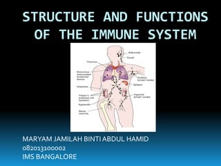
4. Lymphoid Organs
Central Lymphoid Organs
Peripheral Lymphoid
Organs
Thymus Bursa of Fabricius Spleen
Lymph
nodes
Mucosa associated
lymphoid tissue (MALT)
5. CENTRAL LYMPHOID ORGANS
THYMUS
EMBRYOLOGY
3rd & 4th pharyngeal pouches
Mesenchymal stem cells (yolk sac, fetal liver and bone
marrow) reach thymus
Differentiate into thymic lymphoid cells (thymocytes)
Maximally relative size just before birth
Continues to grow about the twelve year
After puberty:
Spontaneous progressive involution
Functions best in early life
7. MICROBIOLOGY PART
Production of thymic lymphocytes
Lymphocyte proliferation in the body
Acquire new surface antigens (‘Thy’ antigens)
Not dependent on antigenic stimulation
Prethymic lymphocytes are not
immunocompetent
‘Educated’ in thymus
Peripheral lymphatic tissue
9. BURSA OF FABRICIUS
Pouch from dorsal part of the cloaca
Day 15: becomes lymphoid organ
Near hatching: full functional ability
7-13 weeks age/puberty: involuting ( normally in
size and functional activity)
Immunocompetent 'bursal lymphocytes' or B cells
Migrate to SPLEEN (mantle, germinal follicles,
perifollicular regions) and LYMPH NODES (cortical
areas and medullary cords)
10. FUNCTION
Humoral immunity
Chicken BURSECTOMISED at hatching failed to form antibodies
when challenged with a bacterial antigen -Glick and Chang (1956)
Competence for Ig M production -early (Day 14 of embryonation)
Ig G late (about Day 21)
Birds bursectomised on Days 18 to 20 synthesize Ig M, but not Ig G
Peyer's patches; 20th week of gestation (spleen and lymph nodes)
Ig M > after birth
Ig G < 3rd month. Adequate by 2-3 years
11. PERIPHERAL LYMPHOID ORGANS
LYMPH NODES
Cortex
Medulla
Cortical follicles and medullary cords
Intermediate zone
Filter for lymph
Proliferation ofT cells and B cells
Local antigenic stimulation -ENLARGED 11
12.
13. SPLEEN
Largest of the lymphoid organs
As a graveyard (effete blood cells)
White pulps and red pulps
Malphigian corpuscles
Germinal centre
'Mantle layer'
Marginal zone
13
16. MUCOSA ASSOCIATED LYMPHOID
TISSUE (MALT)
Mucosa lining of alimentary, respiratory,
genitourinary and other lumina/surfaces that
constantly expose to numerous antigens
Rich lymphoid cells
Peyer's patches
Scattered isolated lymphoid follicles (MALT)
GALT (adenoids, tonsils to follicles of the colon)
BALT (respiratory tract) 16
17. Cells: lymphoid cells, phagocytic cells, B cells andT
cells
Predominantly IgA -from mucosa
Free traffic of antigen-specific effector
lymphocytes between various mucosal and
secretory areas; 'common mucosal or secretory
immune system'
18.
19. Lymphoid Organs
Central Lymphoid Organs
Peripheral Lymphoid
Organs
Thymus Bursa of Fabricius Spleen
Lymph
nodes
Mucosa associated
lymphoid tissue (MALT)
CONCLUSION
20. REFERENCES
Ananthanarayan and Paniker’s,Textbook
of Microbiology, 8th Edition
Dir. Prof. C P Baveja,Textbook of
Microbiology, 4th Edition