Downloaded 235 times
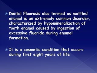
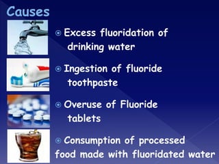

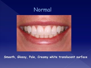
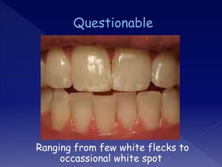
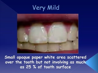
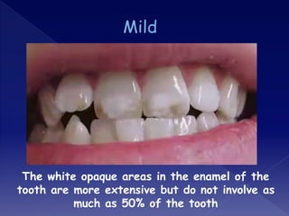
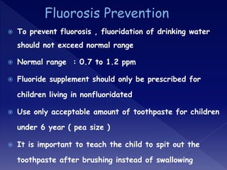
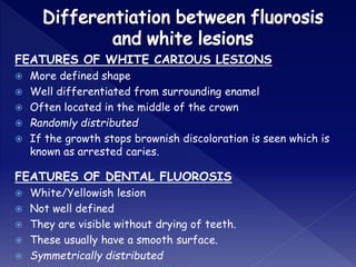


The document discusses dental fluorosis, a cosmetic condition caused by excessive fluoride intake during enamel formation, leading to discolored and hypomineralized enamel. It outlines its causes, symptoms, classification, and treatment options, emphasizing the importance of regulating fluoride levels in drinking water to prevent the condition. Additionally, it compares features of dental fluorosis with white carious lesions and suggests preventive measures for children.


