STPM Form 6 Biology Animal Organs and Tissues
•
22 likes•4,995 views

STPM Form 6 Biology Animal Organs and Tissues

Recommended
More Related Content
What's hot
What's hot (20)
Viewers also liked
Viewers also liked (20)
Similar to STPM Form 6 Biology Animal Organs and Tissues
Similar to STPM Form 6 Biology Animal Organs and Tissues (20)
More from Sook Yen Wong
More from Sook Yen Wong (20)
Recently uploaded
Recently uploaded (20)
STPM Form 6 Biology Animal Organs and Tissues
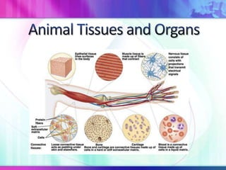
- 3. Epithelial Nerve Muscle Connective • Protective • Coordination of • Body motility • Tissue barrier body functions • Contract and adhesion • Skin • Transmit nerve Relaxes to • Organ • Alimentary impulse allow attachment canals movements • Extracellular • Blood vessels matrix • Ducts • Fill spaces • Protect and Cushion organs • Mechanical support
- 7. a) Form external surfaces of the body b) Closely packed – Adhesion Junctions, Tight Junctions, Gap Junctions c) Apical surface – exposed to external environment d) Basolateral surface – exposed to internal environment e) Attached to basement membrane – Matrix of Glycoprotein and Collagen Fibres (Connective Tissues)
- 11. Thin Flat Attached to basement membrane Blood capillary Alveoli walls Mouth cavity
- 16. Lining kidney tubules, salivary ducts, pancreatic ducts Secretion, excretion, and absorption Associated with goblet cells for secretion
- 22. This is a respiratory epithelium being found in such places as the nasal septum, trachea, and bronchi. Trap and move "pollutants" to the mouth where they are swallowed. Single layered, appreared to be multi-layered due to arrangement of nuclei
- 25. Protective. The multiple layers are too thick for efficient transport of materials (neither secretory or absorptive). The innermost layer continually produces cells (via mitosis) to replace those lost from the outer surface.
- 28. Sweat ducts
- 30. Layered cells Able to modify shapes under different conditions Ureters and Bladders Allow stretching Prevent urine from flowing out to surrounding tissues
- 44. • Nucleus plus cytoplasm Cell body • Mitochondria • Nissls’s granules – ER and polyribosomes for Protein synthesis Dendrons • Branches - dendrites • Conduct impulses towards cell body Axon • Conduct impulses away from cell body Neuroglia • Supporting cells which provide nutrients and oxygen • Schwann cells – Form myelin sheath
- 51. i. Antagonistic pairs ii. Attached to bones by tendons iii. Multinucleated (syncytium) iv. Striated v. Form Muscle Bundles vi. Each muscle fibre is surrounded by connective tissue called endomysium
- 63. Support and protection as internal framework. Provides system of levers with which the skeletal muscles work to move the body. Bones store lipids and minerals (Ca & P). Muscle attachment for motility Site for hematopoiesis (blood cell formation).
- 64. 206 bones in the adult skeleton
- 67. • Relatively heavy Compact • Dense bone • Strong • Long bone • Less dense Spongy • Much lighter bone • Growing bone • Ends of long bone
- 68. 30% Collagen and Glycoprotein fibers 70% inorganic – Calcium hydroxyapatite crystals Magnesium Sodium Hydrogen carbonate Chloride ions
- 69. Cylindrical structure of the bone Called osteon Secrete bone matrix and ground substances
- 70. Haversian Canal 1 artery 1 vein 1 lymph vessel Nerve fibres Lamellae Concentric circles surrounding Haversian Canal
- 74. Bone lamellae Surrounding Haversian canal Lacunae Fluid filling spaces between lamellae Bone cells – osteoblasts are found here
- 75. Canaliculi Fine vessels of cytoplasmic strains connecting lacunae and Haversian canal Connecting each other Supply lacunae with blood Volkman canal Connects 2 Haversian canal together
- 82. • Mature lacunae Osteoblast • Stimulated when • Produce cytoplasmic excessive bone extension into canaliculi • Stimulated substances present • Interconnected with other osteocytes when extra • Reabsorb bone • Allow for exchange of bone substances matrix materials • Digest old bone is needed cells Osteocytes • Form new bones Osteoclast
- 84. Hyaline Yellow Elastic White Fibrous • Trachea • Pinna • Ligamentous • Bronchi • Epiglottis capsules • Skeleton of surrounding cartilageous joints fish – shark • Intervertebral and ray discs • Symphysis pubis
- 86. Cartilage matrix Chondroitin sulphate Secreted by chondroblasts Protein- carbohydrate conjugated with sulphur No blood vessels Diffusion of nutrients and gases
- 87. Chondrocytes Formed from chondroblasts Submerged in small spaces filled with fluid matrix Enclosed in lacunae surrounded by capsule
- 88. Perichondrion Fibrous membrane surrounding cartilage Supply new chondroblasts for maintenance of torn/damaged cartilage Contain blood vessels Allow diffusion of oxygen and nutrients to chondrocytes
- 90. Clear Smooth Glassy Hard Collagen fibre network meshwork Strong but elastic
- 91. Liquid connective tissue About 5 L in adults
- 92. 90% water Buffer 6 – 8 % plasma protein Clotting (Fibrinogen & Protrombin) Transport proteins – 55% Plasma 1% Food cholesterol, Vitamins, Hormones, Iron, Antibodies Variable enzymes and Hormones Blood Variable excretory products RBC 45% Blood cells WBC
- 93. Biconcave No nucleus Erythrocytes Destroyed in spleen 4 months Bone Marrow Granulocyte Leucocytes Agranulocytes Platelets