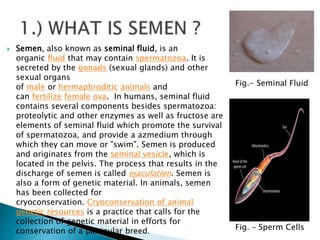
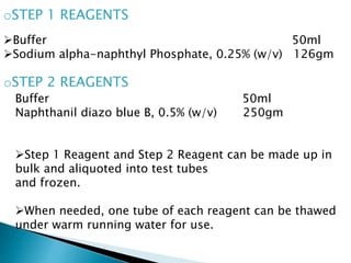
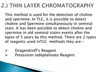
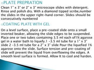
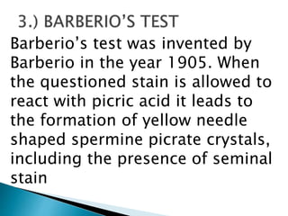
The document is a comprehensive presentation on forensic semen analysis, detailing the composition, morphology, and examination of semen, as well as its significance in forensic science. Key methods for identifying semen stains include the acid phosphate test, cross-over electrophoresis, and various reagent techniques, emphasizing the importance of semen analysis in criminal investigations and infertility assessments. The content also provides insight into chemical properties, physical examination techniques, and the implications of seminal stains in sexual assault cases.


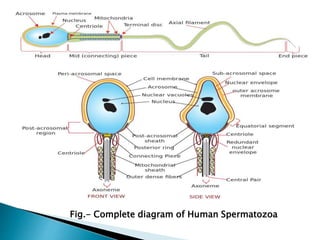
![ During the process of ejaculation, sperm passes
through the ejaculatory ducts and mixes with fluids
from the seminal vesicles, the prostate, and
the bulbourethral glands to form the semen. The
seminal vesicles produce a yellowish viscous fluid
rich in fructose and other substances that makes up
about 70% of human semen. The prostatic secretion,
influenced by dihydrotestosterone, is a whitish
(sometimes clear), thin fluid containing proteolytic
enzymes, citric acid, acid phosphatase and
lipids.[3] The bulbourethral glands secrete a clear
secretion into the lumen of the urethra to lubricate
it.](https://image.slidesharecdn.com/forensicsemenanalysis-181025175838/85/Forensic-semen-analysis-6-320.jpg)


![BIOLOGICAL STAINS semen[Forensic Medicine].pdf](https://cdn.slidesharecdn.com/ss_thumbnails/biologicalstainssemen-250318041913-8535ef17-thumbnail.jpg?width=640&height=640&fit=bounds)


