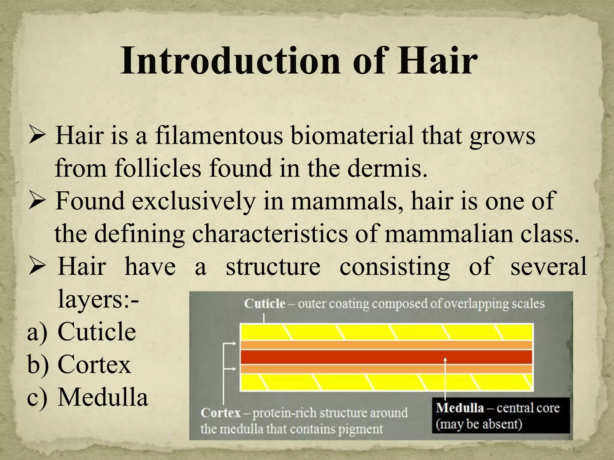
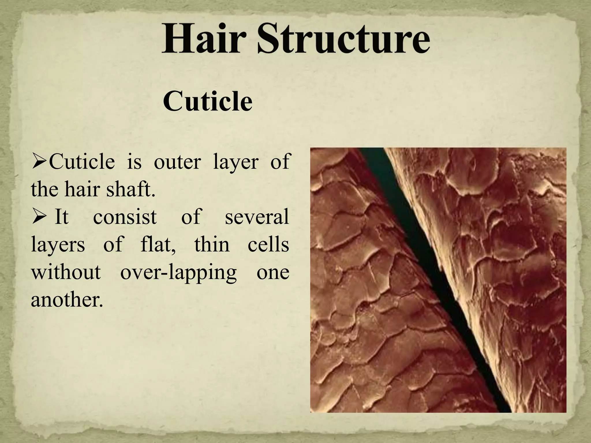
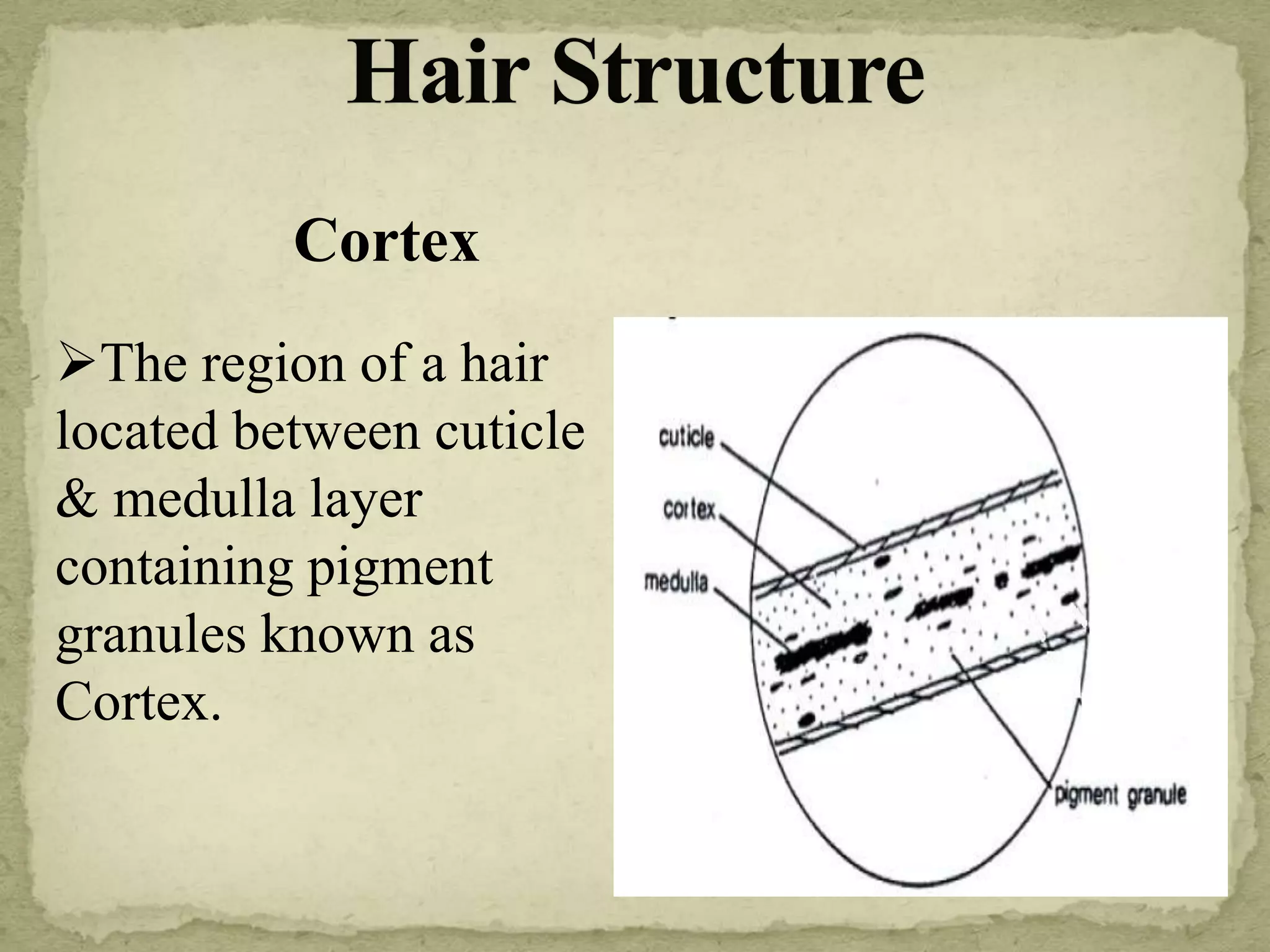
 Hair is a filamentous biomaterial that grows
from follicles found in the dermis.
 Found exclusively in mammals, hair is one of
the defining characteristics of mammalian class.
 Hair have a structure consisting of several
layers:-
a) Cuticle
b) Cortex
c) Medulla
Introduction of Hair
Cuticle is outer layer of
the hair shaft.
 It consist of several
layers of flat, thin cells
without over-lapping one
another.
Cuticle
Cortex
The region of a hair
located between cuticle
& medulla layer
containing pigment
granules known as
Cortex.
Medulla
It is the central part of the hair, made
up of large loosely connected cells
which contain Keratin.
Sometimes medulla may be absent
in some species.
On the basis of appearance of
medulla it is divided into five types:-
1) Continuous
2) Interrupted
3) Fragmented
4) Solid
5) Absent
Human Hair :- Different Sites
Scalp
Pubic
Vulvar
Chest
Beard
Axiliary
Eyebrow
Eyelash
Nose
Limb
Buttocks
Feature Human Hair Animal Hair
Cortex The width of cortex
greater than medulla
Usually less than width of
medulla
Distribution of pigment Towards cuticle Central or denser towards
medulla
Medulla Medulla dis-continuous Medulla continuous
Scales Imbricate Coronal, ring form
Differences between Human &
Animal Hair
Sampling Of Hair
 Spread the exhibit on a clean white surface under proper
illumination.
 With hand magnifier, carefully locate any loose hair and
collect.
 Hair can be collected by forceps or transferred to adhesive
tape.
 Samples collected should be packed in cellophane or paper
folders with proper labeling on them.
 Root bulb and follicle of hair should be carefully preserved for
determination of sex , serological and DNA Examination.
Examination Of
Hair
Temporary
Mount
Scale
Casting
Cellulose
Acetate
Method
Polaroid
Coater
Method
Permanent
Mount
Cross
Sectioning
Micrometry
Temporary Mount
 Make a temporary mount
of the hair sample on a clean
slide with the distilled Water
or glycerin. Cover with cover
slip.
 Observe morphological
characteristics under
microscope.
 On a clean microscopic slide, place a thin layer of
cellulose acetate paste with low viscosity.
 With fine forceps, place hair on to the cellulose
acetate paste and press with another clean slide.
 Allow it to dry for 2-5 min.
Observe scales of hair under microscope.
 Place the hair on a clean microscope slide, securing the
ends with cellophane tape.
 Using a Polaroid film coater, make 2 or 3 passes along the
length of the hair.
 Allow the coating to dry for 23 hours.
 After removing the cellophane tape, gently peel the hair
from the slide.
 With a sharp scalpel, slice away the excess coating
protruding above the flat surface of the scale cast.
 Observe the scale impressions microscopically.
Permanent Mount
Place hair on slide in a
drop of xylene and add
permanent mounting
medium.
 Place a cover slip on the
hair allowing the medium.
 Label the slide
appropriately and allow it
to dry for 48 hours.
Cross Sectioning
 Clean hair in solution of ether and ethanol in ratio 1:1
 Bundle the samples and dip in a block of molten wax
and allow to cool.
 Cross sections can be taken either with a sharp blade
or with a microtone to a thickness of 5-10 microns.
 Place sections on a clean slide and dissolve wax with
a drop of xylene.
 Prepare permanent mount of the sections and examine
under the microscope.
Micrometry
With the help of micrometer measure following distances &
calculate different indices:
Maximum diameter of the shaft.
 Number of scales per unit length.
 Diameter of hair in microns / scale
count.
 Maximum diameter of hair medulla
/ Maximum diameter of hair shaft.
 Minimum diameter of shaft /
Maximum diameter of the shaft x 100
Report Writing
Species of Origin On the basis of morphological
examination
Sex On the basis of microscopic
examination
Site On the basis of morphological
examination
Comparison On the basis of morphological
examination
Forensic examination of hair

Forensic examination of hair

  • 2.
     Hair isa filamentous biomaterial that grows from follicles found in the dermis.  Found exclusively in mammals, hair is one of the defining characteristics of mammalian class.  Hair have a structure consisting of several layers:- a) Cuticle b) Cortex c) Medulla Introduction of Hair
  • 3.
    Cuticle is outerlayer of the hair shaft.  It consist of several layers of flat, thin cells without over-lapping one another. Cuticle
  • 4.
    Cortex The region ofa hair located between cuticle & medulla layer containing pigment granules known as Cortex.
  • 5.
    Medulla It is thecentral part of the hair, made up of large loosely connected cells which contain Keratin. Sometimes medulla may be absent in some species. On the basis of appearance of medulla it is divided into five types:- 1) Continuous 2) Interrupted 3) Fragmented 4) Solid 5) Absent
  • 6.
    Human Hair :-Different Sites Scalp Pubic Vulvar Chest Beard Axiliary Eyebrow Eyelash Nose Limb Buttocks
  • 7.
    Feature Human HairAnimal Hair Cortex The width of cortex greater than medulla Usually less than width of medulla Distribution of pigment Towards cuticle Central or denser towards medulla Medulla Medulla dis-continuous Medulla continuous Scales Imbricate Coronal, ring form Differences between Human & Animal Hair
  • 8.
    Sampling Of Hair Spread the exhibit on a clean white surface under proper illumination.  With hand magnifier, carefully locate any loose hair and collect.  Hair can be collected by forceps or transferred to adhesive tape.  Samples collected should be packed in cellophane or paper folders with proper labeling on them.  Root bulb and follicle of hair should be carefully preserved for determination of sex , serological and DNA Examination.
  • 9.
  • 10.
    Temporary Mount  Makea temporary mount of the hair sample on a clean slide with the distilled Water or glycerin. Cover with cover slip.  Observe morphological characteristics under microscope.
  • 12.
     On aclean microscopic slide, place a thin layer of cellulose acetate paste with low viscosity.  With fine forceps, place hair on to the cellulose acetate paste and press with another clean slide.  Allow it to dry for 2-5 min. Observe scales of hair under microscope.
  • 13.
     Place thehair on a clean microscope slide, securing the ends with cellophane tape.  Using a Polaroid film coater, make 2 or 3 passes along the length of the hair.  Allow the coating to dry for 23 hours.  After removing the cellophane tape, gently peel the hair from the slide.  With a sharp scalpel, slice away the excess coating protruding above the flat surface of the scale cast.  Observe the scale impressions microscopically.
  • 14.
    Permanent Mount Place hairon slide in a drop of xylene and add permanent mounting medium.  Place a cover slip on the hair allowing the medium.  Label the slide appropriately and allow it to dry for 48 hours.
  • 15.
    Cross Sectioning  Cleanhair in solution of ether and ethanol in ratio 1:1  Bundle the samples and dip in a block of molten wax and allow to cool.  Cross sections can be taken either with a sharp blade or with a microtone to a thickness of 5-10 microns.  Place sections on a clean slide and dissolve wax with a drop of xylene.  Prepare permanent mount of the sections and examine under the microscope.
  • 17.
    Micrometry With the helpof micrometer measure following distances & calculate different indices: Maximum diameter of the shaft.  Number of scales per unit length.  Diameter of hair in microns / scale count.  Maximum diameter of hair medulla / Maximum diameter of hair shaft.  Minimum diameter of shaft / Maximum diameter of the shaft x 100
  • 18.
    Report Writing Species ofOrigin On the basis of morphological examination Sex On the basis of microscopic examination Site On the basis of morphological examination Comparison On the basis of morphological examination