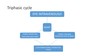
TRIPHASIC CT SCAN
PRESENTED BY;
DR: TAHIRA AGHANI
Principles Of Hepatic Contrast Enhancement
• When contrast agent is administered intravenously, it is rapidly
redistributed from the vascular to the interstitial space while being
continuously excreted by the kidneys.
• In the liver, with its unique dual blood supply this process occurs quite
rapidly
Detection of liver masses
• The conspicuity of a liver lesion depends on the attenuation difference between the lesion
and the normal liver.
• On a non enhanced CT-scan (NECT) liver tumors usually are not visible, because the inherent
contrast between tumor tissue and the surrounding liver parenchyma is too low.
• Only a minority of tumors contain calcifications, cystic components, fat or hemorrage and will
be detected on a NECT.
So i.v. contrast is needed to increase the conspicuity of lesions.
• When we give i.v. contrast, it is important to understand, that there is a dual blood supply to
the liver.
• Normal parenchyma is supplied for 80% by the portal vein and only for 20% by the hepatic
artery, so it will enhance in the portal venous phase.
• All liver tumors however get 100% of their blood supply from the
hepatic artery, so when they enhance it will be in the arterial phase.
• This difference in blood supply results in different enhancement
patterns between liver tumors and normal liver parenchyma in the
various phases of contrast enhancement
Triphasic cycle
HEART
DYE INTRAVENOUSLY
PORTAL VEIN 80%
PERFUSION LATE 50-60SEC
HEPATIC ARTERY 20%
PERFUSION EARLY 20SEC
LIVER PARENCHYMAL INTERSTITIAL
TISSUE
• Three distinct phases of contrast enhancement have been described
in the liver.
1. Hepatic arterial phase(HAP)
2. Portal venous phase(PVP)
3. Equilibrium phase.
• Each of which corresponds to a different portion of the aortic hepatic
time attenuation curve
• Figure showing
• Time-attenuation curve for the
liver showing approximate times
for early, late and equilibrium
phases of hepatic enhancement.
Quoted from
Hepatic Arterial Phase
• In the arterial phase hypervascular tumors will enhance via the
hepatic artery, when normal liver parenchyma does not yet enhances,
because contrast is not yet in the portal venous system.
These hypervascular tumors will be visible as hyperdense lesions in a
relatively hypodense liver.
However when the surrounding liver parenchyma starts to enhance in
the portal venous phase, these hypervascular lesion may become
obscured.
• Small Hepatocellular carcinoma
in cirrhotic liver not visible on
NECT (left), clearly visible in
arterial phase (middle) and not
visible in portal venous phase
(right)
Portal venous phase
• In the portal venous phase hypovascular tumors are detected, when
the normal liver parenchyma enhances maximally.
These hypovascular tumors will be visible as hypodense lesions in a
relatively hyperdense liver.
Equilibrium phase
• In the equilibrium phase at about 10 minutes after contrast injection,
tumors become visible, that either loose their contrast slower than
normal liver, or wash out their contrast faster than normal liver
parenchyma.
These lesions will become either relatively hyperdense or hypodense
to the normal liver.
• Detection of a lesion depends on
difference in attenuation between
liver and lesion.
• LEFT: Arterial phase showing
hypervascular FNH
• MIDDLE: Portal venous phase
showing hypovascular metastasis
• RIGHT: equilibrium phase showing
relatively dense
cholangiocarcinoma
Arterial phase imaging
• Optimal timing and speed of contrast injection are very important for
good arterial phase imaging.
Hypervascular tumors will enhance optimally at 35 sec after contrast
injection (late arterial phase).
This time is needed for the contrast to get from the peripheral vein to
the hepatic artery and to diffuse into the liver tumor.
• On the left a patient who underwent
two phases of arterial imaging at 18
and 35 seconds. In the early arterial
phase we nicely see the arteries, but
we only see some irregular
enhancement within the liver.
• In the late arterial phase we can
clearly identify multiple tumor
masses.
• Notice that in the late arterial phase
there has to be some enhancement of
the portal vein.
• The only time that an early arterial phase is needed is when you need
an arteriogram, for instance as a roadmap for chemoembolization of a
liver tumor.
• Timing of scanning is important, but almost as
important is speed of contrast injection.
• For arterial phase imaging the best results are
with an injection rate of 5ml/sec.
• There are two reasons for this better
enhancement: at 5ml/sec there will be more
contrast delivered to the liver when you start
scanning and this contrast arrives in a higher
concentration.
• On the left a patient with cirrhosis examined
after contrast injection at 2.5ml/sec and at
5ml/sec.
• At 5ml/sec there is far better contrast
enhancement and better tumor detection.
Portal Venous phase
• Portal venous phase imaging works on the opposite
idea.
We image the liver when it is loaded with contrast
through the portal vein to detect hypovascular tumors
• The best moment to start scanning is at about 75
seconds, so this is a late portal venous phase, because
enhancement of the portal vein already starts at 35
sec in the late arterial phase.
This late portal venous phase is also called the hepatic
phase because there already must be enhancement of
the hepatic veins. If you do not seen enhancement of
the hepatic veins, you are too early.
• If you only do portal venous imaging, for instance if
you are only looking for hypovascular metastases in
colorectal cancer, fast contrast injection is not
needed, because in this phase the total amount of
contrast is more important and 3ml/sec will be
sufficient.
• Hypovascular metastases seen as hypodense lesions
in the late portal venous phase. (fig)
Equilibrium Phase
• The equilibrium phase is when contrast is
moving away from the liver and the liver
starts to decrease in density.
This phase begins at about 3-4 minutes
after contrast injection and imaging is
best done at 10 minutes after contrast
injection.
• This phase can be valuable if you're
looking for: fast tumor washout in
hypervascular tumors like HCC or
retention of contrast in the blood pool as
in hemangiomas or the retention of
contrast in fibrous tissue in capsules
(HCC) or scar tissue (FNH, Cholangioca).
Relative hyperdense lesions in the delayed
phase
• Fibrous tissue that's well organized and dense is
very slow to let iodine or gadolineum in.
• Once contrast gets in however, it is equally
slow to get back out in the equilibrium phase.
• So when the normal liver parenchyma washes
out, the fibrous components of a tumor will look
brighter than the background liver tissue.
• Cholangiocarcinoma may have a fibrous stroma
and in the delayed phase it may be the only time
when you see the tumor (figure).
• Small cholangiocarcinoma not visible in portal
venous phase (left), but seen as relative
hyperdense lesion in the delayed phase (right)
Relative hypodense lesions in the delayed
phase
• On the left the importance of the delayed phase in a cirrhotic
patient with an HCC is demonstrated.
• Notice that you do not see the tumor on the non enhanced scan
and also not in the portal venous phase. This is often the case and
demonstrates the importance of the arterial phase.
Now the issue at hand is in small enhancing lesions in a cirrhotic
liver whether it is a benign lesion like a regenerating nodule or a
HCC.
• In the delayed phase we see that the tumor is washed out more
than the surrounding liver parenchyma.
• Benign lesions typically will not show this kind of wash out.
• For instance a FNH or adenoma will show fast enhancement in the
arterial phase, become isodense in the portal venous phase, but it
will stay isodense with liver in the equilibrium phase.
• These benign tumors do not have enough neoplastic neovascularity
to have a fast wash out.
• Especially in cirrhotic patients you have to rely heavily on this
delayed phase to differentiate benign little enhancing lesions from
small HCC's.
• HCC in a cirrhotic liver. Notice fast wash out in equilibrium phase
compared to surrounding liver parenchyma.(fig)
Blood pool and Hemangioma
• Normally when we look at lesions filling with contrast, the density of these
lesions is always compared to the density of the liver parenchyma.
• In hemangiomas however you should not compare the density of the lesion
to the liver, but to the blood pool.
• This means that the areas of enhancement in a hemangioma should match
the attenuation of the appropriate vessels (bloodpool) at all times. So in
the arterial phase the enhancing parts of the lesion must have almost the
same attenuation value as the enhancing aorta , while in the portal venous
phase it must match the enhancement of the portal vein
• If it does not match the bloodpool in every single phase of contrast
enhancement forget the diagnosis of a hemangioma.
• On the left a characteristic hemangioma. Notice that on the
NECT the density of the tumor is the same as the density of
the vessels
• In the arterial phase it is matching the bloodpool and the
attenuation is almost the same as the aorta.
• In the portal venous phase it matches the density of the
portal vein.
• In the equlibrium phase it has the same enhancement as the
vessels.
Eventually the lesion will become iso-attenuating to the liver,
but only because the vessels become iso-attenuating with the
liver. It has nothing to do with the density of the liver
parenchyma itself.
• So think of bloodpool rather than liver if you're thinking of a
hemangioma.
• Hemangioma on NECT, late arterial, late portal venous and
equilibrium phase. Notice that the attenuation of the
hemangioma matches the bloodpool in every single phase
(arrows).(fig)
Use arterial phase imaging in the following
situations:
• Characterisaton of a liver lesion of unknown origin.
• Detection of HCC in patients with a high alpha 1 foetoprotein.
• Screening of cirrhotic patients for HCC.
• Detection of metastases in patients with hypervascular tumors
Characterisation of liver masses
• From a practical point of view, the approach to characterizing a
focal liver lesion seen on CT begins with the determination of its
density.
• If the lesion is of near water density, homogeneous, has sharp
margins and shows no enhancement, then it is a cyst.
• If the lesion does enhance, then the next step is to determine
whether the lesion could be a hemangioma, since this is by far the
most common liver tumor. The enhancement should be
peripheral and nodular, with the same density as the bloodpool in
all phases.
• If it is not a cyst nor a hemangioma, then we further have to study
the lesion.
• Based on the enhancement pattern, we divide masses into
hypervascular and hypovascular lesions.
• Usually a combination of the enhancement pattern and gross
pathologic features, like the presence of fat, blood, calcifications,
cystic or fibrotic components, in combination with the clinical
history will limit the differential diagnosis (figure).
Hypervascular lesions
• Arterially enhancing lesions are mostly
benign lesions and include primary liver
tumors as FNH, adenoma and small
hemangiomas that fill rapidly with
contrast.
• These benign tumors have to be
differentiated from the most common
hypervascular malignant liver tumor,
which is HCC and metastases from
hypervascular tumors like melanoma,
renal cell carcinoma, breast, sarcoma and
neuroendocrine tumors (islet cell tumors,
carcinoid, pheochromocytoma).
• Hypervascular lesions may look very similar in the arterial
phase (figure).
• Differentiation is done by looking at the enhancement
pattern in the other phases and additional gross
pathologic features together with clinical findings.
• Hypervascular metastases will be considered in patients
with a known primary tumor.
• In general HCC is considered when there is a setting of
cirrhosis, while FNH is considered in young women and
hepatic adenoma in patients on oral contraceptives,
anabolic steroids or with a history of glycogen storage
disease.
• Four different tumors with enhancement in the late
arterial phase. From left to right: HCC in cirrhotic liver;
FNH with central scar in adolescent; adenoma in young
woman on contraceptives and finally a hemangioma with
typical enhancement in the other phases (not shown)
Hypovascular lesions
• Hypovascular liver tumors are more common than hypervascular
tumors.
• Most hypovascular lesions are malignant and metastases are by
far the most common.
• Although primary liver tumors are mostly hypervascular, there are
exceptions. 10% of HCC is hypovascular. Cholangioca is
hypovascular, but may show delayed enhancement
• On the left a hypovascular mass with irregular enhancement in
the late arterial and late portal venous phase. This is a sign of
malignancy.
• On the delayed images a relative dense structure is seen centrally,
which looses its contrast slower compared to normal liver.
• This means that this tumor is mainly composed of fibrous tissue.
The fibrous tissue has also retracted the liver capsule.
• These imaging findings are very suggestive of a
cholangiocarcinoma.
• Cholangiocarcinoma with relative hyperdense scar tissue in the
equilibrium phase (arrow).fig
Scar
• Liver lesions which may have a central scar are
FNH, fibrolamellar carcinoma, cholangiocarcinoma,
hemangioma and hepatocellular carcinoma.
• On CT a scar is sometimes visible as a hypodense
structure.
• On MR scar tissue is hypointense on both T1WI
and T2WI due to intense fibrotic changes. An
example is the central scar of fibrolamellar
carcinoma (FLC)
• An exception to this rule is the central scar in FNH
which is hyperintense on T2WI due to edema.
• T2WI can be very helpfull if there is a problem in
differentiating FNH from FLC.
• Both on CT and MRI scar tissue will enhance in the
delayed phase.
• On the left a lesion with a typical central scar.
It has a hypodense centre on the NECT.
• In the portal venous phase there is
homogeneus enhancement of the lesion
except for the scar.
• Enhancement of the fibrous tissue of the
central scar is seen only on the delayed phase
images.
• The combination of homogeneous
enhancement and central scar is typical for
the diagnosis of FNH
• FNH with central scar seen in NECT, portal
venous phase and equilibrium phase.(fig)
Capsule
• Liver lesions which may have a capsule are
Adenoma, HCC and cystadenoma or
cystadenocarcinoma.
• The most common tumor with a capsule is
HCC.
• The capsule will not enhance in the arterial
phase and even in the portal venous phase it
will be hypodense, because the fibrous tissue
enhances very slowly.
• A capsule is usually best seen in the delayed
phase as a relative hyperdense structure.
• NECT and Arterial phase image showing
hypodense capsule of HCC(fig)
• Adenoma frequently has a thin
fibrous capsule seen in 30% of
cases.
• It has a well defined contour and
subcapsular feeding arteries.
• Capsule in Adenoma not seen in
portal venous phase and well
appreciated in delayed
phase(fig)
• On the left a different patient with HCC.
• Only in the equilibrium phase a relatively
bright capsule was seen.
The image on the left was taken 8
minutes after contrast injection.
• Notice that the tumor itself is relative
hypodense in the equilibrium phase.
• So it has a fast wash out.
• LEFT: Bright enhancing capsule of HCC in
equilibrium phase.RIGHT: Pathologic
specimen.(fig)
Calcifications
Central calcifications are seen in:
• Metastases (especially in colorectal tumors)
• Fibrolamellar carcinoma (FLC)
• Cholangiocarcinomas
• Hemangiomas
• These calcifications are hyperdense on CT
and hypointense on T1 and T2 MR images.
In FLC these calcifications are located within
the central scar as seen on the left
• NECT of a Fibrolamellar carcinoma with
central calcifications(fig)
Fat
• Fat within liver tumors is seen in:
• Adenoma
• HCC
• Metastatic liposarcoma
• Angiomyolipoma
• The case on the left shows an
adenoma with fat depositions
within the tumor.
• Low density area due to fat in
Adenoma(fig)
Hemorrhage
• Hemorrhage in liver tumors is seen in:
• Adenoma
• HCC
• Hemorrhage is most commonly seen
in adenomas.
• The case on the left shows a well
circumscribed lesion with
hemorrhage.
At resection the lesion proved to be
an adenoma.
• Hemorrhage within an adenoma.(fig)
Cystic components
• If a lesion has a near water density
in the centre and does not show
enhancement in the centre, we
usually will call it a cystic lesion.
• You have to realize, that it still can
be a tumor as in cystic metastases
or metastases with central
necrosis.
• Secondly you always have to add
abscess to the differential
diagnosis.
• On the left a patient with hypovascular lesions with
a low density, so it may be cystic i.e fluid
containing.
• These lesions are multiple, but not spread out
through the liver, so we describe them as clustered
or satelite lesions.
• This is a typical finding which makes the lesions
suspective for liver abcesses.
This was a case of diverticulitis.
• The common route is through the portal vein as a
result of abdominal infection.
• The bacteria enter the slow flow portal system,
where they layer within the vessel and finally these
bacteria 'fall down' into the dependent portion of
the right lobe.
• On the left a typical case of a
echinococcus cyst with
'daughter cysts' within the large
cyst.
Most cases of echinococcus
cysts however are not that
typical.
• CT and T2W MR-image of
echinococcus cyst.(fig)
• If you look at the CT image on the left,
the first impression might be that there
are only simple cysts within the liver.
• However, if you look more carefully, you
will notice that some of the hypodense
lesions show vague rim enhancement.
• And although you might think that these
could be cystic metastases, the US-
findings clearly show, that these lesions
are hyperechoic solid masses.
• So you have to be very carefull in calling a
lesion cystic, because you might end up
missing metastases or looking in the
wrong file for a differential diagnosis.
Retraction of liver capsule
• Most liver tumors will present as a mass.
Some tumors however have an infiltrative growth
pattern with a lot of fibrous tissue and do not
cause mass effect.
• As the fibrous stroma matures, the tissue will
contract and cause retraction of the liver capsule
(figure).
• Breast cancer metastases can be infiltrative. When
they shrink they can cause multiple retractions.
• This will give a pseudo-cirrhosis appearance.
• Hepatic and delayed phase in a patient with breast
metastases causing retraction of liver capsule.
Notice delayed enhancement of the fibrotic tissue
(arrows)
• The most common tumor however to cause
retraction is cholangiocarcinoma.
The delayed image on the left shows a large
cholangiocarcinoma with dense enhancing
fibrous tissue and retraction of the liver
capsule.
Notice the resemblance with the case above.
• Another cause of local retraction is atrophy
due to biliary obstruction or chronic portal
venous obstruction.
• Delayed phase image of a cholangiocarcinoma
with relative dense fibrous stroma with
retraction of liver capsule(fig)
• On the left another case of
cholangiocarcinoma with
multifocal lesions.
• Notice the retraction and the
delayed enhancement of the
fibrotic component of the tumor.
• Hepatic and delayed phase in a
patient with multifocal
cholangiocarcinoma causing
retraction of liver capsule.(fig)
Peripheral enhancement and progressive fill in
• Many will regard 'peripheral
enhancement and progressive fill in' as a
typical feature of hemangioma, but it is
not.
• Peripheral rim enhancement is a typical
feature of malignant lesions and only
discontinuous nodular peripheral
enhancement that matches bloodpool is
a typical feature of hemangioma.
• LEFT: rim enhancement in metastasis.
RIGHT: hemangioma with discontinuous
nodular peripheral enhancement(fig)
• Many lesions will show progressive fill in.
In hemangiomas this progressive fill in must have
the same density as the bloodpool.
• Many hypovascular metastases will show contrast
diffusion into a lesion starting on the outside.
Usually the center does not fill in.
• Cholangiocarcinomas will show progressive fill in
because the fibrous centre will enhance slowly.
• You will see it enhance in the delayed phase
So if you want to make the diagnosis of a
hemangioma you have to look at all the other
phases to see if the enhancement matches the
bloodpool.
• Progressive fill in in a hemangioma (left),
cholangiocarcinoma (middle) and metastases
(right).(fig)
Triphasic CT scan

Triphasic CT scan

  • 1.
    TRIPHASIC CT SCAN PRESENTEDBY; DR: TAHIRA AGHANI
  • 2.
    Principles Of HepaticContrast Enhancement • When contrast agent is administered intravenously, it is rapidly redistributed from the vascular to the interstitial space while being continuously excreted by the kidneys. • In the liver, with its unique dual blood supply this process occurs quite rapidly
  • 3.
    Detection of livermasses • The conspicuity of a liver lesion depends on the attenuation difference between the lesion and the normal liver. • On a non enhanced CT-scan (NECT) liver tumors usually are not visible, because the inherent contrast between tumor tissue and the surrounding liver parenchyma is too low. • Only a minority of tumors contain calcifications, cystic components, fat or hemorrage and will be detected on a NECT. So i.v. contrast is needed to increase the conspicuity of lesions. • When we give i.v. contrast, it is important to understand, that there is a dual blood supply to the liver. • Normal parenchyma is supplied for 80% by the portal vein and only for 20% by the hepatic artery, so it will enhance in the portal venous phase.
  • 4.
    • All livertumors however get 100% of their blood supply from the hepatic artery, so when they enhance it will be in the arterial phase. • This difference in blood supply results in different enhancement patterns between liver tumors and normal liver parenchyma in the various phases of contrast enhancement
  • 5.
    Triphasic cycle HEART DYE INTRAVENOUSLY PORTALVEIN 80% PERFUSION LATE 50-60SEC HEPATIC ARTERY 20% PERFUSION EARLY 20SEC LIVER PARENCHYMAL INTERSTITIAL TISSUE
  • 6.
    • Three distinctphases of contrast enhancement have been described in the liver. 1. Hepatic arterial phase(HAP) 2. Portal venous phase(PVP) 3. Equilibrium phase. • Each of which corresponds to a different portion of the aortic hepatic time attenuation curve
  • 7.
    • Figure showing •Time-attenuation curve for the liver showing approximate times for early, late and equilibrium phases of hepatic enhancement. Quoted from
  • 8.
    Hepatic Arterial Phase •In the arterial phase hypervascular tumors will enhance via the hepatic artery, when normal liver parenchyma does not yet enhances, because contrast is not yet in the portal venous system. These hypervascular tumors will be visible as hyperdense lesions in a relatively hypodense liver. However when the surrounding liver parenchyma starts to enhance in the portal venous phase, these hypervascular lesion may become obscured.
  • 9.
    • Small Hepatocellularcarcinoma in cirrhotic liver not visible on NECT (left), clearly visible in arterial phase (middle) and not visible in portal venous phase (right)
  • 10.
    Portal venous phase •In the portal venous phase hypovascular tumors are detected, when the normal liver parenchyma enhances maximally. These hypovascular tumors will be visible as hypodense lesions in a relatively hyperdense liver.
  • 11.
    Equilibrium phase • Inthe equilibrium phase at about 10 minutes after contrast injection, tumors become visible, that either loose their contrast slower than normal liver, or wash out their contrast faster than normal liver parenchyma. These lesions will become either relatively hyperdense or hypodense to the normal liver.
  • 12.
    • Detection ofa lesion depends on difference in attenuation between liver and lesion. • LEFT: Arterial phase showing hypervascular FNH • MIDDLE: Portal venous phase showing hypovascular metastasis • RIGHT: equilibrium phase showing relatively dense cholangiocarcinoma
  • 13.
    Arterial phase imaging •Optimal timing and speed of contrast injection are very important for good arterial phase imaging. Hypervascular tumors will enhance optimally at 35 sec after contrast injection (late arterial phase). This time is needed for the contrast to get from the peripheral vein to the hepatic artery and to diffuse into the liver tumor.
  • 14.
    • On theleft a patient who underwent two phases of arterial imaging at 18 and 35 seconds. In the early arterial phase we nicely see the arteries, but we only see some irregular enhancement within the liver. • In the late arterial phase we can clearly identify multiple tumor masses. • Notice that in the late arterial phase there has to be some enhancement of the portal vein.
  • 15.
    • The onlytime that an early arterial phase is needed is when you need an arteriogram, for instance as a roadmap for chemoembolization of a liver tumor.
  • 16.
    • Timing ofscanning is important, but almost as important is speed of contrast injection. • For arterial phase imaging the best results are with an injection rate of 5ml/sec. • There are two reasons for this better enhancement: at 5ml/sec there will be more contrast delivered to the liver when you start scanning and this contrast arrives in a higher concentration. • On the left a patient with cirrhosis examined after contrast injection at 2.5ml/sec and at 5ml/sec. • At 5ml/sec there is far better contrast enhancement and better tumor detection.
  • 17.
    Portal Venous phase •Portal venous phase imaging works on the opposite idea. We image the liver when it is loaded with contrast through the portal vein to detect hypovascular tumors • The best moment to start scanning is at about 75 seconds, so this is a late portal venous phase, because enhancement of the portal vein already starts at 35 sec in the late arterial phase. This late portal venous phase is also called the hepatic phase because there already must be enhancement of the hepatic veins. If you do not seen enhancement of the hepatic veins, you are too early. • If you only do portal venous imaging, for instance if you are only looking for hypovascular metastases in colorectal cancer, fast contrast injection is not needed, because in this phase the total amount of contrast is more important and 3ml/sec will be sufficient. • Hypovascular metastases seen as hypodense lesions in the late portal venous phase. (fig)
  • 18.
    Equilibrium Phase • Theequilibrium phase is when contrast is moving away from the liver and the liver starts to decrease in density. This phase begins at about 3-4 minutes after contrast injection and imaging is best done at 10 minutes after contrast injection. • This phase can be valuable if you're looking for: fast tumor washout in hypervascular tumors like HCC or retention of contrast in the blood pool as in hemangiomas or the retention of contrast in fibrous tissue in capsules (HCC) or scar tissue (FNH, Cholangioca).
  • 19.
    Relative hyperdense lesionsin the delayed phase • Fibrous tissue that's well organized and dense is very slow to let iodine or gadolineum in. • Once contrast gets in however, it is equally slow to get back out in the equilibrium phase. • So when the normal liver parenchyma washes out, the fibrous components of a tumor will look brighter than the background liver tissue. • Cholangiocarcinoma may have a fibrous stroma and in the delayed phase it may be the only time when you see the tumor (figure). • Small cholangiocarcinoma not visible in portal venous phase (left), but seen as relative hyperdense lesion in the delayed phase (right)
  • 20.
    Relative hypodense lesionsin the delayed phase • On the left the importance of the delayed phase in a cirrhotic patient with an HCC is demonstrated. • Notice that you do not see the tumor on the non enhanced scan and also not in the portal venous phase. This is often the case and demonstrates the importance of the arterial phase. Now the issue at hand is in small enhancing lesions in a cirrhotic liver whether it is a benign lesion like a regenerating nodule or a HCC. • In the delayed phase we see that the tumor is washed out more than the surrounding liver parenchyma. • Benign lesions typically will not show this kind of wash out. • For instance a FNH or adenoma will show fast enhancement in the arterial phase, become isodense in the portal venous phase, but it will stay isodense with liver in the equilibrium phase. • These benign tumors do not have enough neoplastic neovascularity to have a fast wash out. • Especially in cirrhotic patients you have to rely heavily on this delayed phase to differentiate benign little enhancing lesions from small HCC's. • HCC in a cirrhotic liver. Notice fast wash out in equilibrium phase compared to surrounding liver parenchyma.(fig)
  • 21.
    Blood pool andHemangioma • Normally when we look at lesions filling with contrast, the density of these lesions is always compared to the density of the liver parenchyma. • In hemangiomas however you should not compare the density of the lesion to the liver, but to the blood pool. • This means that the areas of enhancement in a hemangioma should match the attenuation of the appropriate vessels (bloodpool) at all times. So in the arterial phase the enhancing parts of the lesion must have almost the same attenuation value as the enhancing aorta , while in the portal venous phase it must match the enhancement of the portal vein • If it does not match the bloodpool in every single phase of contrast enhancement forget the diagnosis of a hemangioma.
  • 22.
    • On theleft a characteristic hemangioma. Notice that on the NECT the density of the tumor is the same as the density of the vessels • In the arterial phase it is matching the bloodpool and the attenuation is almost the same as the aorta. • In the portal venous phase it matches the density of the portal vein. • In the equlibrium phase it has the same enhancement as the vessels. Eventually the lesion will become iso-attenuating to the liver, but only because the vessels become iso-attenuating with the liver. It has nothing to do with the density of the liver parenchyma itself. • So think of bloodpool rather than liver if you're thinking of a hemangioma. • Hemangioma on NECT, late arterial, late portal venous and equilibrium phase. Notice that the attenuation of the hemangioma matches the bloodpool in every single phase (arrows).(fig)
  • 23.
    Use arterial phaseimaging in the following situations: • Characterisaton of a liver lesion of unknown origin. • Detection of HCC in patients with a high alpha 1 foetoprotein. • Screening of cirrhotic patients for HCC. • Detection of metastases in patients with hypervascular tumors
  • 24.
    Characterisation of livermasses • From a practical point of view, the approach to characterizing a focal liver lesion seen on CT begins with the determination of its density. • If the lesion is of near water density, homogeneous, has sharp margins and shows no enhancement, then it is a cyst. • If the lesion does enhance, then the next step is to determine whether the lesion could be a hemangioma, since this is by far the most common liver tumor. The enhancement should be peripheral and nodular, with the same density as the bloodpool in all phases. • If it is not a cyst nor a hemangioma, then we further have to study the lesion. • Based on the enhancement pattern, we divide masses into hypervascular and hypovascular lesions. • Usually a combination of the enhancement pattern and gross pathologic features, like the presence of fat, blood, calcifications, cystic or fibrotic components, in combination with the clinical history will limit the differential diagnosis (figure).
  • 25.
    Hypervascular lesions • Arteriallyenhancing lesions are mostly benign lesions and include primary liver tumors as FNH, adenoma and small hemangiomas that fill rapidly with contrast. • These benign tumors have to be differentiated from the most common hypervascular malignant liver tumor, which is HCC and metastases from hypervascular tumors like melanoma, renal cell carcinoma, breast, sarcoma and neuroendocrine tumors (islet cell tumors, carcinoid, pheochromocytoma).
  • 26.
    • Hypervascular lesionsmay look very similar in the arterial phase (figure). • Differentiation is done by looking at the enhancement pattern in the other phases and additional gross pathologic features together with clinical findings. • Hypervascular metastases will be considered in patients with a known primary tumor. • In general HCC is considered when there is a setting of cirrhosis, while FNH is considered in young women and hepatic adenoma in patients on oral contraceptives, anabolic steroids or with a history of glycogen storage disease. • Four different tumors with enhancement in the late arterial phase. From left to right: HCC in cirrhotic liver; FNH with central scar in adolescent; adenoma in young woman on contraceptives and finally a hemangioma with typical enhancement in the other phases (not shown)
  • 27.
    Hypovascular lesions • Hypovascularliver tumors are more common than hypervascular tumors. • Most hypovascular lesions are malignant and metastases are by far the most common. • Although primary liver tumors are mostly hypervascular, there are exceptions. 10% of HCC is hypovascular. Cholangioca is hypovascular, but may show delayed enhancement • On the left a hypovascular mass with irregular enhancement in the late arterial and late portal venous phase. This is a sign of malignancy. • On the delayed images a relative dense structure is seen centrally, which looses its contrast slower compared to normal liver. • This means that this tumor is mainly composed of fibrous tissue. The fibrous tissue has also retracted the liver capsule. • These imaging findings are very suggestive of a cholangiocarcinoma. • Cholangiocarcinoma with relative hyperdense scar tissue in the equilibrium phase (arrow).fig
  • 28.
    Scar • Liver lesionswhich may have a central scar are FNH, fibrolamellar carcinoma, cholangiocarcinoma, hemangioma and hepatocellular carcinoma. • On CT a scar is sometimes visible as a hypodense structure. • On MR scar tissue is hypointense on both T1WI and T2WI due to intense fibrotic changes. An example is the central scar of fibrolamellar carcinoma (FLC) • An exception to this rule is the central scar in FNH which is hyperintense on T2WI due to edema. • T2WI can be very helpfull if there is a problem in differentiating FNH from FLC. • Both on CT and MRI scar tissue will enhance in the delayed phase.
  • 29.
    • On theleft a lesion with a typical central scar. It has a hypodense centre on the NECT. • In the portal venous phase there is homogeneus enhancement of the lesion except for the scar. • Enhancement of the fibrous tissue of the central scar is seen only on the delayed phase images. • The combination of homogeneous enhancement and central scar is typical for the diagnosis of FNH • FNH with central scar seen in NECT, portal venous phase and equilibrium phase.(fig)
  • 30.
    Capsule • Liver lesionswhich may have a capsule are Adenoma, HCC and cystadenoma or cystadenocarcinoma. • The most common tumor with a capsule is HCC. • The capsule will not enhance in the arterial phase and even in the portal venous phase it will be hypodense, because the fibrous tissue enhances very slowly. • A capsule is usually best seen in the delayed phase as a relative hyperdense structure. • NECT and Arterial phase image showing hypodense capsule of HCC(fig)
  • 31.
    • Adenoma frequentlyhas a thin fibrous capsule seen in 30% of cases. • It has a well defined contour and subcapsular feeding arteries. • Capsule in Adenoma not seen in portal venous phase and well appreciated in delayed phase(fig)
  • 32.
    • On theleft a different patient with HCC. • Only in the equilibrium phase a relatively bright capsule was seen. The image on the left was taken 8 minutes after contrast injection. • Notice that the tumor itself is relative hypodense in the equilibrium phase. • So it has a fast wash out. • LEFT: Bright enhancing capsule of HCC in equilibrium phase.RIGHT: Pathologic specimen.(fig)
  • 33.
    Calcifications Central calcifications areseen in: • Metastases (especially in colorectal tumors) • Fibrolamellar carcinoma (FLC) • Cholangiocarcinomas • Hemangiomas • These calcifications are hyperdense on CT and hypointense on T1 and T2 MR images. In FLC these calcifications are located within the central scar as seen on the left • NECT of a Fibrolamellar carcinoma with central calcifications(fig)
  • 34.
    Fat • Fat withinliver tumors is seen in: • Adenoma • HCC • Metastatic liposarcoma • Angiomyolipoma • The case on the left shows an adenoma with fat depositions within the tumor. • Low density area due to fat in Adenoma(fig)
  • 35.
    Hemorrhage • Hemorrhage inliver tumors is seen in: • Adenoma • HCC • Hemorrhage is most commonly seen in adenomas. • The case on the left shows a well circumscribed lesion with hemorrhage. At resection the lesion proved to be an adenoma. • Hemorrhage within an adenoma.(fig)
  • 36.
    Cystic components • Ifa lesion has a near water density in the centre and does not show enhancement in the centre, we usually will call it a cystic lesion. • You have to realize, that it still can be a tumor as in cystic metastases or metastases with central necrosis. • Secondly you always have to add abscess to the differential diagnosis.
  • 37.
    • On theleft a patient with hypovascular lesions with a low density, so it may be cystic i.e fluid containing. • These lesions are multiple, but not spread out through the liver, so we describe them as clustered or satelite lesions. • This is a typical finding which makes the lesions suspective for liver abcesses. This was a case of diverticulitis. • The common route is through the portal vein as a result of abdominal infection. • The bacteria enter the slow flow portal system, where they layer within the vessel and finally these bacteria 'fall down' into the dependent portion of the right lobe.
  • 38.
    • On theleft a typical case of a echinococcus cyst with 'daughter cysts' within the large cyst. Most cases of echinococcus cysts however are not that typical. • CT and T2W MR-image of echinococcus cyst.(fig)
  • 39.
    • If youlook at the CT image on the left, the first impression might be that there are only simple cysts within the liver. • However, if you look more carefully, you will notice that some of the hypodense lesions show vague rim enhancement. • And although you might think that these could be cystic metastases, the US- findings clearly show, that these lesions are hyperechoic solid masses. • So you have to be very carefull in calling a lesion cystic, because you might end up missing metastases or looking in the wrong file for a differential diagnosis.
  • 40.
    Retraction of livercapsule • Most liver tumors will present as a mass. Some tumors however have an infiltrative growth pattern with a lot of fibrous tissue and do not cause mass effect. • As the fibrous stroma matures, the tissue will contract and cause retraction of the liver capsule (figure). • Breast cancer metastases can be infiltrative. When they shrink they can cause multiple retractions. • This will give a pseudo-cirrhosis appearance. • Hepatic and delayed phase in a patient with breast metastases causing retraction of liver capsule. Notice delayed enhancement of the fibrotic tissue (arrows)
  • 41.
    • The mostcommon tumor however to cause retraction is cholangiocarcinoma. The delayed image on the left shows a large cholangiocarcinoma with dense enhancing fibrous tissue and retraction of the liver capsule. Notice the resemblance with the case above. • Another cause of local retraction is atrophy due to biliary obstruction or chronic portal venous obstruction. • Delayed phase image of a cholangiocarcinoma with relative dense fibrous stroma with retraction of liver capsule(fig)
  • 42.
    • On theleft another case of cholangiocarcinoma with multifocal lesions. • Notice the retraction and the delayed enhancement of the fibrotic component of the tumor. • Hepatic and delayed phase in a patient with multifocal cholangiocarcinoma causing retraction of liver capsule.(fig)
  • 43.
    Peripheral enhancement andprogressive fill in • Many will regard 'peripheral enhancement and progressive fill in' as a typical feature of hemangioma, but it is not. • Peripheral rim enhancement is a typical feature of malignant lesions and only discontinuous nodular peripheral enhancement that matches bloodpool is a typical feature of hemangioma. • LEFT: rim enhancement in metastasis. RIGHT: hemangioma with discontinuous nodular peripheral enhancement(fig)
  • 44.
    • Many lesionswill show progressive fill in. In hemangiomas this progressive fill in must have the same density as the bloodpool. • Many hypovascular metastases will show contrast diffusion into a lesion starting on the outside. Usually the center does not fill in. • Cholangiocarcinomas will show progressive fill in because the fibrous centre will enhance slowly. • You will see it enhance in the delayed phase So if you want to make the diagnosis of a hemangioma you have to look at all the other phases to see if the enhancement matches the bloodpool. • Progressive fill in in a hemangioma (left), cholangiocarcinoma (middle) and metastases (right).(fig)