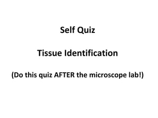
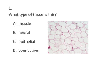
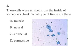
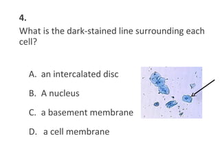
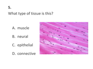
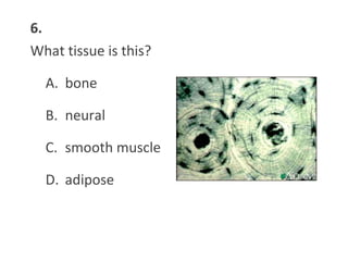
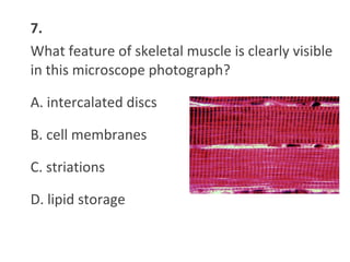
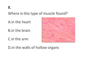
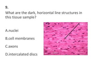
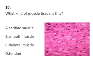
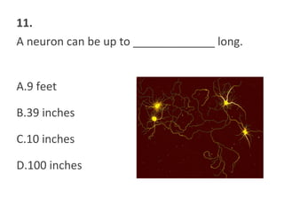
Self Quiz
Tissue Identification
(Do this quiz AFTER the microscope lab!)
1.
What type of tissue is this?
A. muscle
B. neural
C. epithelial
D. connective
2.
These cells were scraped from the inside of
someone’s cheek. What type of tissue are they?
A. muscle
B. neural
C. epithelial
D. connective
3.
What are the dark-stained structures visible in
the centers of these cells?
A. mitochondria
B. nuclei
C. food
D. intercalated discs
4.
What is the dark-stained line surrounding each
cell?
A. an intercalated disc
B. A nucleus
C. a basement membrane
D. a cell membrane
5.
What type of tissue is this?
A. muscle
B. neural
C. epithelial
D. connective
6.
What tissue is this?
A. bone
B. neural
C. smooth muscle
D. adipose
7.
What feature of skeletal muscle is clearly visible
in this microscope photograph?
A. intercalated discs
B. cell membranes
C. striations
D. lipid storage
8.
Where is this type of muscle found?
A.in the heart
B.in the brain
C.in the arm
D.in the walls of hollow organs
9.
What are the dark, horizontal line structures in
this tissue sample?
A.nuclei
B.cell membranes
C.axons
D.intercalated discs
10.
What kind of muscle tissue is this?
A.cardiac muscle
B.smooth muscle
C.skeletal muscle
D.tendon
11.
A neuron can be up to _____________ long.
A.9 feet
B.39 inches
C.10 inches
D.100 inches
12.
The longest part of a neuron is called ______.
A. the axon
B. the cell body
C. a dendrite
D. an intercalated disc
13.
The smallest muscle cells in the body are:
A. adipose
B. skeletal muscle
C. smooth muscle
D. up to 10” long
14.
This connective tissue does not usually divide
after puberty:
A. adipose
B. bone
C. cartilage
D. cardiac muscle

Tissue microscope mini-quiz

  • 1.
    Self Quiz Tissue Identification (Dothis quiz AFTER the microscope lab!)
  • 2.
    1. What type oftissue is this? A. muscle B. neural C. epithelial D. connective
  • 3.
    2. These cells werescraped from the inside of someone’s cheek. What type of tissue are they? A. muscle B. neural C. epithelial D. connective
  • 4.
    3. What are thedark-stained structures visible in the centers of these cells? A. mitochondria B. nuclei C. food D. intercalated discs
  • 5.
    4. What is thedark-stained line surrounding each cell? A. an intercalated disc B. A nucleus C. a basement membrane D. a cell membrane
  • 6.
    5. What type oftissue is this? A. muscle B. neural C. epithelial D. connective
  • 7.
    6. What tissue isthis? A. bone B. neural C. smooth muscle D. adipose
  • 8.
    7. What feature ofskeletal muscle is clearly visible in this microscope photograph? A. intercalated discs B. cell membranes C. striations D. lipid storage
  • 9.
    8. Where is thistype of muscle found? A.in the heart B.in the brain C.in the arm D.in the walls of hollow organs
  • 10.
    9. What are thedark, horizontal line structures in this tissue sample? A.nuclei B.cell membranes C.axons D.intercalated discs
  • 11.
    10. What kind ofmuscle tissue is this? A.cardiac muscle B.smooth muscle C.skeletal muscle D.tendon
  • 12.
    11. A neuron canbe up to _____________ long. A.9 feet B.39 inches C.10 inches D.100 inches
  • 13.
    12. The longest partof a neuron is called ______. A. the axon B. the cell body C. a dendrite D. an intercalated disc
  • 14.
    13. The smallest musclecells in the body are: A. adipose B. skeletal muscle C. smooth muscle D. up to 10” long
  • 15.
    14. This connective tissuedoes not usually divide after puberty: A. adipose B. bone C. cartilage D. cardiac muscle