Downloaded 206 times


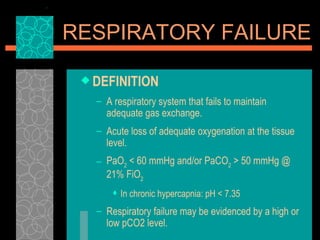


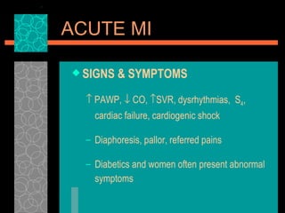


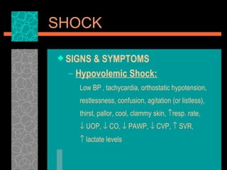


This document provides an overview of several common critical medical conditions including respiratory failure, ARDS, acute MI, CHF, GI bleed, DKA, shock, and sepsis. It defines each condition and discusses signs and symptoms, causes, complications, treatments, and nursing interventions. Respiratory failure can result from ventilation-perfusion mismatching or intrapulmonary shunting. ARDS causes damage to the alveolar-capillary interface leading to pulmonary edema. Acute MI is caused by coronary artery obstruction from thrombus or plaque. CHF occurs when the heart cannot pump sufficient blood to meet metabolic needs.


