
Recommended
More Related Content
What's hot
What's hot (20)
Similar to Fasciola hepatic.- complete life cycle, Different larva stages
Similar to Fasciola hepatic.- complete life cycle, Different larva stages (20)
More from SoniaBajaj10
More from SoniaBajaj10 (20)
Recently uploaded
Recently uploaded (20)
Fasciola hepatic.- complete life cycle, Different larva stages
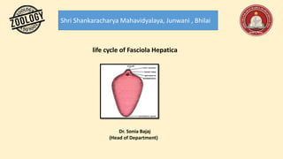
- 1. Shri Shankaracharya Mahavidyalaya, Junwani , Bhilai life cycle of Fasciola Hepatica Dr. Sonia Bajaj (Head of Department)
- 2. Life History of Fasciola Hepatica: (i) Copulation and Fertilization of Fasciola Hepatica: F. hepatica is hermaphrodite even then cross- fertilization is of common occurrence. Hence, before fertilization copulation occurs; during copulation, which occurs in the bile duct of the sheep, the Cirrus of one Fasciola is inserted into the Lauer's canal of other Fasciola and the sperms are deposited into the oviduct, so that cross-fertilization takes place. During self- fertilization, which occurs only when cross-fertilization does not take place, the sperms from the same Fasciola enter the female genital aperture and pass down the uterus to fertilize the eggs in the oviduct. (ii) Formation of Egg Capsules in Fasciola Hepatica: The eggs are brownish in colour, oval in shape and measure about 130 to 150 µ in length and 63 to 90 µ in width. the eggs are fertilized in the oviduct, the fertilized eggs receive yolk cells from vitelline glands and they get enclosed in a chitinous shell formed by granules in the yolk cells giving out droplets, the shell hardens and becomes brownish yellow; the shell has an operculum or lid. Mehlis’s glands play no role in the formation of the shell. The completed ‘eggs’ are called capsules which are large in size and they pass into the uterus where development starts. Capsules come out of the gonopore into the bile duct of the sheep, they reach the intestine and are passed out with the faeces. The capsules which fall in water or damp places will develop at about 75°F. Capsules are produced throughout the year, and one fluke may produce 500,000 capsules.
- 3. (iii) Development of Fasciola Hepatica: Development starts in the uterus and is continued on the ground. The fertilized egg divides into a small propagatory cell and a larger somatic cell. The somatic cell divides and forms the ectoderm of the larva. Later the propagatory cell divides into two cells, one of which forms the endoderm and mesoderm of the larva, and the other forms a mass of germ cells at the posterior end of the larva. This method of development takes place in the formation of all larval stages during the life history. In two weeks time, a small ciliated miracidium larva is formed and it comes out of the shell by forcing the operculum. The miracidium produces a proteolytic enzyme which erodes the lower surface of the operculum.
- 4. Miracidium Larva: Miracidium larva is a minute, oval and elongated, free-swimming stage, it is covered with 18 to 21 flat ciliated epidermal cells lying in five rings. The first ring is made of Sex plates (two dorsal, two lateral and two ventral), second ring has again six plates (three dorsal and three ventral), third ring has three plates (one dorsal and two ventrolateral), fourth ring has four plates (two right and two left) and fifth ring has two plates (one left and one right). Anteriorly it has a conical apical papilla, and attached to it is a glandular sac with an opening called apical gland.. There are two pigmented X-shaped eye spots and a nervous system. There is a pair of protonephridia, each with two flame cells. The miracidium does not feed, it swims about in water or moisture film, but it dies in eight hours unless it can reach a suitable intermediate host, which is some species of amphibious snail of genus Limnaea . After getting a suitable host the miracidium adheres to it by its apical papilla and enters the pulmonary sac of the snail, from where it penetrates into the body tissues with the aid of penetration glands and finally reaches to snail’s digestive gland. In the tissues the miracidium casts off its ciliated epidermis, loses its sense organs and it swells up and changes in shape to form a sporocyst.
- 5. Sporocyst: The sporocyst is an elongated germinal sac about 0.7 mm long and covered with a thin cuticle, below which are mesenchyme cells and some muscles. The glands, nerve tissue, apical papilla and eye spots of miracidium disappear. The hollow interior of sporocyst has a pair of protonephridia each with two flame cells it has germ cells and germ balls. The germ cells have descended in a direct line from the original ovum from the miracidium developed. The sporocyst moves about in the host tissues and its germ cells develop into a third type of larva called redia larva. A sporocyst forms 5 to 8 rediae. The rediae larvae pass out of the sporocyst by rupture of its body wall into the snail tissues with the aid of the muscular collar and ventral processes, then the rediae migrate to the liver of the snail.
- 6. Redia: The redia is elongated about 1.3 mm to 1.6 mm in length with two ventral processes called lappets or procruscula near the posterior end and a birth pore near the anterior end. Body wall has cuticle, mesenchyme and muscles, and near the anterior end, just in front of the birth pore, the muscles form a circular ridge, the collar used for locomotion. Redia has an anterior mouth, pharynx in which numerous pharyngeal glands open, sac-like intestine and there is a pair of protonephridia with two pairs of flame cells. Its cavity has germ cells and germ balls. The germ cells of redia give rise during summer months to a second generation of daughter rediae, but in winter they produce the fourth larval stage, the cercaria larva. Thus, either the primary redia or daughter redia produce cercaria larvae which escape from the birth pore of the redia into the snail tissues. Each redia forms about 14 to 20 cercariae.
- 7. Cercaria: The cercaria has an oval body about 0.25 mm to 0.35 mm long and a simple long tail. Its epidermis is soon shed and replaced by cuticle; below the cuticle are muscles and cystogenous glands. It has rudiments of organs of an adult; there are two suckers (oral sucker and ventral sucker) and an alimentary canal consisting of mouth, buccal cavity, pharynx, oesophagus and a bifurcated intestine. There is an excretory bladder with a pair of protonephridial canals (excretory tubules) with a number of flame cells. An excretory duct originates from the bladder, travels through the tail and bifurcates to open out through a pair of nephridiopores. There are two large penetration glands, but they are non-functional in the cercaria of Fasciola. It also has the rudiments of reproductive organs formed from germ cells. The cercariae escape from the birth pore of the redia, then migrate from the digestive gland of the snail into the pulmonary sac from where they pass out into surrounding water. The time taken in snail from the entry of miracidia to the exit of cercariae is five to six weeks.
- 8. Metacercaria: The cercariae swim about in water for 2 to 3 days; they then lose their tails and get enclosed in a cyst secreted by cystogenous glands. The encysted cercaria is called a metacercaria which is about 0.2 mm in diameter and it is in fact a juvenile fluke. If the metacercariae are formed in water they can live for a year, but if they are formed on grass or vegetation then they survive only for a few weeks, they can withstand short periods of drying. The various larval stages (the miracidium, sporocyst, redia, and cercaria) are all formed in the same way from germ cells which are set aside at the first division. There is, thus, a distinction between germ cells and somatic cells, and germ cells alone form the various larval stages.
- 9. Adult flukes in liver → copulation and fertilization → laying of capsules in the bile ducts → capsules in the intestine (stages in sheep’s body) → capsules out in faces → miracidia escape from capsules (stages in open) → miracidia → sporocysts → rediae → cercariae → (stages in snail’s body) → cercariae → metacercariae (stages in open) → metacercariae young flukes → adult flukes (stages in a fresh sheep’s body).
- 10. References- Modern text book – R.L.Kotpal Jantu Vigyan- S.M. Sexsena Jantu Vigyan- Dr.H.N. Baijal