
Recommended
More Related Content
What's hot
What's hot (20)
Similar to General feature of male & female Ascaris, comparison of Ascaris ,Life cycle of Ascaris,
Similar to General feature of male & female Ascaris, comparison of Ascaris ,Life cycle of Ascaris, (20)
More from SoniaBajaj10
More from SoniaBajaj10 (20)
Recently uploaded
Recently uploaded (20)
General feature of male & female Ascaris, comparison of Ascaris ,Life cycle of Ascaris,
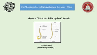
- 1. Shri Shankaracharya Mahavidyalaya, Junwani , Bhilai General Characters & life cycle of Ascaris Dr. Sonia Bajaj (Head of Department)
- 2. Ascaris General Characters Nematodes are also known as roundworms. Ascaris lumbricoides is also known as the common roundworm. The roundworms are different from the flatworms and tapeworms as they have cylindrical body, pseudocoelom and a complete digestive tract lined by endodermal epithelium. Most of these round worms are free living but some parasitic worms also exist. The species belonging to the genus Ascaris are large-sized and they inhabit in the intestines of the vertebrate hosts. Ascaris Lumbricoides is the most common roundworm which is the gastro- intestine parasite of the man. Distribution Ascaris is cosmopolitan in distribution but it is chiefly found in India, China, Korea, Philippines and Pacific islands. Habit and habitat Ascaris is one of the most familiar endoparasites of humans. It has been reported in the intestines of pig, cattle, sheep, man and squirrels. It frequently inhabits the small intestine of children than adults.
- 3. External features Size and shape of the body: The body of the roundworms is elongated and cylindrical. It gradually tapers at both ends. The anterior end is more slender than that of the posterior end. The sex of the roundworms is separate with sexual dimorphism. The body of these worms is covered by cuticle, which has minute striations which imparts a pseudo segmented appearance to the worms. • Female roundworm measures about 20-40 cm in length and 4-6 mm in diameter, the posterior end of the female round worm is straight compared to that of the male. • Male roundworms measure up to 20 cm in length and 2-4 mm in diameter. The males are smaller compared to the females. The posterior end of the male roundworms is curved. Body color: The fresh specimens are light yellow to light pink in color. The semitransparency of the body wall enables visibility of some of the internal organs. Longitudinal streaks: Along the entire length of the body of the roundworms, four longitudinal streaks are present namely one mid dorsal, one mid ventral and two lateral. The dorsal and ventral lines appear pure white whereas the lateral lines are more visible and appear brown in color. These longitudinal streaks are the impressions of the syncytial epidermis which is much thick along the median dorsal, median ventral and lateral positions.
- 4. Anterior end: The anterior end of the roundworms is round and bears a triradiate mouth guarded by three broad lips or labia. One of the lips is situated mid-dorsally and the remaining two are situated sub-ventrally. The inner margin of each lip is forked, fleshy core made up of two anterior extensions of labial parenchyma and bears minute tentacles. The outer surface of the lips bears minute sensory papillae. Posterior end: Sexual dimorphism is clearly visible at the posterior end of these roundworms. The posterior end is straight in the female worms whereas it is curved in the male worms. In females, just before the tail ending, anus is present. This anus is guarded by lips. Only digestive tube opens outside through the anus. In males, anus is replaced by cloaca. Cloaca is the common aperture for the digestive tract ad genital tubes. Two chitinous spicules of equal size are seen protruding out of the cloacal aperture. These spicules are called as penial setae. These spicules help in the transfer of the sperms into the female vagina during the process of copulation. Also the tail end of the male Ascaris has numerous genital papillae which have a role in copulation. Excretory pore: A single excretory pore lies mid-ventrally at a distance of about 2mm from the anterior end. Female gonopore: The genital pore of the female lies mid-ventrally at about one-third distance from the anterior end. On the other hand the genital pore of the male opens into cloaca. The genital pore and anus open separately in the female roundworms.
- 6. Life history of Ascaris The life history of Ascaris includes many stages namely, • Copulation, • Fertilization, • Formation of Zygote, • Cleavage and early development, infection to new host, later development in the new host and finally migration. All the stages are described hereunder: Copulation The copulation takes place in the small intestine of humans in which the adult roundworms reside. The male worm orients its body at right angles to that of the female worm so that its cloacal aperture opposes the vulva of the female. The penial spicules of the male worm help in opening the vulva of the female worm. After the opening of the vulva, the sperms are transferred into the vagina from where the sperms are passed on to the proximal end of the uteri. Fertilization After the sperms reach the proximal end of the uteri, the process of fertilization begins. After fertilization, the ova undergo second maturation division. The zygote The eggs which are not fertilized contain glycogen and fat. After fertilization, the glycogen globules migrate to the surface and form a fertilization membrane which later hardens to form thick, clear and chitinous shell. Now the fat globules form a thin lipoid layer below the shell.
- 7. As the zygote moves down the uterus, the uterine wall secretes a thick hard, yellow or brown aluminous coat with a typical wavy texture. The fertilized eggs at this stage are elliptical in shape. Under appropriate conditions of temperature, moisture and oxygen the fertilized eggs undergo cleavage to develop into infective stage.
- 8. Cleavage and early development The cleavage in roundworms is determinate and spiral type. The first division results in two cells namely dorsal (AB) and ventral (P1). Next the dorsal cell divides into anterior A and posterior B, whereas the ventral cell divides into upper EMST and the lower P2. The four-celled embryo thus formed is initially in T-shape but soon it becomes rhomboidal in shape as the P2 comes to lie posterior to EMST. In the next cleavage, * A and B further divide in to right and left cells, * EMST divides into E and MST, * P2 divides into P3 and C Further, * E divides into E1 and E2 * P3 divides into p4 and D Finally, P4 divides into G1 and G2 The fate of various cells is pre-determined and fixed as follows, * The descendants of A and B give rise to ectoderm * MST gives rise to mesoderm and ectoderm of foregut * E1 and E2 gives rise to endoderm * G1 and G2 form germ cell primordium * C and D together give rise to ectoderm and mesoderm
- 10. After all the divisions, embryo attains 16 cells and forms a hollow ball like structure called as blastula. The cavity of blastula is called as blastocoel. Blastula undergoes invagination process also called as gastrulation to form gastrula. The gastrula grows in size and gets transformed into an active juvenile with alimentary canal, nerve ring and a larval excretory system. This juvenile closely resembles the nematode genus Rhabditis, found in soil and human faeces. Thus this juvenile is also known as rhabditiform larva of first stage. This stage is not infective. In a week time this larva undergoes molting within the egg shell to form second stage larva which is infective. Infection to the new host As there is no intermediate host in the life cycle of Ascaris, humans acquire infection by a) directly ingesting Ascaris eggs, with second stage larva b) contaminated food or water. After ingestion of the eggs, due to the action of the digestive juices of the small intestine, the egg shell of the larva is dissolved and the larva hatch out.
- 11. Later development and migration Just hatched out juvenile measures about 0.25 to 0.3 mm in length and 13 to 15µ in diameter. This juvenile performs active thrashing movements to bore through the intestinal epithelium and begins to migrate to other parts of the host body. The larva migrates the hepatic portal circulation and then to the liver and then to heart through post-caval vein. From the heart it migrates to the lungs via pulmonary artery. In the lungs it remains for few days and increases in size. It ruptures the blood capillaries and bores its way to alveoli. Here this second larval stage undergoes moulting to become third stage larva. This third stage larva further undergoes another moulting to transform into fourth stage larva which is about 2-3 mm in length. This fourth stage larva migrates from the alveoli to the pharynx through trachea. From the pharynx this larva reenters the gut when coughed and swallowed back. This fourth stage larva undergoes final moulting to become adult. These adult worms attain sexual maturity within 8-10 week time. Some larvae do not follow the above said usual pattern of migration but reach the brain or spinal cord. The larva cannot survive in these organs and thus a calcareous cyst is formed around it.
- 13. References- Modern text book – R.L.Kotpal Jantu Vigyan- S.M. Sexsena Jantu Vigyan- Dr.H.N. Baijal