
Recommended
More Related Content
What's hot
What's hot (20)
Similar to Cardiovascular system
Similar to Cardiovascular system (20)
Recently uploaded
Recently uploaded (20)
Cardiovascular system
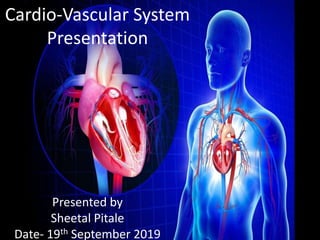
- 1. Cardio-Vascular System Presentation Presented by Sheetal Pitale Date- 19th September 2019
- 2. Contents • What is cardiovascular system (CVS) • Important components of CVS – Blood – Heart – Lungs – Lymphatic system • Anatomy of CVS • Physiology of heart – Double circulation system – Cardiac cycle
- 3. What is cardiovascular system(CVS) Cardio + Vascular = CVS Oxy- Artery Deoxy- Veins CVS is responsible for •Converting Deoxygenated blood into oxygenated blood •Supply oxygenated blood to body cells •Generates energy (ATP) packets for running body functions like muscle contraction etc
- 4. Important components of (CVS) • Blood: There is about 6litres of blood continuously circulating in our body through the heart. It is made up of liquid(plasma) and solids(the blood cells). • Heart: The heart takes deoxygenated blood from different parts of body and transport oxygenated blood to parts of body at cell level • Lungs: There are pairs of lungs in thoracic cavity. They helps to convert deoxygenated blood to oxygenated blood and transported to heart through pulmonary vein. • Lymphatic system: Lymphatic System is special type of circulatory system which consist of lymph, lymph nodes or glands and lymphatic vessels for immune system of the body
- 5. Anatomy of CVS- Blood Composition of Blood • Total volume of Blood: 5-6 litres in our body • Specific Gravity of Blood: 1050- 1060 • Viscosity of Blood: 4-5 times of water • pH of Blood: 7.4 +/- 0.05 alkaline The blood is made up of plasma and cells. Cells represents 45% of blood volume and consist of • Red Blood cells / Erythrocytes (RBC): Normal count = 5million per cumm • White Blood cells / Leukocytes (WBC): Normal count = 4000- 11000 per cumm • Platelets / Thrombocytes : Normal count = 1.5-4 lacs /cumm Plasma represents 55% of total blood volume in which there is 91% of water and 9% of solids.
- 6. Anatomy of CVS- Blood Cells of blood RBC/ Erythrocytes • Structure: circular biconcave non nucleated disc • Normal count: – At birth: 6-7 million/cumm – Male : 5-6 million/cumm – Female : 4.5-5.5 million/ cumm • life span: 120 days • Functions of RBC – It contain haemoglobin which carry oxygen to different parts of body at tissue level. – Helps in indentifying the blood groups
- 7. Anatomy of CVS- Blood Cells of blood WBC/ leukocytes : • Normal count : – At birth: 20000/cumm – Adult : 4000-11000/cumm • Contents of WBC – Neutrophils : It is first line of defence against bacterial attack in the body – Eosinophils : It is mild phagocytes – Basophils : It liberates histamine which is responsible for allergic reaction manifestation – Lymphocytes : produces antibodies against antigens – Monocytes : It second line of defence against bacterial attack after neutrophils in our body
- 8. Anatomy of CVS- Blood Platelets/ Thrombocytes • Normal count = 1.5-4 lacs/cumm • Life span = 8-12days • Location= spleen • Functions of Thrombocytes – Helps in haemostasis of blood – Help in blood coagulation – Help in phagocytosis of viruses in blood
- 9. Anatomy of CVS- Blood • Functions of blood – Carries oxygen to the tissues – Carries nutrients to the tissues – Carries away waste products from tissues to the excretory organ – Carries hormones from glands to the target tissues – Fights against antigens/ foreign particles in the body – Helps in maintaining the body temperature – Maintains water balance in body
- 10. Anatomy of CVS – Heart • Introduction • Layers • Chambers • Valves • Muscles • Nodal cells • Great vessels ( arteries and veins)
- 11. Anatomy of CVS- Heart Introduction • The heart is hallow muscular organ which pumps the blood to various parts of the body. • It is situated obliquely in thoracic cavity (middle mediastinum) between the two lungs and behind the sternum. • The normal adult heart measures 12cm vertically and 6cm antero- posterior. • The weight of male is 300gms and female heart is 250gms. • The size of heart is about the fist of the individual. • The heart consist of apex and base.
- 12. Anatomy of CVS – Heart Layers of heart The heart is covered by fibro-serous membrane called pericardium. Pericardium has three layers 1. Epicardium: outermost layer composed of lose connective tissues 2. Myocardium: middle layer which opens blood vessel (arteries) supplying blood to heart muscles 3. Endocardium: innermost layer for protecting the heart chambers .
- 13. Anatomy of CVS- Heart Chambers • Atria – Right Atrium: This chamber carries deoxygenated blood from body through superior venacava (SVC ) and inferior venacava (IVC) – Left Atrium: This chamber carries oxygenated blood from body through pulmonary veins • Ventricles – Right Ventricle : This chamber carries deoxygenated blood received from right atrium through tricuspid valve – Left Ventricle : This chamber carries oxygenated blood from left atrium through mitral valve ( bicuspid)
- 14. Anatomy of CVS- Heart Valves The valves are required to avoid the regurgitation of blood flow. The valves are one directional. There are four valves in heart. 1. Tricuspid valve: It has three leaflets that opens and closes allowing deoxygenated blood from right atrium to right ventricle 2. Mitral valve (bicuspid): It has two leaflets that opens and closes allowing oxygenated blood from left atrium to left ventricle 3. Pulmonary valve: It is the semi lunar (tricuspid) valve that connects right ventricle to pulmonary artery carrying deoxygenated blood to lungs 4. Aortic valve: It is the semi lunar (tricuspid) valve that connects left ventricle to aorta carrying oxygenated blood to the body
- 15. Anatomy of CVS- Heart Muscles Cardiac muscles (myocardium): It is involuntary striated muscles constitutes tissues of heart wall and responsible for contractibility Papillary muscles: This are the smooth muscles inside the ventricles for the opening of valves Bundle of His and Purkenje fibres: These are special muscles for faster conduction of electrical signals during systole of ventricles Purkenje fibres papillary muscles
- 16. Anatomy of CVS- Heart Nodal cells The Nodal cells are the specialised cells present in the wall of heart which helps in the conduction of the electrical signals to heart muscles causing in contraction . There are five nodal cells they are as follows 1. Sinoatrial node (SA node): SA node is the major element in conduction system which controls the heart rate 2. Atrioventricular node (AV node): AV node is the relay station between the upper and the lower chambers of heart 3. Bundle of His: It is an important part of conduction system of heart as it transmits impulses from AV node to the septum and the ventricles of heart 4. Bundle braches : This are divided in right and left bundle branch which transmits impulse to right and left ventricles 5. Purkinje fibres : It has specialised bunch of muscles which conducts faster impulses to the heart
- 17. Anatomy of CVS- Heart Great vessels The heart consist of following great vessels 1. Superior venacava (SVC): SVC carry deoxygenated blood from upper parts of the body to the right atrium. 2. Inferior venacava (IVC): IVC carries the deoxygenated blood from the lower parts of the body to the right atrium 3. Pulmonary artery (PA): It gets divded into right and left branch which carries deoxygenated blood from right ventricle and gives to the both lungs for oxygenation of blood 4. Pulmonary veins(PV): It carries oxygenated blood from lungs and brings to left atrium 5. Aorta : It is the great vessel which carries oxygenated blood from left ventricle and supplies all parts of the body
- 18. Anatomy of CVS- Heart Blood supply and venous drainage 1. Coronary arteries: It supplies oxygenated blood to entire myocardium the blood is received through 2 ostium (sinuses) located at aortic root. There are 4 main coronary artery – Right coronary artery (RCA) – Left coronary (LCA) – Left Circumflex artery (LCX) – Left anterior descending artery (LAD) 2. Coronary veins : It collects deoxygenated blood from entire myocardium the blood is drain through sinuses located at right atrium .
- 19. Anatomy of CVS- lungs • Lungs are pair of spongy air filled organs located on either side of chest • Internally lungs are made of small units called alveoli which responsible for exchanging inhaled oxygen into deoxygenated received pulmonary artery, converting oxygenated blood and sending back to heart through pulmonary veins this is called respiration
- 20. Anatomy of CVS- Lymphatic system Lymphatic system: Lymphatic System is special type of circulatory system for immune system of the body which consist of • Lymph: It is a fluid like plasma and the tissues fluids. It may contain bacteria in any infections • Lymphatic vessel : Lymphatic vessels starts in tissues spaces between the cells and starts like a veins. This lymph flows in large lymphatic vessels • Lymph glands or nodes: They are the small bean shaped structure situated near neck, axilla, groin and also in pelvic
- 21. Physiology of Heart Double circulation Systematic Circulation • IVC & SVC carries deoxygenated blood from body to right atrium. • The blood pass into right ventricle through atrioventricular valve (AV) • Then the blood is pumped into lungs through pulmonary artery (deoxygenated blood) Pulmonary Circulation • Lungs undertake gas exchange to convert deoxy blood into oxy blood • Then this Oxygenated blood is passed through pulmonary vein to left atrium • The oxygenated blood is then passed into left ventricles through mitral valve. Then Oxy blood is pumped into body back through aorta and it’s valve
- 22. Physiology of Heart cardiac cycle The sequence of changes in the pressure and flow in the heart chambers and blood vessels in the two subsequent cardiac contraction is known as cardiac cycle. There are phases of Cardiac Cycle. Normal cardiac cycle takes 0.8secs • Systole ( Contraction ) of chambers • Diastole ( relaxation ) of chambers Events in cardiac cycle • Atrial systole: Both Atria are emptied into respective ventricles by opening their valves. The duration is about 0.1secs. • Ventricular systole: Left ventricle on contraction forces oxygenated blood into aorta to body. Right ventricle on contraction forces deoxygenated blood into pulmonary artery to lung The duration is about 0.3secs. First heart sound is heard (HS1) • Ventricular diastole: The duration is about 0.5secs the semilunar valve close which cause the second heart sound (HS2). This a passive filling of ventricles from respective atrium. During this phase tricuspid and mitral valves are open. • Atrial diastole: The duration is about 0.7secs during this phase the muscles relaxes and receives blood from Lung and Body