Lecture_Unit1_ Transport systems in Animals complete.pptx
•Download as PPTX, PDF•
0 likes•7 views

Life Sciences Grade 11

Recommended
Recommended
More Related Content
Similar to Lecture_Unit1_ Transport systems in Animals complete.pptx
Similar to Lecture_Unit1_ Transport systems in Animals complete.pptx (20)
Recently uploaded
Recently uploaded (20)
Lecture_Unit1_ Transport systems in Animals complete.pptx
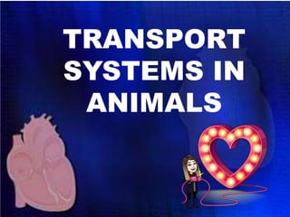
- 2. Welcome to the new chapter on Transport Systems in Animals! This is a super fun unit especially because you can relate to all the topics to be discussed. You are going to learn so much about your own body! Lets dive straight in! Ready, set, go!! ALL THE PICS OF Ms P WILL HAVE LINKS TO VIDEOS OR SUPPORTIVE MATERIAL Click me
- 4. FUNCTIONS OF ARTERIES • Arteries: transports oxygenated (O2) blood except for pulmonary artery. • Main artery Aorta pumping oxygenated blood AWAY from heart rest of body. • Other arteries: • Renal artery (kidney) • Hepatic artery (liver)
- 6. FUNCTIONS OF VEINS • Veins: transports deoxygenated blood except for the pulmonary vein. • Main vein is called the vena cava – pumping deoxygenated blood towards heart from rest of body. • Other veins: • Renal vein (kidney) • Hepatic vein (liver)
- 7. FUNCTIONS OF CAPILLARIES Capillaries: Connect arteries & veins structurally suited exchange of substances between blood in capillaries & interstitial tissue fluid (between cells)
- 8. DIFFERENT BLOOD VESSELS: ARTERIES, VEINS AND CAPILLARIES
- 9. THE HUMAN CIRCULATORY SYSTEM: HEART AND ASSOCIATED VESSELS
- 10. DOUBLE CIRCULATION • Circulatory systems 2 distinct circuits. • The pumps of the 2 circuits serve different tissues but combined into single organ, the heart. • The 2 circuits are called: 1. Pulmonary circuit 2. Systemic circuit
- 11. DOUBLE CIRCULATION 1. Pulmonary circuit Heart pumps deoxygenated (CO2) blood to lungs & oxygenated (O2) blood back to heart. 2. Systemic circuit Heart pumps oxygenated (O2) blood to body cells & deoxygenated (CO2) blood back to heart.
- 13. BLOOD VESSELS TRANSPORT GAS, NUTRIENTS & WASTE PRODUCTS THROUGH BODY
- 14. THE HUMAN CIRCULATORY SYSTEM: HEART AND ASSOCIATED VESSELS Mammals have 4-chambered heart with 2 atria & 2 ventricles Left side of heart pumps & receives only oxygen- rich blood, Right side receives & pumps only oxygen-poor blood The mammalian cardiovascular system meets the body’s continuous demand for O2. Blood begins its flow when deoxygenated blood flow from the body into the right atrium.
- 15. Blood then flows from right atrium into right ventricle through tricuspid valve The blood is then pumped into lungs, through semilunar valve via pulmonary artery. In lungs, blood loads O2 and unloads CO2. Oxygen-rich blood from lungs enters heart via 4 x pulmonary vein at left atrium Blood then flows into left ventricle through bicuspid valve. This blood is then pumped through semilunar valve into aorta with takes blood to entire body.
- 16. Deoxygenated blood returns to heart through superior vena cava (blood from head, neck, forelimbs) & inferior vena cava (blood from trunk & hind limbs) Superior vena cava & inferior vena cava flow into the right atrium. The atrioventricular (AV) valves (tricuspid and bicuspid valves) separate each atrium & ventricle The semilunar valves control blood flow to the aorta & the pulmonary artery.
- 19. THE INTERNAL STRUCTURE:HUMAN HEART
- 20. BLOOD FLOW THROUGH THE HEART RED: OXYGENATED BLOOD - BLUE: DEOXYGENATED BLOOD
- 21. CIRCULATORY SYSTEMS IN ANIMALS Many invertebrates do not have a circulatory system at all. diffusion. E.G. Hydra
- 22. CIRCULATORY SYSTEMS IN ANIMALS • In animals with multiple layers of cells - cells are too far from external environment for simple osmosis/ diffusion. • Higher animals - 2 primary types of circulatory systems - open & closed.
- 23. CIRCULATORY SYSTEMS IN ANIMALS OPEN- • The circulatory fluid – Hemolymph (is same as interstitial tissue fluid) • heart pumps hemolymph through vessels into sinuses (open fluid-filled spaces) • Certain substances are exchanged between hemolymph and cells. • Hemolymph returns to heart through pores. • The heart is a tubular structure.
- 24. CIRCULATORY SYSTEMS IN ANIMALS OPEN-
- 25. CIRCULATORY SYSTEMS IN ANIMALS CLOSED- • Circulate blood entirely within vessels. • Blood is distinct from interstitial fluid. • Chemical exchange occurs between blood & interstitial fluid & between interstitial fluid & body cells.
- 26. CIRCULATORY SYSTEMS IN ANIMALS CLOSED-
- 27. CARDIAC CYCLE The heart contracts and relaxes in a rhythmic cycle called the cardiac cycle The contraction / pumping, systole The relaxation / filling diastole The heart rate pulse (number of beats per minute
- 28. CARDIAC CYCLE RED: OXYGENATED BLOOD - BLUE: DEOXYGENATED BLOOD ARTRIAL SYSTOLE VENTRICULAR DIASTOLE VENTRICULAR SYSTOLE ARTRIAL DIASTOLE
- 29. MAINTAINING THE HEART’S RHYTHMIC BEAT Some cardiac muscle cells are self-excitable, contract without any signal from nervous system The sinoatrial (SA) node (pacemaker, sets rate & timing at which cardiac muscle cells contract Impulses from SA node travel to atrioventricular (AV) node At AV node, impulses are delayed & then travel to Purkinje fibres that make ventricles contract
- 30. MAINTAINING THE HEART’S RHYTHMIC BEAT Fig. 42-9-5 Signals spread throughout ventricles. 4 Purkinje fibers Pacemaker generates wave of signals to contract. 1 SA node (pacemaker) ECG Signals are delayed at AV node. 2 AV node Signals pass to heart apex. 3 Bundle branches Heart apex
- 31. LYMPHATIC SYSTEM Lymphatic system returns fluid that leaks out in capillary beds This system aids in body defence (lymphocytes) Fluid, called lymph, re-enters circulation directly at venous end of capillary bed & indirectly through lymphatic system The lymphatic system drains into veins in neck. Lymph nodes are organs that filter lymph & play an important role in the body’s defence Edema is swelling caused by disruptions in flow of lymph.
- 32. LYMPHATIC SYSTEM VS. CIRCULATORY -
- 33. DISEASES OF THE HEART AND CIRCULATORY SYSTEM Cardiovascular diseases are disorders of the heart and the blood vessels They account for more than half the deaths in the South Africa One type of cardiovascular disease, atherosclerosis, is caused by the build-up of plaque (fat) deposits within arteries.
- 34. A heart attack is the death of cardiac muscle tissue resulting from blockage of one or more coronary arteries A stroke is the death of nervous tissue in the brain, usually resulting from rupture or blockage of arteries in the head. Hypertension, or high blood pressure, promotes atherosclerosis and increases the risk of heart attack and stroke. Hypertension can be reduced by dietary changes, exercise, and/or medication