Urine Analysis and Kidney function tests
•
9 likes•827 views

Urine Analysis and Kidney function tests

Recommended
More Related Content
What's hot
What's hot (20)
Similar to Urine Analysis and Kidney function tests
Similar to Urine Analysis and Kidney function tests (20)
More from Amany Elsayed
More from Amany Elsayed (20)
Recently uploaded
Recently uploaded (20)
Urine Analysis and Kidney function tests
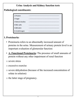
- 1. 1 Urine Analysis and Kidney function tests Pathological constituents: 1.Proteinuria: • Proteinuria refers to an abnormally increased amount of proteins in the urine. Measurement of urinary protein level is an important evaluation of glomerular function: • A. Functional Proteinuria: The presence of small amounts of protein without any other impairment of renal function o severe stress o excessive exercise o severe dehydration (because of the increased concentration of solute in solution) o the latter stage of pregnancy.
- 2. 2 o B. Organic Proteinuria: o Pre-renal: This is caused by a general disease, which affects the kidneys and is an indication of renal damage, such as essential hypertension. o Renal: All types of renal diseases will cause proteinuria (acute or chronic nephritis and nephrotic syndrome). o Post-renal: - This case may be due to lesions in the renal pelvis, bladder, prostate and urethra. - It may also caused by bacterial infection of the urinary tract. These patients usually have pain on passing urine (dysuria). - When a patient has any kind of urinary infection, protein and pus cells (pyuria) can be usually found in the urine. 2.Glucosuria: • A 24 hours specimen of urine from a normal subject contains a small amount of reducing substances, generally less than 1 g, of which 20-200 mg is glucose. • Glucosuria implies that sufficient glucose is present to be detectable by a simple clinical test. • The traditional test employed is Benedict's solution, which is blue because of the presence of Cu+3 ions. • On reduction of Benedict's solution by glucose or other reducing substances, the blue colour disappears and an orange-red precipitate of Cu+2 oxide is formed.
- 3. 3 • When glucose in tubular fluid exceeds the transport maximum (180 mg/100 ml), it appears in urine (glycosuria). • Glucose in tubular fluid hinders water reabsorption by osmosis, causing polyuria. ♦The causes of glucosuria 1. Low renal threshold: • This leads to renal glucosuria; occurs with normal blood glucose level but tubular reabsorption of glucose is below normal thus permitting some glucose to spill into the urine. Glucose reabsorption • The transporter for glucose on the basolateral membrane has a limited capacity to carry glucose back into the blood. • If blood glucose rises above 180 mg/dl, some of the glucose fails to be reabsorbed and remains in the urine glucosuria
- 4. 4
- 5. 5 2. Hyperglycemia with impaired glucose tolerance: • This leads to Diabetes mellitus; a pathologic state associated with increased blood sugar and polyuria. • The urine is colourless due to diluted polyuria and of high specific gravity due to extra load of dissolved solids (glucose). • Hyperglycemia without glucosuria may be found if there is a raised threshold due to diminished renal plasma flow, this is quite often seen in elderly diabetic. Diabetes mellitus - caused by either 1) deficiency of insulin (Type I) or 2) deficiency of insulin receptors (Type II). - Diabetes mellitus features high glucose in the blood (hyperglycemia)
- 6. 6 Diabetes insipidus - is caused by inadequeate ADH secretion. - Due to the shortage of ADH, water reabsorption in CD is compromised, leading to polyuria. Other sugars • Lactose: This is often present in o the urine of pregnancy after 20 weeks. o in nursing mothers (during lactation). o occasionally found in patients on a milk diet (in case of liver cirrhosis). • Galactose: Galactosuria occurs usually inferior to Galactosemia, which occurs as a congenital abnormality. It is due to deficiency of the hepatic enzyme galactose-1-phosphate uridyl transferase, which is concerned, with the conversion of galactose to glucose. Accumulation of galactose-1-phosphate leads to renal tubular damage with aminoaciduria, proteinuria, acidosis and liver damage with jaundice. The high blood galactose leads to cataracts. • Fructose: Congenital fructosuria is usually due to the deficiency of an isoenzyme of aldolase. Fructose-1-phosphate accumulates after ingestion of fructose or sucrose and this causes hypoglycemia and liver damage.
- 7. 7 • Pentose: Pentosuria occurs usually after ingestion of large amounts of pentose-containing fruits (berries and pears). Congenital pentosuria due to L-xylulose reductase deficiency is less rare. • Identification of sugars other than glucose is performed by thin- layer chromatographic separation methods 3. Ketonuria: • The body normally metabolizes fat completely to CO2 and H2O, so that only 3-15 mg of ketones is excreted daily which cannot be qualitatively detected in urine. • Whenever there is inadequate carbohydrate in the diet or a defect in carbohydrate metabolism (e.g. in diabetes mellitus) the body metabolizes increasing amounts of fatty acids. • When this increase is large, fatty acid oxidation is incomplete and intermediates products of fat metabolism appear in the blood (ketonemia) and are excreted in the urine (ketonuria). • These intermediary products are acetoacetic acid, acetone and B-hydroxybutyric acid. • Ketonemia and ketonuria also develop when the patient is suffering from carbohydrate deficiency. • Thus ketonuria is found in starvation or on a badly balanced reducing diet.
- 8. 8 • Postoperative ketonuria is commonly seen. It is due to a combination of: • the starvation that precedes the operation, • the vomiting that often follows it and, • possibly to the effect of anaesthia which depletes liver glycogen. 4. Hemoglobinuria: • Intravascular hemolysis (lysis of red blood corpuscles) from any cause liberates hemoglobin into the circulation. • Haptoglobins are specific 2-globulins that bind hemoglobin at the globin. • The function of haptoglobins is to conserve iron after intrvascular hemolysis by binding to hemoglobin. • Haptoglobin bound to hemoglobin is taken up mainly in the liver; the haptoglobin is slowly resynthesized and the iron reticulates from hemoglobin that is then released. • When the plasma hemolysis exceeds 100 mg %, hemoglobin is released into the surrounding medium, enters the glomeruli and appears in the urine.
- 9. 9
- 10. 10 5. Hematuria: • It is characterized by the presence of hemoglobin and unruptured red blood corpuscles. • It is caused when blood passed into the urine through some lesions of the kidney or the urinary tract. • When the red blood cells enter the urine hemolysis usually occurs after a short time. 6. Bile pigments: • The main pigments excreted in the urine are bilirubin and urobilinogen. 7. Bile salts: • Human bile contains 4 bile acids, cholic acid, deoxycholic acid, chemodeoxycholic acid and lithocholic acid. • They are synthesized in the liver from cholesterol. • Only small amounts of bile acids are found in the free state. • The major part is conjugated through amide linkages with glycine and taurine. • The conjugated bile acids are neutralized by Na+ , K+ giving glycocholates and taurocholates (bile salts). • Bile salts in the intestine are necessary for absorption of fats.
- 11. 11 • Bile salts have a hydrotropic behavior; have a powerful capacity of lowering surface tension rendering certain substances soluble in water in which otherwise they would be insoluble, facilitating the close contact between fat and water- soluble lipase.
- 12. 12 ◘ Microscopical examination: 1. Red blood cells (erythrocyte): • Red cells do not contain granules, and so can be distinguished from pus cells. • They also have a clearly seen outline, and are usually smaller. • Red cells are not normally seen in urine, but may be present in urinary infections, tumors and calculi. • Red blood cells can come from any part of the urinary system from the glomerulus to the uretheral meatus and may be a contaminant during menstruation in the urine of women. • Therefore, the significance of red blood cells (hematuria) must be related to the other findings in the urinary sediment. • If there is enough blood in the urine it will be turbid (smoky). Red cells are sometimes destroyed in the circulation and hemoglobin rather than red cells may be passed into the urine (hemoglobinuria). • The urine will be clear and red.
- 13. 13 Red blood cells 2. Pus cells: • Pus cells are polymorphs leukocytes which have come from the blood to fight bacteria invading the urinary tract. • They have a nucleus with several segments, which can often be seen with the high power objective. • As pus cells are broken down in the urine, their nuclei disappear and only the cell membranes remain. • Five pus cells per mm3 is a common figure. • Patients with pus cells more than 5 per mm3 are thought to have a urinary tract infection. • Also the patients who have infections in the vagina and urethra have discharge from these organs called pus (Pyuria). • These discharges contain protein and may contain pus cells and red cells.
- 14. 14 White blood cells 3. Epithelial cells: • These are flat large cells with a nucleus that can usually be seen quite easily. • They come from the epithelial (skin) on the side of the ureters, bladder, vagina and urethra. • Epithelial cells are usually singles that is there is one cell at a time. • A few epithelial cells are found in normal urine.
- 15. 15 Epithelial cells 4. Casts: • These are solid substances (proteins or remains of the dead cells in kidney diseases) blocking the kidney tubule and becomes loose and goes down into the urine. • Because it was found inside the tubule a cast has the shape of the tubule from which it came.
- 16. 16 ♦ Can be different types of casts : • Cellular casts: - which are often seen in the urine of jaundiced patient due to increased red cell destruction. - They are yellow-brown. • Granular casts: - when seen, indicate renal dysfunction. - They are usually dark in colour and contain small granules. Granular casts consist of antigenic protein granules. - These protein granules appear to represent fragments of parent serum proteins. - They are found when there is protein in the urine. - All urine showing ++ proteinuria should be examined for granular casts. • Hyaline casts: - they are clear, colourless and have no granules in them. - They dissolve in alkaline urine. - They are the proteins left when water is reabsorbed in the tubule. - Normal urine often has some of these casts, and they do not mean that the kidney is diseased.
- 17. 17 Granular Cast Cellular Cast
- 18. 18 Hyaline Cast 5. Crystals: a) Acidic urine:
- 19. 19 Uric acid Calcium Oxalate Crystals
- 20. 20 Urate Crystals b) Alkaline urine:
- 21. 21 Amorphous Phosphate Crystals Triple Phosphate Crystals
- 22. 22 6. Bacteria: • Normal urine contains no bacteria. • However, bacteria often grow in normal urine if it is left in warm room. • They got into the urine from a dirty jar, from the air or from the patient's skin. • Finding bacteria in old urine means nothing but in fresh urine means that the patient has a urinary infection. • Usually there will be pus cells and protein present also. • A common kind of bacteria found in the urine is called E. coli. It is a thin motile rod like a very small pencil.
- 23. 23 Bacteria 7. Yeast and fungi: • the threads or mycelium of a mould or a fungus are longer and thicker than a bacterium and they branch. • When yeasts grow, a parent cell often makes a daughter cell, which is smaller and grows from one end.
- 24. 24 8. Spermatozoa: • they are the male sex cells. • They are often found in the man's urine and are quite normal. • After sexual intercourse they may be found in a woman's urine. • Spermatozoa should be reported when present in large amounts, which may suggest a lesion in genito-urinary tract.
- 25. 25 9. Parasites: • in some parts of the world especially tropical countries, one of the most parasites are the ova of a worm called Schistosoma haematobium which lives in the veins of the walls of bladder and ureters. • The worm lays eggs that go through the side of the bladder into the urine. • The disease that it causes is called Schistosomiasis or bilharziasis. • With the ova in the urine there are usually also protein, red cells and pus cells. • When you are looking for the ova of S. haematobium you should take a specimen that has been passed by the patient between midday and two in the afternoon. Schistosoma haematobium Schistosoma mansoni
- 26. 26
- 27. 27 ◘ Three kinds of movement: 1-Motility • You will see three kinds of movement in a wet film of urine, stool or blood. • The first kind is the movement of microorganisms themselves, called motility. • Motile bacteria like E. coli can be seen swimming from one place in the field to another; while everything else stays still. 2-Brownian movement • The second kind of movement is called Brownian movement. • If you look at any very small particle lying free in a liquid, you will see that it is always moving and is never quite still. • Their movements are very small and quite fast, the particles seen to shake about only. 3-streaming • The third kind of movement is called streaming. • You will see a stream or river of particles moving in the same direction.
- 28. 28 Renal Function Tests Steps of urine formation
- 29. 29 ◘ Glomerular filtration rate (GFR): • It is the number of milliliters of plasma, which are filtered at the glomeruli every minute (ml/minute). • Renal function tests may be divided into those which: o measure glomerular filtration and o those which study tubular function. • The common renal function tests involve the determination of a number of different substances either in plasma or urine or both to calculate the clearance. • Clearance varies with body size and is proportional to the surface area. • GFR Is the amount of filtrate formed per minute by the two kidneys combined (Filtrate = plasma - proteins) • For the average adult male, GFR is about 125 ml/min. • This amounts to a rate of 180 L/day. • An average of 99% of the filtrate is reabsorbed, so that only 1-2 L of urine per day is excreted.
- 30. 30 • Renal plasma flow (RPF), which is the volume of blood plasma delivered to the kidneys per unit time (625 mL/min) • The glomerular filtration rate (GFR) = volume of plasma filtered per unit of time = 125 ml/min 180 liters per day • Filtration fraction = GFR/renal plasma flow = 20%
- 31. 31 GFR must be precisely controlled. a. If GFR is too high - increase in urine output - threat of dehydration and electrolyte depletion. b. If GFR is too low - insufficient excretion of wastes. c. The only way to adjust GFR from moment to moment is to change glomerular blood pressure.
- 32. 32 The nephron has two ways to prevent drastic changes in GFR when blood pressure rises: 1) Constriction of the afferent arteriole to reduce blood flow into the glomerulus 2) Dilation of the efferent arteriole to allow the blood to flow out more easily. Change in an opposite direction if blood pressure falls Diuretics - are chemicals that increase urine volume. - They are used for treating hypertension and congestive heart failure because they reduce overall fluid volume. - work by either increasing glomerular filtration or reducing tubular reabsorption. - Caffeine falls into the former category; alcohol into the latter (alcohol suppresses the release of ADH).
- 33. 33 • Many diuretics produce osmotic diuresis by inhibiting sodium reabsorption ◘ Clearance There are three ways in which a substance can be handled by the kidney: • Filtration at the glomeruli and reabsorption by the tubules. • Such reabsorption may be either active against concentration and osmotic gradients or passive when the tubular epithelium is permeable to a substance which had been absorbed such as urea. • Filtration at the glomeruli and secretion by the tubule such as creatinine. • Filtration at the glomeruli without reabsorption or secretion in the tubules such as inulin. • Is the number of milliliters of plasma (blood), which contain the amount of that substance excreted in a minute by the kidney. • Creatinine clearance (ml/min.) = U x V/ P • Where U = Concentration of creatinine in urine (g/L). V = Volume of urine in ml/min. P = Concentration of creatinine in plasma (g/L).
- 34. 34 • The clearance of a substance filtered at glomeruli and neither reabsorbed nor secreted by the tubules and not metabolized in the body give the GFR. • When a substance is partly reabsorbed by the tubules, its clearance is less than GFR like glucose, which is almost completely reabsorbed (clearance is nil), substances which are not reabsorbed but secreted by the tubules, its clearance is greater than GFR. How to carry out a clearance test? • The test is best carried in the morning. • Allow the patient to have a breakfast as usual then carry out the test over a two hours period. • Give a drink of water about a cup shortly before beginning of the test. • Instruct the patient to empty the urinary bladder completely as possible. • Note the time and discard this specimen. Get the patient again to empty the bladder as completely as possible after an interval of an hour and again an hour later. • Keep these two specimens. After collecting the first urine sample take a specimen of blood. • Determine the concentration of the substance content in blood and urine and measure accurately the volumes of urine for the determination of rate of urine flow in ml/min.
- 35. 35 Types of clearance (a) Exogenous inulin clearance: • The polysaccharide inulin is filtered at the glomeruli but neither secreted nor reabsorbed by the tubule. (b) Endogenous clearance: 1-Creatinine clearance: • Creatinine is not a component of the diet but is formed in the body at a relatively constant rate which is dependent upon the mass of the muscles. • Its clearance has been much used as an approximate measure of GFR. • It does not require the intravenous administration of a test substance as in the case with an exogenous clearance study using inulin. • Normal range: o 85-140 ml/min for male. o 85-125 ml/min for female. 2-Urea clearance: o Urea is derived from general protein metabolism. o Both dietary protein and endogenous tissue protein contribute to the amount of urea produced but the former is often more important.
- 36. 36 o Urea is reabsorbed passively in the renal tubule, thus its clearance cannot be used as an approximate measure to the GFR. o Normal range:40-65 ml/min. Clinical application: the glomerular filtration rate • GFR: important value for estimating the kidney function. • Calculated by using molecules which are filtered but not secreted nor reabsorbed. • P X GFR = U X V • P = plasma concentration of A, in mg/mL • GFR = glomerular filtration rate of plasma, in mL/min • U = urine concentration of A, in mg/mL • V = rate of urine production in, in mL/min • Solving the equation for GFR will give: • GFR = (U X V)/P
- 37. 37 Clinical application: the glomerular filtration rate • Best molecule to use: inulin but not occurring naturally in the body • Second best: creatinine • Urea: cannot be used since it is both secreted and reabsorbed