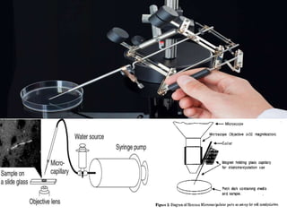
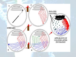
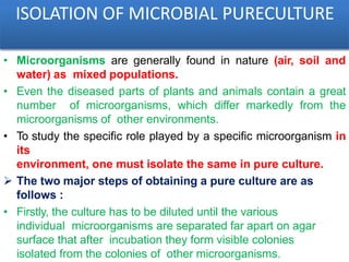
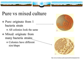
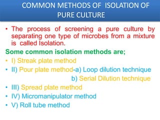
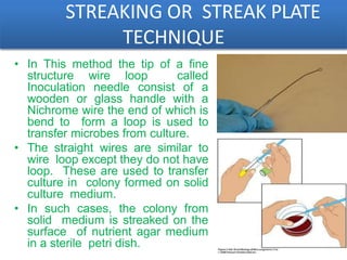
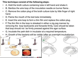
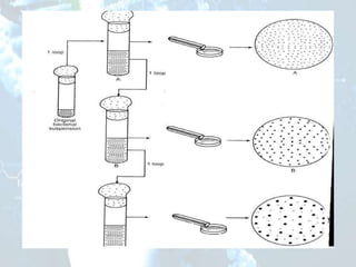
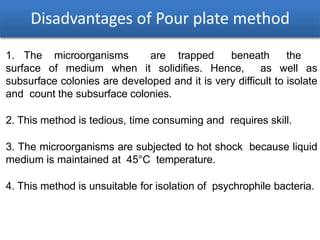
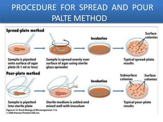
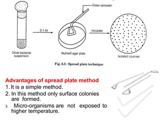
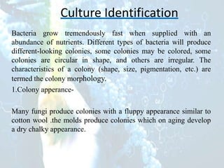
9953056974 Young Call Girls In Mahavir enclave Indian Quality Escort service
Isolation & Identification of Microorganism
1. Isolation & Identification ofMicroorganism
Prepared by:
Dr. Sandhya Hora
Assistant Professor
Department of Biotechnology
HIMT Group of Institutions, Greater Noida
2.
3. ISOLATION OF MICROBIAL PURECULTURE
• Microorganisms are generally found in nature (air, soil and
water) as mixed populations.
• Even the diseased parts of plants and animals contain a great
number of microorganisms, which differ markedly from the
microorganisms of other environments.
• To study the specific role played by a specific microorganism in
its
environment, one must isolate the same in pure culture.
The two major steps of obtaining a pure culture are as
follows :
• Firstly, the culture has to be diluted until the various
individual microorganisms are separated far apart on agar
surface that after incubation they form visible colonies
isolated from the colonies of other microorganisms.
4.
5. COMMON METHODS OF ISOLATION OF
PURE CULTURE
• The process of screening a pure culture by
separating one type of microbes from a mixture
is called Isolation.
Some common isolation methods are;
• I) Streak plate method
• II) Pour plate method-a) Loop dilution technique
b) Serial Dilution technique
• III) Spread plate method
• IV) Micromanipulator method
• V) Roll tube method
6. STREAKING OR STREAK PLATE
TECHNIQUE
• In This method the tip of a fine
structure wire loop called
Inoculation needle consist of a
wooden or glass handle with a
Nichrome wire the end of which is
bend to form a loop is used to
transfer microbes from culture.
• The straight wires are similar to
wire loop except they do not have
loop. These are used to transfer
culture in colony formed on solid
culture medium.
• In such cases, the colony from
solid medium is streaked on the
surface of nutrient agar medium
in a sterile petri dish.
7. This technique consist of the following steps-
• A. Hold the broth culture containing tube in left hand and shake it.
• B. Sterilize the wire loop of the inoculation needle on burner flame .
• C. Remove the cotton plug of the broth culture tube by little finger of right
hand.
• D. Flame the mouth of the test tube immediately.
• E. Insert the wire loop to form a thin film and replace the cotton plug.
• F. The thin film in the loop is streaked in either a zig-zag manner by
removing the loop backwards and forwards firmly. Care should be taken
that loop should not be firmly pressed against the agar surface.
• G. Incubate the petri dish in incubator at a required temperature.
• H. Growth of the bacteria will be visible (after an overnight incubation)on
the
streaked marks.
8. II) Pour plate method
• The bacterial culture and liquid agar
medium are mixed together.
• After mixing the medium, the medium
containing the culture poured into sterilized
petridishes (petriplates), allowed solidifying
and then incubated.
• After incubation colonies appear on the
surface.
• Link: https://microbeonline.com/pour-
plate- method-principle-procedure-uses-
dis- advantages/
9.
10. Disadvantages of Pour plate method
1. The microorganisms are trapped beneath the
surface of medium when it solidifies. Hence, as well as
subsurface colonies are developed and it is very difficult to isolate
and count the subsurface colonies.
2. This method is tedious, time consuming and requires skill.
3. The microorganisms are subjected to hot shock because liquid
medium is maintained at 45°C temperature.
4. This method is unsuitable for isolation of psychrophile bacteria.
12. II) SERIAL DILUTION
This method is commonly used to obtain pure cultures of those
microorganisms that have not yet been successfully cultivated on solid
media and grow only in liquid media.
A microorganism that predominates in a mixed culture can be isolated
in pure
form by a series of dilutions.
The inoculum is subjected to serial dilution in a sterile liquid medium,
and a large number of tubes of sterile liquid medium are inoculated with
aliquots of each successive dilution.
13. • If we take out 1 ml of this medium and mix it with 9 ml of fresh
sterile liquid medium, we would then have 100 microorganisms
in 10 ml or 10 microorganisms/ ml.
• If we add 1 ml of this suspension to another 9 ml. of fresh
sterile liquid medium, each ml would now contain a single
microorganism.
• If this tube shows any microbial growth, there is a very high
probability that this growth has resulted from the introduction of
a single microorganism in the medium and represents the pure
14. III) Spread plate method
• Links for further
study:https://vlab.amrita.edu/?sub=3&brch=73&sim=213&
cnt
=1
• https://jcm.asm.org/content/jcm/29/7/1462.full.pdf
15. Advantages of spread plate method
1.It is a simple method.
2. In this method only surface colonies
are formed.
3. Micro-organisms are not exposed to
higher temperature.
16. IV-MICROMANIPULATOR METHOD
• Micromanipulators have been built, which permit one to pick
out a single cell from a mixed culture. This instrument is
used in conjunction with a microscope to pick a single cell
(particularly bacterial cell) from a hanging drop preparation.
ADVANTAGES OF MICROMANIPULATOR METHOD
• The advantages of this method are that one can be
reasonably sure that the cultures come from a single cell
and one can obtain strains with in the species.
DISADVANTAGES
• Disadvantages are that the equipment is expensive,
• Its manipulation is very tedious, and it requires a skilled
operator.
17.
18. Culture Identification
Bacteria grow tremendously fast when supplied with an
abundance of nutrients. Different types of bacteria will produce
different-looking colonies, some colonies may be colored, some
colonies are circular in shape, and others are irregular. The
characteristics of a colony (shape, size, pigmentation, etc.) are
termed the colony morphology.
1.Colony apperance-
Many fungi produce colonies with a fluppy appearance similar to
cotton wool .the molds produce colonies which on aging develop
a dry chalky appearance.
19. .A. Yeasts-
Yeast, a type of fungi (plural for fungus), is found in many
places from nature, to research labs and even everyday kitchens
for baking. Yeast colonies generally look similar to bacterial
colonies. Some species, such as candida, can grow as white
patches with a glossy surface.
For example:
ROUND YEAST COLONIES PINK YEAST COLONIES
20. B. BACTERIA-
1.Bacillus subtilis 2.Proteus vulgaris 3.Staphyloccus aures
Each distinct circular colony should represent an individual
bacterial cell or group that has divided repeatedly. Being kept
in one place, the resulting cells have accumulated to form a
visible patch. Most bacterial colonies appear white, cream, or
yellow in color, and fairly circular in shape.
21. 2.COLONY FORMS-
The colony shape may be circular, filamentous, rhizidoidal,
punctiform(dot like),irregular, or spindle shape.
3.COLONY ELEVATION-This form is used to describe the
depth of the colony developed by microbes.A colony may be
flat(thin film over the agar surface), raised, convex or umbonate
or with papillae surface.
4.COLONY MARGINS-The margins may be entire,
undulate(wavy), crenate, dentate , lobate, rhizoidal or
filamentous.
5.OPTICAL DENSITY-The colony may be transparent or
transluscent (foggy in appearance) or opaque(not permitting light
to pass through it ) or irrediscent (rainbow colour).
6.COLOUR-Many microbes develop colonies which are
pigmented.Such coloured substances are either water soluble or
insoluble.The soluble pigments diffuse out in the medium.