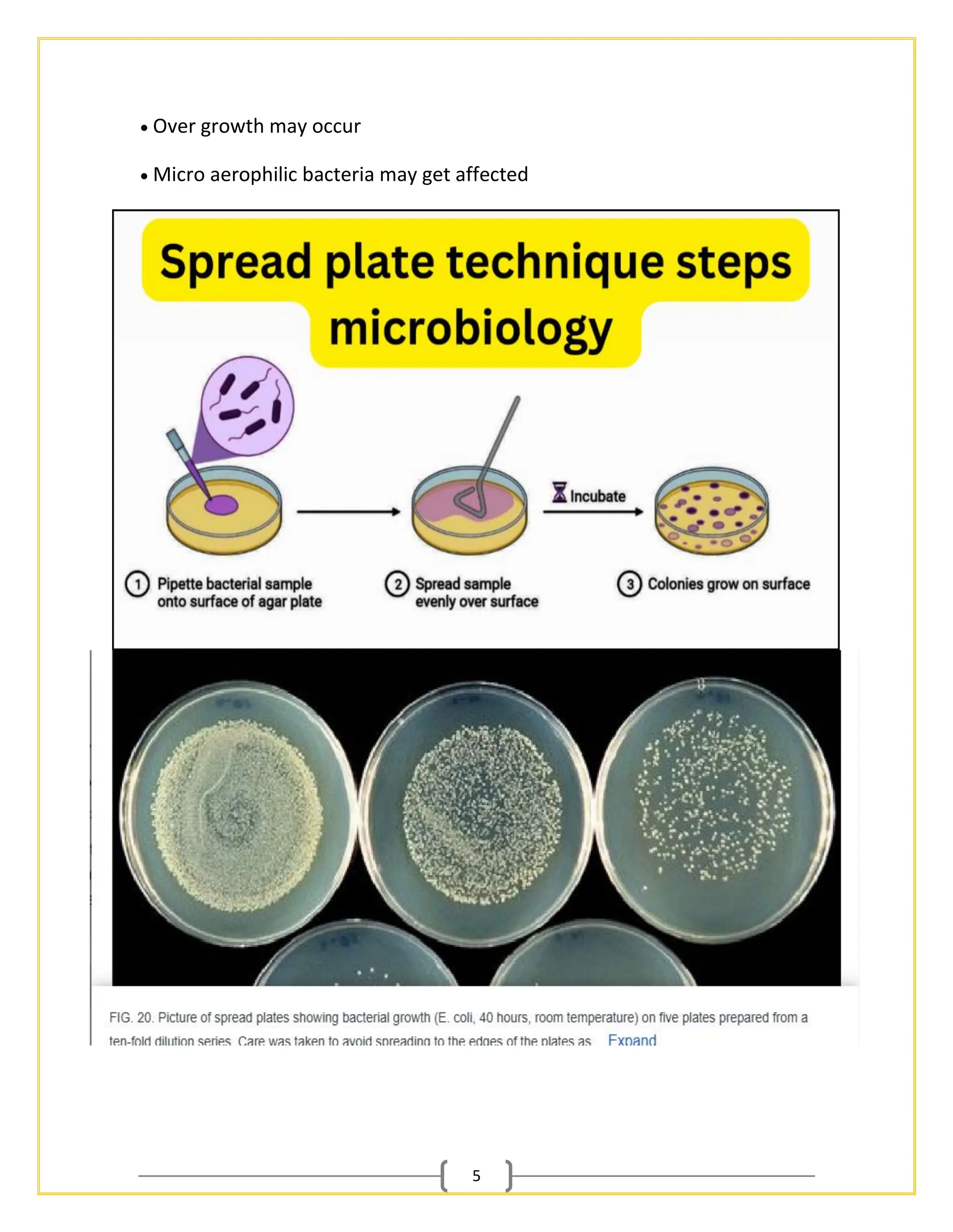
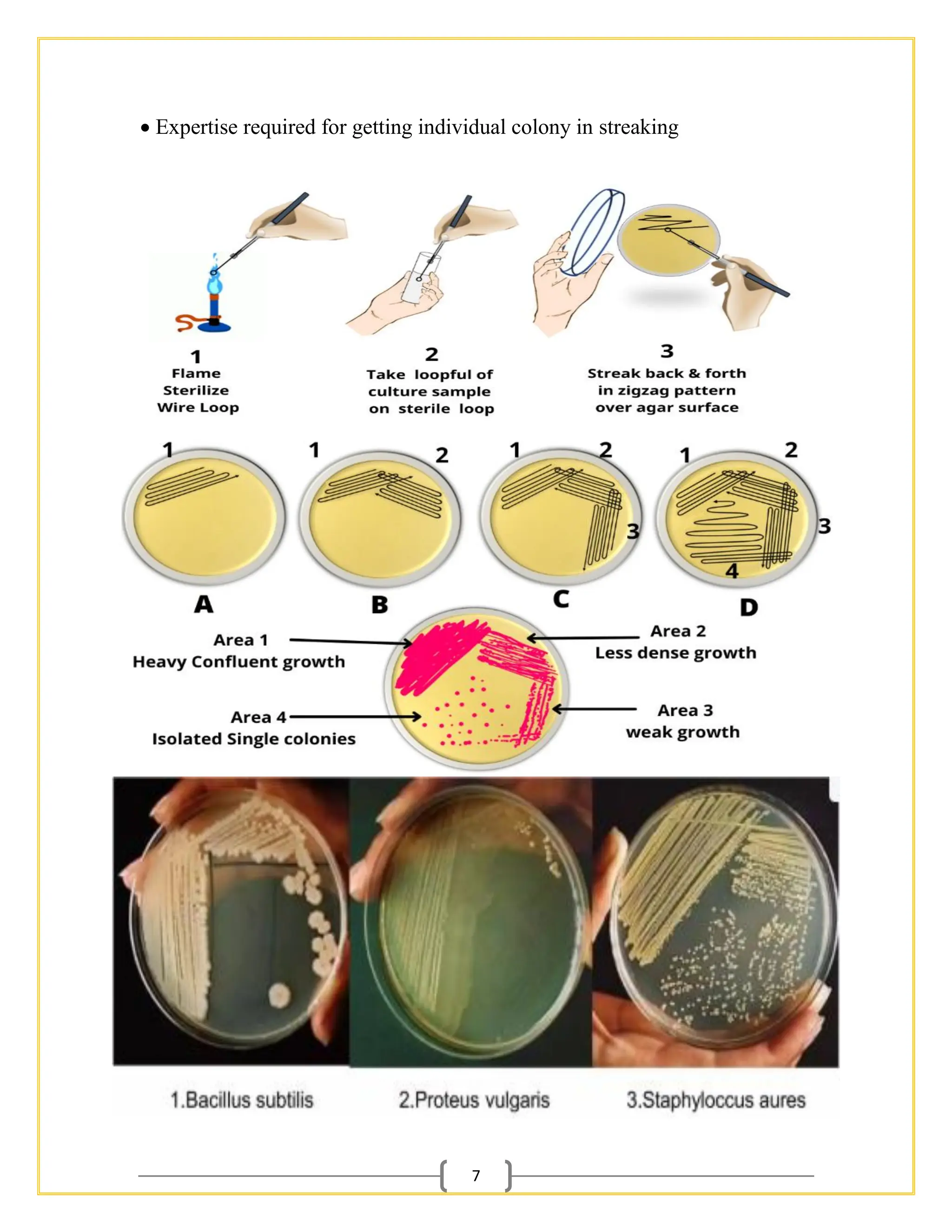
1
Introduction
Microorganisms are present on all inanimate surfaces creating ubiquitous sources of
possible contamination in the laboratory. Experimental success relies on the ability
of a scientist to sterilize work surfaces and equipment as well as prevent contact of
sterile instruments and solutions with non-sterile surfaces. Study of microorganism
needs accurate handling or it adversely affects the handlers. Standard operating
procedures are the key step in performing the microbiology study. This not only
gives the reliable result but also ensure the safety of the laboratory technicians.
Plating is the common technique employed and the petriplates of different sizes can
be used for different purposes. It is recommended that non-pathogenic strains be
used when learning the various plating methods. By following the procedures
described in this protocol:
 Perform plating procedures for enumeration of bacteria without contaminating
media and self.
 Isolate single bacterial colonies by the streak-plating method.
 Use pour-plating and spread-plating method for variety of applications like
desired bacterial screening General instructions
 Sterile workspace and premises is essential for microbial works
 Sterilize all instruments, solutions, and media prior to using them for plating
procedures.
 Clean work area with phenol or 70 %alcohol to minimize possible contamination.
 Keep burner with flame prior to work to create a sterile field.
 In all techniques sterilization of glass wares in hot air oven and the Medias in
prescribed manner should be done prior to plating.
 Media which are autoclaved and glassware should be cooled to sufficient levels
before plating
 Marking of the petriplate should be done in base of the plate.
2
1) Pour plate technique Method
This method often is used to count the number of microorganisms in a mixed
sample, which is added to a molten agar medium prior to its solidification. Molten
agar should be cooled to 44˚C before plating otherwise it may lead to death of
the desired organism. The process results in colonies uniformly distributed
throughout the solid medium when the appropriate sample dilution is plated. This
technique is used to perform viable plate counts, in which the total 17 number of
colony forming units within the agar and on surface of the agar on a single plate
is enumerated. Viable plate counts provide scientists a standardized means to
generate growth curves, to calculate the concentration of cells in the tube from
which the sample was plated, and to investigate the effect of various
environments or growth conditions on bacterial cell survival or growth rate. This
method is advantageous when our organism is environment bacteria and the
prevalence is less.
3
Materials required Sample, sterilized petri plates, sterilized nutrient media,
flame, glass marker Procedure of Pour plate technique
• With the help of serial dilution technique, the sample should be prepared. The
good dilution is one which gives colonies in between 30 to 300.
• Label the petri dishes in the bottom of plate
• Put 1 ml prepared dilution sample in the petri plate near the flame
• Cool the media and pour it in the plate. 100 ml media can be poured to 4 plates
• Mix the plate well for uniform spreading and allow it to solidify and incubate
Limitations
• Some colonies may be hidden inside agar
• Heat labile organism will die
Advantages of Pour Plate Technique
1. It is easy to perform and doesn’t require extra tools or materials for
inoculation.
2. Don’t require previously solidified agar medium.
3. Don’t have a risk of gouging during inoculation as like in streaking and
spreading.
4. Even a very low microbial load can be detected.
5. Along with the isolation of microorganisms, their isolated colonies can be
obtained. The number of CFU/mL can be calculated as well.
6. It can use any type of specimen like clinical or environmental samples,
liquid or solid (it can be dissolved).
7. It is suitable for isolating facultative and anaerobic microorganisms.
Aerobes can also be isolated by this method.
Limitations of Pour Plate Technique
1. Solid or semisolid samples must be suspended prior to inoculation. It is
very difficult if the sample is not easily soluble.
2. There is a need for serial dilution of the sample otherwise too numerous
colonies will be formed that can’t be counted or identified as discrete.
3. Heat-sensitive organisms can be affected by molten media at 40-45°C.
4
4. It requires some time for the growth of organisms and the formation of
colonies. There will be less supply of oxygen to microbial cells at the
bottom of the solidified media, hence growing slowly.
5. Colonies may be smaller than in streaking or spreading which increases the
chance of overlooking them.
6. Obligate aerobes may face difficulty in growing at the bottom of the plates.
Some don’t even grow.
7. The method is time-consuming as it needs dissolving the solid samples,
serial dilutions, and melting of the media at a certain temperature (42 –
45°C).
2) Spread plate technique Method
• The spread plate technique is used for enumeration, enrichment, screening
and selection of microorganism. In this the culture is uniformly spread over the
surface of an agar plate, resulting in the formation of isolated colonies
distributed evenly across the agar surface if the appropriate concentration of
cells is plated. Materials required Sample, sterilized petri plates, sterilized
nutrient media, flame, glass marker, glass rod (alternatively sterile plastic rod
also can be used), beaker with alcohol
Procedure
• Sterilize the petri plate and nutrient medium. Cool it to 56∘C. pour in the plate
and allow it to settle.
• Then prepare the sample. Serial dilute if necessary. Add 0.1 ml of sample in
the surface of dried agar plate
• Dip the spreader in alcohol, flame and cool it
• Spread the sample uniformly near the flame
• Incubate the plate in inverted position 18 Advantage over other methods
• Colony morphology can be seen clearly
• Can be used for screening and selection Limitations
5
• Over growth may occur
• Micro aerophilic bacteria may get affected
6
3) Streaking Method
This method is used for obtaining pure culture from the mixed culture. Quadrant
streaking is done in the petri plate in such way that all four corners are used for
isolating a single bacterial colony.(best method for isolation of micro-organisms)
Materials required Sample, sterilized petri plates, sterilized nutrient media,
flame, glass marker, metal loop
Procedure
1. Media should be poured in petri plate and allowed to settle. Then it is dried till
the condensed water becomes dry
2. Flame the loop until it becomes red hot and allows it to cool. Then pick the
colony aseptically near the flame
3. Place loop with culture in petri plate and take it to other quadrat without
touching the edge of the petri plate. Then flame the loop to sterilize
4. From the previous line draw another line perpendicular to the old line with
sterile loop. This line also should not touch the corner.
5. Then sterilize the loop and draw another line from the previous quadrant
perpendicular to the old line with sterile loop. This line also should not touch the
corner
6. Then sterilize the loop and from the old line draw another line with the loop
and stop in the half quadrant. This way we can get individual colony without
contamination in one plate.
7. The bacteria grown in single colony are assumed to have formed from the
single bacteria and they are called as clone .
Advantage over other method
• Pure culture can be obtained. If colony morphology is known contaminated
cultures can be purified Limitations
7
• Expertise required for getting individual colony in streaking
8
4) Antibiotic sensitivity testing using petri plate
Antibiotic sensitivity of desired organism also can be tested with the
plating procedure. For this known amount of culture with the same OD or
McFarland unit concentration should be checked every time to get uniform
results. The known concentration of bacteria should be inoculated as
spread plate to form uniform lawn. Then the discs should be equally placed
with uniform concentration of antibiotic. With the help of the ruler the zone
of inhibition should be checked
Materials required Fresh culture of bacteria to be tested, Inoculation loop ,
Burner, McFarland solution, Saline solution, Muller Hinton agar plate ,
Antibiotic disc to be tested , Incubator , Ruler , Forceps and beaker with
alcohol , glass rod
Procedure
1. Take pure culture of the organism to be tested (fresh culture of 12-24
hrs. desired)
2. This should be uniformly mixed with saline and compared with
McFarland standard OD
9
3. Alternatively, the bacteria to be tested should be well studied and
compared with the OD. So the concentration to be checked for different
antibiotic will be always uniform 4. Aseptically spread the colony with the
sterile glass spreader to the Muller Hinton plate 5. Allow the plate for dry
5 minutes 6. Place the antibiotic disc with the help of sterile forceps and
gently press 7. Incubate the plate without inverting 8. After incubation
measure the zone of inhibition. 20 9. Compare the measurement obtained
from the individual antibiotics with the standard table to determine the
sensitivity zone.

Isolation of Microorganisms for B.Sc. Biotechnology & Botany Sem-3

  • 1.
    1 Introduction Microorganisms are presenton all inanimate surfaces creating ubiquitous sources of possible contamination in the laboratory. Experimental success relies on the ability of a scientist to sterilize work surfaces and equipment as well as prevent contact of sterile instruments and solutions with non-sterile surfaces. Study of microorganism needs accurate handling or it adversely affects the handlers. Standard operating procedures are the key step in performing the microbiology study. This not only gives the reliable result but also ensure the safety of the laboratory technicians. Plating is the common technique employed and the petriplates of different sizes can be used for different purposes. It is recommended that non-pathogenic strains be used when learning the various plating methods. By following the procedures described in this protocol:  Perform plating procedures for enumeration of bacteria without contaminating media and self.  Isolate single bacterial colonies by the streak-plating method.  Use pour-plating and spread-plating method for variety of applications like desired bacterial screening General instructions  Sterile workspace and premises is essential for microbial works  Sterilize all instruments, solutions, and media prior to using them for plating procedures.  Clean work area with phenol or 70 %alcohol to minimize possible contamination.  Keep burner with flame prior to work to create a sterile field.  In all techniques sterilization of glass wares in hot air oven and the Medias in prescribed manner should be done prior to plating.  Media which are autoclaved and glassware should be cooled to sufficient levels before plating  Marking of the petriplate should be done in base of the plate.
  • 2.
    2 1) Pour platetechnique Method This method often is used to count the number of microorganisms in a mixed sample, which is added to a molten agar medium prior to its solidification. Molten agar should be cooled to 44˚C before plating otherwise it may lead to death of the desired organism. The process results in colonies uniformly distributed throughout the solid medium when the appropriate sample dilution is plated. This technique is used to perform viable plate counts, in which the total 17 number of colony forming units within the agar and on surface of the agar on a single plate is enumerated. Viable plate counts provide scientists a standardized means to generate growth curves, to calculate the concentration of cells in the tube from which the sample was plated, and to investigate the effect of various environments or growth conditions on bacterial cell survival or growth rate. This method is advantageous when our organism is environment bacteria and the prevalence is less.
  • 3.
    3 Materials required Sample,sterilized petri plates, sterilized nutrient media, flame, glass marker Procedure of Pour plate technique • With the help of serial dilution technique, the sample should be prepared. The good dilution is one which gives colonies in between 30 to 300. • Label the petri dishes in the bottom of plate • Put 1 ml prepared dilution sample in the petri plate near the flame • Cool the media and pour it in the plate. 100 ml media can be poured to 4 plates • Mix the plate well for uniform spreading and allow it to solidify and incubate Limitations • Some colonies may be hidden inside agar • Heat labile organism will die Advantages of Pour Plate Technique 1. It is easy to perform and doesn’t require extra tools or materials for inoculation. 2. Don’t require previously solidified agar medium. 3. Don’t have a risk of gouging during inoculation as like in streaking and spreading. 4. Even a very low microbial load can be detected. 5. Along with the isolation of microorganisms, their isolated colonies can be obtained. The number of CFU/mL can be calculated as well. 6. It can use any type of specimen like clinical or environmental samples, liquid or solid (it can be dissolved). 7. It is suitable for isolating facultative and anaerobic microorganisms. Aerobes can also be isolated by this method. Limitations of Pour Plate Technique 1. Solid or semisolid samples must be suspended prior to inoculation. It is very difficult if the sample is not easily soluble. 2. There is a need for serial dilution of the sample otherwise too numerous colonies will be formed that can’t be counted or identified as discrete. 3. Heat-sensitive organisms can be affected by molten media at 40-45°C.
  • 4.
    4 4. It requiressome time for the growth of organisms and the formation of colonies. There will be less supply of oxygen to microbial cells at the bottom of the solidified media, hence growing slowly. 5. Colonies may be smaller than in streaking or spreading which increases the chance of overlooking them. 6. Obligate aerobes may face difficulty in growing at the bottom of the plates. Some don’t even grow. 7. The method is time-consuming as it needs dissolving the solid samples, serial dilutions, and melting of the media at a certain temperature (42 – 45°C). 2) Spread plate technique Method • The spread plate technique is used for enumeration, enrichment, screening and selection of microorganism. In this the culture is uniformly spread over the surface of an agar plate, resulting in the formation of isolated colonies distributed evenly across the agar surface if the appropriate concentration of cells is plated. Materials required Sample, sterilized petri plates, sterilized nutrient media, flame, glass marker, glass rod (alternatively sterile plastic rod also can be used), beaker with alcohol Procedure • Sterilize the petri plate and nutrient medium. Cool it to 56∘C. pour in the plate and allow it to settle. • Then prepare the sample. Serial dilute if necessary. Add 0.1 ml of sample in the surface of dried agar plate • Dip the spreader in alcohol, flame and cool it • Spread the sample uniformly near the flame • Incubate the plate in inverted position 18 Advantage over other methods • Colony morphology can be seen clearly • Can be used for screening and selection Limitations
  • 5.
    5 • Over growthmay occur • Micro aerophilic bacteria may get affected
  • 6.
    6 3) Streaking Method Thismethod is used for obtaining pure culture from the mixed culture. Quadrant streaking is done in the petri plate in such way that all four corners are used for isolating a single bacterial colony.(best method for isolation of micro-organisms) Materials required Sample, sterilized petri plates, sterilized nutrient media, flame, glass marker, metal loop Procedure 1. Media should be poured in petri plate and allowed to settle. Then it is dried till the condensed water becomes dry 2. Flame the loop until it becomes red hot and allows it to cool. Then pick the colony aseptically near the flame 3. Place loop with culture in petri plate and take it to other quadrat without touching the edge of the petri plate. Then flame the loop to sterilize 4. From the previous line draw another line perpendicular to the old line with sterile loop. This line also should not touch the corner. 5. Then sterilize the loop and draw another line from the previous quadrant perpendicular to the old line with sterile loop. This line also should not touch the corner 6. Then sterilize the loop and from the old line draw another line with the loop and stop in the half quadrant. This way we can get individual colony without contamination in one plate. 7. The bacteria grown in single colony are assumed to have formed from the single bacteria and they are called as clone . Advantage over other method • Pure culture can be obtained. If colony morphology is known contaminated cultures can be purified Limitations
  • 7.
    7 • Expertise requiredfor getting individual colony in streaking
  • 8.
    8 4) Antibiotic sensitivitytesting using petri plate Antibiotic sensitivity of desired organism also can be tested with the plating procedure. For this known amount of culture with the same OD or McFarland unit concentration should be checked every time to get uniform results. The known concentration of bacteria should be inoculated as spread plate to form uniform lawn. Then the discs should be equally placed with uniform concentration of antibiotic. With the help of the ruler the zone of inhibition should be checked Materials required Fresh culture of bacteria to be tested, Inoculation loop , Burner, McFarland solution, Saline solution, Muller Hinton agar plate , Antibiotic disc to be tested , Incubator , Ruler , Forceps and beaker with alcohol , glass rod Procedure 1. Take pure culture of the organism to be tested (fresh culture of 12-24 hrs. desired) 2. This should be uniformly mixed with saline and compared with McFarland standard OD
  • 9.
    9 3. Alternatively, thebacteria to be tested should be well studied and compared with the OD. So the concentration to be checked for different antibiotic will be always uniform 4. Aseptically spread the colony with the sterile glass spreader to the Muller Hinton plate 5. Allow the plate for dry 5 minutes 6. Place the antibiotic disc with the help of sterile forceps and gently press 7. Incubate the plate without inverting 8. After incubation measure the zone of inhibition. 20 9. Compare the measurement obtained from the individual antibiotics with the standard table to determine the sensitivity zone.