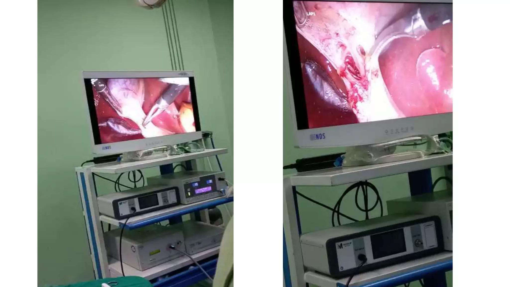
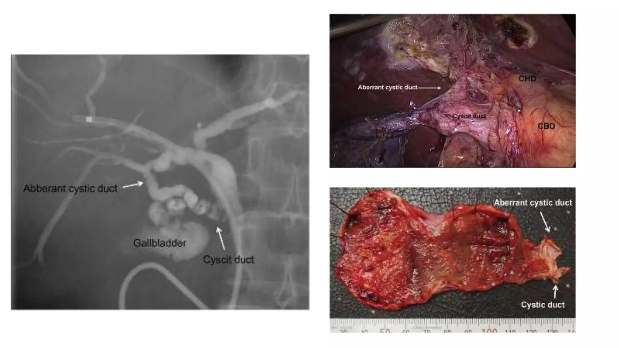
5. Figure 1. Drawing illustrates the normal biliary tract. The cystic duct
(arrows) connects the gallbladder to the extrahepatic bile duct and
usually enters from the right approximately halfway between the porta
hepatis and the ampulla of Vater. It also contains the valves of Heister.
Normal Anatomy
The cystic duct attaches the gallbladder to the extrahepatic bile duct; its point of
insertion into the extrahepatic bile duct marks the division between the common
hepatic duct and the common bile duct.
6. The cystic duct usually measures 2–4 cm in length and contains
prominent concentric folds known as the spiral valves of Heister.
The cystic duct frequently exhibits a tortuous or serpentine course.
The normal diameter of the cystic duct is variable, ranging from 1 to 5
mm.
The cystic duct usually joins the extrahepatic bile duct approximately
halfway between the porta hepatis and the ampulla of Vater.
The cystic duct enters the extrahepatic bile duct from the right lateral
aspect in 49.9% of cases, from the medial aspect in 18.4%, and from
an anterior or posterior position in 31.7%.
7. Figure 2. Normal cystic duct anatomy. ERCP image shows a normal-caliber cystic duct (solid
arrow). Note the undulating contour of the duct produced by the valves of Heister. An air bubble
(open arrow) is noted in the common bile duct.
Absence of filling of the cystic duct at ERCP is
usually related to patient positioning rather than
cystic duct obstruction.
8. Figure 5. Coronal oblique MR cholangiopancreatogram demonstrates the normal cystic duct (arrow) connecting the
gallbladder to the extrahepatic bile duct (arrowhead). Gallbladder calculi are also present.
In most cases, the normal cystic duct is not seen at US.
However, with optimal technique, the normal cystic duct can be
visualized in up to 50% of cases as an anechoic tubular
structure connecting the gallbladder and bile duct. A cystic
duct that runs parallel to the distal extrahepatic bile duct may be
confused with a vessel; however, differentiation is possible
with Doppler US.
The cystic duct is not routinely visualized at CT. The cystic duct
appears as a low-attenuation tubular structure with thin, enhancing
walls.
The cystic duct is routinely seen at MRCP and can be traced to its
junction with the extrahepatic bile duct in most cases.
9. Figure 6. Anatomic variants in the cystic duct. Drawings illustrate how the cystic duct may
insert into the extrahepatic bile duct with a shows right lateral insertion (A), anterior spiral
insertion (B), posterior spiral insertion (C), low lateral insertion with a common sheath (D),
proximal insertion (E), or low medial insertion (F).
Anatomic Variants
Variations in Cystic Duct Insertion
10. Figure 7a. (7a) Cholangiogram shows a right lateral insertion of the cystic duct (arrows) into the extrahepatic bile duct.
(7b) Cholangiogram shows a medial insertion of the cystic duct (arrows) into the extrahepatic bile duct. (7c) Coronal
oblique MR cholangiopancrea-togram shows a low, medially inserting cystic duct (straight arrows) that parallels the bile
duct (curved arrow).
11. Figure 9. Low medial insertion of the cystic duct into the ampulla of Vater in an 11-year-old girl with recurrent pancreatitis.
ERCP image shows direct filling of the cystic duct (single straight arrow) from an ampullary injection of contrast material.
The cystic duct and bile duct (double arrows) join at the ampulla. Stones are identified in the cystic duct (curved arrow),
and air is noted in the gallbladder (g). Recurrent pancreatitis in the patient was attributed to this abnormal anatomy.
12. Figure 10a. Calculus in a low, medially inserting cystic duct mimicking a distal common bile duct calculus in a 69-year-old man. (a) ERCP
image shows a low, medially inserting cystic duct remnant (straight arrows) mimicking the distal bile duct. The calculus (curved arrow) in the
cystic duct remnant was initially presumed to lie in the distal bile duct. (b) ERCP image obtained after rotating the patient demonstrates a
catheter in the cystic duct remnant (straight arrows) and shows that the calculus (curved arrow) lies in the cystic duct remnant. No calculus is
seen in the distal bile duct (arrowheads).
13. Anomalous or aberrant bile ducts are usually of no clinical significance, unless they lead to diagnostic confusion on
imaging studies or result in increased potential for iatrogenic injury.
Ducts at greatest risk for injury at cholecystectomy are those that course near the cystic duct or gallbladder or empty
directly into these structures. Anomalous ducts that empty directly into the cystic duct (cysticohepatic ducts) are
found in 1%–2% of individuals.
Accessory bile ducts, especially those arising from the right lobe, may join the common hepatic duct at its junction with the
cystic duct or may insert directly into the cystic duct.
Up to 5% of patients will have a major right segmental bile duct joining the extrahepatic bile duct at or near the
cystic duct.
This anatomic variant creates a risk of inadvertent ligation or transection of the aberrant duct near the cystic duct insertion
at cholecystectomy.
Rare anomalies of the cystic duct include insertion into the right hepatic duct, double cystic ducts with or
without a duplicated gallbladder, and absence of the cystic duct with the gallbladder emptying directly
into the common bile duct.
Anatomic Variants
Anomalous Bile Ducts
14. Figure 12a. Aberrant junction of the bile duct and cystic duct. (a) ERCP image shows an aberrant right hepatic duct (small
arrows) entering the cystic duct (large arrow). (b) Coronal MR cholangiopancreatogram obtained in a different patient
demonstrates an aberrant right hepatic duct (small arrows) draining a circumscribed portion of the liver and entering the
common hepatic duct 4 cm distal to the confluence of the right and left hepatic ducts. The cystic duct (large arrow) drains
into the aberrant right hepatic duct. (Figure 12 reprinted, with permission, from reference 20.)
15.
16. Laparoscopic cholecystectomy (LC) is the
most common elective laparoscopic
procedure performed globally and is the gold
standard treatment for gallstone disease.
The bile duct injury (BDI) remains the most
dreaded complication, with an incidence rate
of 0.3%.
Clear identification of biliary anatomy and
establishing the critical view of safety is
essential in avoiding BDI.
However, this can be challenging, with
variations in biliary anatomy in ~47% of
patients.
An extremely rare variant is the
duplicated cystic duct.
17. Case Presentation
A 57-year-old woman was admitted to our hospital due to epigastric abdominal pain. A physical
examination revealed no remarkable findings.
Laboratory studies showed an elevated white blood cell count (10400/μL), aspartate
aminotransferase (AST: 84 U/L), alanine aminotransferase (ALT: 39 U/L), alkaline phosphatase (ALP:
466 U/L), γ-glutamyl transpeptidase 205 U/L). Ultrasonography and computed tomography (CT)
revealed small gallstones in the gallbladder and some stones in the common bile duct. A large liver
cyst was also detected in S4. Thus, the patient was diagnosed with cholecysto-choledocholithiasis.
Laparoscopic cholecystectomy was planned after the complete removal of the gallstones in the
common bile duct following endoscopic sphincterotomy (EST).
However, a cystic duct which communicates with the intrahepatic bile duct of the posterior segmental
branch was suspected based on the gross X-ray images obtained after the extraction of the bile duct
stones. Therefore, the magnetic resonance cholangiopancreatography (MRCP) was performed.
MRCP showed strong suspicion of a single gallbladder with a double cystic duct.
18. Thus, to confirm this rare condition, endoscopic retrograde cholangiography (ERC) was additionally
performed for a second time. This time, a normal cystic duct was found to arise from the neck of the
gallbladder, from which it descended and joined the common bile duct.
In addition, the aberrant cystic duct arose from the cystic duct and communicated with the
intrahepatic bile duct of the posterior segmental branch.
Thus, we determined that the patient had a single gallbladder with a double cystic duct.
An endoscopic nasobiliary drainage (ENBD) tube was placed for intraoperative cholangiography in
order to protect the common bile and hepatic duct from injury during surgery. Laparoscopic
cholecystectomy was performed under general anesthesia.
During the procedure, the gallbladder was divided from the gallbladder bed in the fundus- to- hilar
direction, in order to explore the double bile ducts. The main and aberrant cystic ducts and the
common hepatic duct were identified by meticulous dissection and intraoperative cholangiography.
These cystic ducts were then ligated and precisely divided.
Laparoscopic cholecystectomy was finished and no drain was placed.
The excised specimen showed a duplicated cystic duct containing debris and chronic cholecystitis
19.
20. With more than half a million cholecystectomies performed annually, variations in
biliary anatomy present a significant challenge for laparoscopic surgeons.
Duplication of the cystic duct is extremely rare, with only 20 previous cases
reported in the literature.
Three types of duplicated cystic ducts have been previously described:
1) The ‘Y’ type, in which two cystic ducts meet to form a common channel.
2) the ‘H’ type, in which the accessory duct enters into the right, left or common
hepatic duct.
3) the trabecular type, in which the accessory duct enters the liver substance
directly.
While preoperative MRCP or endoscopic retrograde cholangiopancreatography
(ERCP) may identify abnormal anatomy, this is not guaranteed to prevent
encountering unexpected variations.
21.
22. Of the seven reported cases of cystic duct duplication where a preoperative ERCP was performed, only
three had the anomaly detected.
In most cases, the second cystic duct was an intraoperative finding.
This emphasizes the importance of constant vigilance during LC even when preoperative imaging yields
no abnormality.
In conclusion, the duplicated cystic duct is a highly uncommon variant of biliary anatomy, which may pose
a significant dilemma to the laparoscopic surgeon.
Constant vigilance for anatomical variance is essential to avoid adverse outcomes, with early involvement
of a hepatobiliary specialist if there is unexplained bile leakage.
Unremarkable preoperative imaging does not exclude the presence of abnormal anatomy.
However, the point at which the cystic duct joins the extrahepatic bile duct is variable, ranging from high at the level of the porta hepatis to low at the level of the ampulla.