A 43-year-old female patient presented with symptomatic cholelithiasis. Imaging and labs were normal. During her planned laparoscopic cholecystectomy, surgeons discovered an extremely rare anatomical variant - the patient had a single gallbladder but with two cystic ducts. One cystic duct connected to the common bile duct as normal, but the other connected to an intrahepatic bile duct branch. Pre-operative imaging did not detect this duplication. The operation was successfully completed with identification and ligation of both cystic ducts to remove the gallbladder.
Imaging of the Biliary System and its DisordersAbhineet Dey
Clinical data such as history, physical examination, and laboratory tests are useful in identifying patients with biliary obstruction and biliary sources of infection. However, if intervention is planned, noninvasive imaging is needed to confirm the presence, location, and extent of the disease process.
Currently, the most commonly available and used noninvasive modalities are ultrasound (US), computed tomography (CT), magnetic resonance (MR), and nuclear medicine hepatobiliary scintigraphy (HIDA).
Acute scrotum is a general term referring to an emergency condition affecting the contents or the wall of the scrotum.
There are a number of conditions that present acutely, predominantly with pain and/or swelling
A careful and detailed history and examination, and in some cases, investigations allow differentiation between these diagnoses. A prompt diagnosis is essential as the patient may require urgent surgical intervention
Testicular torsion refers to twisting of the spermatic cord, causing ischaemia of the testicle.
Testicular torsion results from inadequate fixation of the testis to the tunica vaginalis producing ischemia from reduced arterial inflow and venous outflow obstruction.
The prevalence of testicular torsion in adult patients hospitalized with acute scrotal pain is approximately 25 to 50 percent
Couples presenting to the infertility clinic- Do they really have infertility...Sujoy Dasgupta
Dr Sujoy Dasgupta presented the study on "Couples presenting to the infertility clinic- Do they really have infertility? – The unexplored stories of non-consummation" in the 13th Congress of the Asia Pacific Initiative on Reproduction (ASPIRE 2024) at Manila on 24 May, 2024.
More Related Content
Similar to Cystic Duct Normal Anatomy and Anatomic Variants.pptx
Acute scrotum is a general term referring to an emergency condition affecting the contents or the wall of the scrotum.
There are a number of conditions that present acutely, predominantly with pain and/or swelling
A careful and detailed history and examination, and in some cases, investigations allow differentiation between these diagnoses. A prompt diagnosis is essential as the patient may require urgent surgical intervention
Testicular torsion refers to twisting of the spermatic cord, causing ischaemia of the testicle.
Testicular torsion results from inadequate fixation of the testis to the tunica vaginalis producing ischemia from reduced arterial inflow and venous outflow obstruction.
The prevalence of testicular torsion in adult patients hospitalized with acute scrotal pain is approximately 25 to 50 percent
Couples presenting to the infertility clinic- Do they really have infertility...Sujoy Dasgupta
Dr Sujoy Dasgupta presented the study on "Couples presenting to the infertility clinic- Do they really have infertility? – The unexplored stories of non-consummation" in the 13th Congress of the Asia Pacific Initiative on Reproduction (ASPIRE 2024) at Manila on 24 May, 2024.
Tom Selleck Health: A Comprehensive Look at the Iconic Actor’s Wellness Journeygreendigital
Tom Selleck, an enduring figure in Hollywood. has captivated audiences for decades with his rugged charm, iconic moustache. and memorable roles in television and film. From his breakout role as Thomas Magnum in Magnum P.I. to his current portrayal of Frank Reagan in Blue Bloods. Selleck's career has spanned over 50 years. But beyond his professional achievements. fans have often been curious about Tom Selleck Health. especially as he has aged in the public eye.
Follow us on: Pinterest
Introduction
Many have been interested in Tom Selleck health. not only because of his enduring presence on screen but also because of the challenges. and lifestyle choices he has faced and made over the years. This article delves into the various aspects of Tom Selleck health. exploring his fitness regimen, diet, mental health. and the challenges he has encountered as he ages. We'll look at how he maintains his well-being. the health issues he has faced, and his approach to ageing .
Early Life and Career
Childhood and Athletic Beginnings
Tom Selleck was born on January 29, 1945, in Detroit, Michigan, and grew up in Sherman Oaks, California. From an early age, he was involved in sports, particularly basketball. which played a significant role in his physical development. His athletic pursuits continued into college. where he attended the University of Southern California (USC) on a basketball scholarship. This early involvement in sports laid a strong foundation for his physical health and disciplined lifestyle.
Transition to Acting
Selleck's transition from an athlete to an actor came with its physical demands. His first significant role in "Magnum P.I." required him to perform various stunts and maintain a fit appearance. This role, which he played from 1980 to 1988. necessitated a rigorous fitness routine to meet the show's demands. setting the stage for his long-term commitment to health and wellness.
Fitness Regimen
Workout Routine
Tom Selleck health and fitness regimen has evolved. adapting to his changing roles and age. During his "Magnum, P.I." days. Selleck's workouts were intense and focused on building and maintaining muscle mass. His routine included weightlifting, cardiovascular exercises. and specific training for the stunts he performed on the show.
Selleck adjusted his fitness routine as he aged to suit his body's needs. Today, his workouts focus on maintaining flexibility, strength, and cardiovascular health. He incorporates low-impact exercises such as swimming, walking, and light weightlifting. This balanced approach helps him stay fit without putting undue strain on his joints and muscles.
Importance of Flexibility and Mobility
In recent years, Selleck has emphasized the importance of flexibility and mobility in his fitness regimen. Understanding the natural decline in muscle mass and joint flexibility with age. he includes stretching and yoga in his routine. These practices help prevent injuries, improve posture, and maintain mobilit
Ozempic: Preoperative Management of Patients on GLP-1 Receptor Agonists Saeid Safari
Preoperative Management of Patients on GLP-1 Receptor Agonists like Ozempic and Semiglutide
ASA GUIDELINE
NYSORA Guideline
2 Case Reports of Gastric Ultrasound
New Drug Discovery and Development .....NEHA GUPTA
The "New Drug Discovery and Development" process involves the identification, design, testing, and manufacturing of novel pharmaceutical compounds with the aim of introducing new and improved treatments for various medical conditions. This comprehensive endeavor encompasses various stages, including target identification, preclinical studies, clinical trials, regulatory approval, and post-market surveillance. It involves multidisciplinary collaboration among scientists, researchers, clinicians, regulatory experts, and pharmaceutical companies to bring innovative therapies to market and address unmet medical needs.
Prix Galien International 2024 Forum ProgramLevi Shapiro
June 20, 2024, Prix Galien International and Jerusalem Ethics Forum in ROME. Detailed agenda including panels:
- ADVANCES IN CARDIOLOGY: A NEW PARADIGM IS COMING
- WOMEN’S HEALTH: FERTILITY PRESERVATION
- WHAT’S NEW IN THE TREATMENT OF INFECTIOUS,
ONCOLOGICAL AND INFLAMMATORY SKIN DISEASES?
- ARTIFICIAL INTELLIGENCE AND ETHICS
- GENE THERAPY
- BEYOND BORDERS: GLOBAL INITIATIVES FOR DEMOCRATIZING LIFE SCIENCE TECHNOLOGIES AND PROMOTING ACCESS TO HEALTHCARE
- ETHICAL CHALLENGES IN LIFE SCIENCES
- Prix Galien International Awards Ceremony
263778731218 Abortion Clinic /Pills In Harare ,sisternakatoto
263778731218 Abortion Clinic /Pills In Harare ,ABORTION WOMEN’S CLINIC +27730423979 IN women clinic we believe that every woman should be able to make choices in her pregnancy. Our job is to provide compassionate care, safety,affordable and confidential services. That’s why we have won the trust from all generations of women all over the world. we use non surgical method(Abortion pills) to terminate…Dr.LISA +27730423979women Clinic is committed to providing the highest quality of obstetrical and gynecological care to women of all ages. Our dedicated staff aim to treat each patient and her health concerns with compassion and respect.Our dedicated group ABORTION WOMEN’S CLINIC +27730423979 IN women clinic we believe that every woman should be able to make choices in her pregnancy. Our job is to provide compassionate care, safety,affordable and confidential services. That’s why we have won the trust from all generations of women all over the world. we use non surgical method(Abortion pills) to terminate…Dr.LISA +27730423979women Clinic is committed to providing the highest quality of obstetrical and gynecological care to women of all ages. Our dedicated staff aim to treat each patient and her health concerns with compassion and respect.Our dedicated group of receptionists, nurses, and physicians have worked together as a teamof receptionists, nurses, and physicians have worked together as a team wwww.lisywomensclinic.co.za/
These simplified slides by Dr. Sidra Arshad present an overview of the non-respiratory functions of the respiratory tract.
Learning objectives:
1. Enlist the non-respiratory functions of the respiratory tract
2. Briefly explain how these functions are carried out
3. Discuss the significance of dead space
4. Differentiate between minute ventilation and alveolar ventilation
5. Describe the cough and sneeze reflexes
Study Resources:
1. Chapter 39, Guyton and Hall Textbook of Medical Physiology, 14th edition
2. Chapter 34, Ganong’s Review of Medical Physiology, 26th edition
3. Chapter 17, Human Physiology by Lauralee Sherwood, 9th edition
4. Non-respiratory functions of the lungs https://academic.oup.com/bjaed/article/13/3/98/278874
- Video recording of this lecture in English language: https://youtu.be/lK81BzxMqdo
- Video recording of this lecture in Arabic language: https://youtu.be/Ve4P0COk9OI
- Link to download the book free: https://nephrotube.blogspot.com/p/nephrotube-nephrology-books.html
- Link to NephroTube website: www.NephroTube.com
- Link to NephroTube social media accounts: https://nephrotube.blogspot.com/p/join-nephrotube-on-social-media.html
Recomendações da OMS sobre cuidados maternos e neonatais para uma experiência pós-natal positiva.
Em consonância com os ODS – Objetivos do Desenvolvimento Sustentável e a Estratégia Global para a Saúde das Mulheres, Crianças e Adolescentes, e aplicando uma abordagem baseada nos direitos humanos, os esforços de cuidados pós-natais devem expandir-se para além da cobertura e da simples sobrevivência, de modo a incluir cuidados de qualidade.
Estas diretrizes visam melhorar a qualidade dos cuidados pós-natais essenciais e de rotina prestados às mulheres e aos recém-nascidos, com o objetivo final de melhorar a saúde e o bem-estar materno e neonatal.
Uma “experiência pós-natal positiva” é um resultado importante para todas as mulheres que dão à luz e para os seus recém-nascidos, estabelecendo as bases para a melhoria da saúde e do bem-estar a curto e longo prazo. Uma experiência pós-natal positiva é definida como aquela em que as mulheres, pessoas que gestam, os recém-nascidos, os casais, os pais, os cuidadores e as famílias recebem informação consistente, garantia e apoio de profissionais de saúde motivados; e onde um sistema de saúde flexível e com recursos reconheça as necessidades das mulheres e dos bebês e respeite o seu contexto cultural.
Estas diretrizes consolidadas apresentam algumas recomendações novas e já bem fundamentadas sobre cuidados pós-natais de rotina para mulheres e neonatos que recebem cuidados no pós-parto em unidades de saúde ou na comunidade, independentemente dos recursos disponíveis.
É fornecido um conjunto abrangente de recomendações para cuidados durante o período puerperal, com ênfase nos cuidados essenciais que todas as mulheres e recém-nascidos devem receber, e com a devida atenção à qualidade dos cuidados; isto é, a entrega e a experiência do cuidado recebido. Estas diretrizes atualizam e ampliam as recomendações da OMS de 2014 sobre cuidados pós-natais da mãe e do recém-nascido e complementam as atuais diretrizes da OMS sobre a gestão de complicações pós-natais.
O estabelecimento da amamentação e o manejo das principais intercorrências é contemplada.
Recomendamos muito.
Vamos discutir essas recomendações no nosso curso de pós-graduação em Aleitamento no Instituto Ciclos.
Esta publicação só está disponível em inglês até o momento.
Prof. Marcus Renato de Carvalho
www.agostodourado.com
Title: Sense of Taste
Presenter: Dr. Faiza, Assistant Professor of Physiology
Qualifications:
MBBS (Best Graduate, AIMC Lahore)
FCPS Physiology
ICMT, CHPE, DHPE (STMU)
MPH (GC University, Faisalabad)
MBA (Virtual University of Pakistan)
Learning Objectives:
Describe the structure and function of taste buds.
Describe the relationship between the taste threshold and taste index of common substances.
Explain the chemical basis and signal transduction of taste perception for each type of primary taste sensation.
Recognize different abnormalities of taste perception and their causes.
Key Topics:
Significance of Taste Sensation:
Differentiation between pleasant and harmful food
Influence on behavior
Selection of food based on metabolic needs
Receptors of Taste:
Taste buds on the tongue
Influence of sense of smell, texture of food, and pain stimulation (e.g., by pepper)
Primary and Secondary Taste Sensations:
Primary taste sensations: Sweet, Sour, Salty, Bitter, Umami
Chemical basis and signal transduction mechanisms for each taste
Taste Threshold and Index:
Taste threshold values for Sweet (sucrose), Salty (NaCl), Sour (HCl), and Bitter (Quinine)
Taste index relationship: Inversely proportional to taste threshold
Taste Blindness:
Inability to taste certain substances, particularly thiourea compounds
Example: Phenylthiocarbamide
Structure and Function of Taste Buds:
Composition: Epithelial cells, Sustentacular/Supporting cells, Taste cells, Basal cells
Features: Taste pores, Taste hairs/microvilli, and Taste nerve fibers
Location of Taste Buds:
Found in papillae of the tongue (Fungiform, Circumvallate, Foliate)
Also present on the palate, tonsillar pillars, epiglottis, and proximal esophagus
Mechanism of Taste Stimulation:
Interaction of taste substances with receptors on microvilli
Signal transduction pathways for Umami, Sweet, Bitter, Sour, and Salty tastes
Taste Sensitivity and Adaptation:
Decrease in sensitivity with age
Rapid adaptation of taste sensation
Role of Saliva in Taste:
Dissolution of tastants to reach receptors
Washing away the stimulus
Taste Preferences and Aversions:
Mechanisms behind taste preference and aversion
Influence of receptors and neural pathways
Impact of Sensory Nerve Damage:
Degeneration of taste buds if the sensory nerve fiber is cut
Abnormalities of Taste Detection:
Conditions: Ageusia, Hypogeusia, Dysgeusia (parageusia)
Causes: Nerve damage, neurological disorders, infections, poor oral hygiene, adverse drug effects, deficiencies, aging, tobacco use, altered neurotransmitter levels
Neurotransmitters and Taste Threshold:
Effects of serotonin (5-HT) and norepinephrine (NE) on taste sensitivity
Supertasters:
25% of the population with heightened sensitivity to taste, especially bitterness
Increased number of fungiform papillae
New Directions in Targeted Therapeutic Approaches for Older Adults With Mantl...i3 Health
i3 Health is pleased to make the speaker slides from this activity available for use as a non-accredited self-study or teaching resource.
This slide deck presented by Dr. Kami Maddocks, Professor-Clinical in the Division of Hematology and
Associate Division Director for Ambulatory Operations
The Ohio State University Comprehensive Cancer Center, will provide insight into new directions in targeted therapeutic approaches for older adults with mantle cell lymphoma.
STATEMENT OF NEED
Mantle cell lymphoma (MCL) is a rare, aggressive B-cell non-Hodgkin lymphoma (NHL) accounting for 5% to 7% of all lymphomas. Its prognosis ranges from indolent disease that does not require treatment for years to very aggressive disease, which is associated with poor survival (Silkenstedt et al, 2021). Typically, MCL is diagnosed at advanced stage and in older patients who cannot tolerate intensive therapy (NCCN, 2022). Although recent advances have slightly increased remission rates, recurrence and relapse remain very common, leading to a median overall survival between 3 and 6 years (LLS, 2021). Though there are several effective options, progress is still needed towards establishing an accepted frontline approach for MCL (Castellino et al, 2022). Treatment selection and management of MCL are complicated by the heterogeneity of prognosis, advanced age and comorbidities of patients, and lack of an established standard approach for treatment, making it vital that clinicians be familiar with the latest research and advances in this area. In this activity chaired by Michael Wang, MD, Professor in the Department of Lymphoma & Myeloma at MD Anderson Cancer Center, expert faculty will discuss prognostic factors informing treatment, the promising results of recent trials in new therapeutic approaches, and the implications of treatment resistance in therapeutic selection for MCL.
Target Audience
Hematology/oncology fellows, attending faculty, and other health care professionals involved in the treatment of patients with mantle cell lymphoma (MCL).
Learning Objectives
1.) Identify clinical and biological prognostic factors that can guide treatment decision making for older adults with MCL
2.) Evaluate emerging data on targeted therapeutic approaches for treatment-naive and relapsed/refractory MCL and their applicability to older adults
3.) Assess mechanisms of resistance to targeted therapies for MCL and their implications for treatment selection
Flu Vaccine Alert in Bangalore Karnatakaaddon Scans
As flu season approaches, health officials in Bangalore, Karnataka, are urging residents to get their flu vaccinations. The seasonal flu, while common, can lead to severe health complications, particularly for vulnerable populations such as young children, the elderly, and those with underlying health conditions.
Dr. Vidisha Kumari, a leading epidemiologist in Bangalore, emphasizes the importance of getting vaccinated. "The flu vaccine is our best defense against the influenza virus. It not only protects individuals but also helps prevent the spread of the virus in our communities," he says.
This year, the flu season is expected to coincide with a potential increase in other respiratory illnesses. The Karnataka Health Department has launched an awareness campaign highlighting the significance of flu vaccinations. They have set up multiple vaccination centers across Bangalore, making it convenient for residents to receive their shots.
To encourage widespread vaccination, the government is also collaborating with local schools, workplaces, and community centers to facilitate vaccination drives. Special attention is being given to ensuring that the vaccine is accessible to all, including marginalized communities who may have limited access to healthcare.
Residents are reminded that the flu vaccine is safe and effective. Common side effects are mild and may include soreness at the injection site, mild fever, or muscle aches. These side effects are generally short-lived and far less severe than the flu itself.
Healthcare providers are also stressing the importance of continuing COVID-19 precautions. Wearing masks, practicing good hand hygiene, and maintaining social distancing are still crucial, especially in crowded places.
Protect yourself and your loved ones by getting vaccinated. Together, we can help keep Bangalore healthy and safe this flu season. For more information on vaccination centers and schedules, residents can visit the Karnataka Health Department’s official website or follow their social media pages.
Stay informed, stay safe, and get your flu shot today!
5. Figure 1. Drawing illustrates the normal biliary tract. The cystic duct
(arrows) connects the gallbladder to the extrahepatic bile duct and
usually enters from the right approximately halfway between the porta
hepatis and the ampulla of Vater. It also contains the valves of Heister.
Normal Anatomy
The cystic duct attaches the gallbladder to the extrahepatic bile duct; its point of
insertion into the extrahepatic bile duct marks the division between the common
hepatic duct and the common bile duct.
6. The cystic duct usually measures 2–4 cm in length and contains
prominent concentric folds known as the spiral valves of Heister.
The cystic duct frequently exhibits a tortuous or serpentine course.
The normal diameter of the cystic duct is variable, ranging from 1 to 5
mm.
The cystic duct usually joins the extrahepatic bile duct approximately
halfway between the porta hepatis and the ampulla of Vater.
The cystic duct enters the extrahepatic bile duct from the right lateral
aspect in 49.9% of cases, from the medial aspect in 18.4%, and from
an anterior or posterior position in 31.7%.
7. Figure 2. Normal cystic duct anatomy. ERCP image shows a normal-caliber cystic duct (solid
arrow). Note the undulating contour of the duct produced by the valves of Heister. An air bubble
(open arrow) is noted in the common bile duct.
Absence of filling of the cystic duct at ERCP is
usually related to patient positioning rather than
cystic duct obstruction.
8. Figure 5. Coronal oblique MR cholangiopancreatogram demonstrates the normal cystic duct (arrow) connecting the
gallbladder to the extrahepatic bile duct (arrowhead). Gallbladder calculi are also present.
In most cases, the normal cystic duct is not seen at US.
However, with optimal technique, the normal cystic duct can be
visualized in up to 50% of cases as an anechoic tubular
structure connecting the gallbladder and bile duct. A cystic
duct that runs parallel to the distal extrahepatic bile duct may be
confused with a vessel; however, differentiation is possible
with Doppler US.
The cystic duct is not routinely visualized at CT. The cystic duct
appears as a low-attenuation tubular structure with thin, enhancing
walls.
The cystic duct is routinely seen at MRCP and can be traced to its
junction with the extrahepatic bile duct in most cases.
9. Figure 6. Anatomic variants in the cystic duct. Drawings illustrate how the cystic duct may
insert into the extrahepatic bile duct with a shows right lateral insertion (A), anterior spiral
insertion (B), posterior spiral insertion (C), low lateral insertion with a common sheath (D),
proximal insertion (E), or low medial insertion (F).
Anatomic Variants
Variations in Cystic Duct Insertion
10. Figure 7a. (7a) Cholangiogram shows a right lateral insertion of the cystic duct (arrows) into the extrahepatic bile duct.
(7b) Cholangiogram shows a medial insertion of the cystic duct (arrows) into the extrahepatic bile duct. (7c) Coronal
oblique MR cholangiopancrea-togram shows a low, medially inserting cystic duct (straight arrows) that parallels the bile
duct (curved arrow).
11. Figure 9. Low medial insertion of the cystic duct into the ampulla of Vater in an 11-year-old girl with recurrent pancreatitis.
ERCP image shows direct filling of the cystic duct (single straight arrow) from an ampullary injection of contrast material.
The cystic duct and bile duct (double arrows) join at the ampulla. Stones are identified in the cystic duct (curved arrow),
and air is noted in the gallbladder (g). Recurrent pancreatitis in the patient was attributed to this abnormal anatomy.
12. Figure 10a. Calculus in a low, medially inserting cystic duct mimicking a distal common bile duct calculus in a 69-year-old man. (a) ERCP
image shows a low, medially inserting cystic duct remnant (straight arrows) mimicking the distal bile duct. The calculus (curved arrow) in the
cystic duct remnant was initially presumed to lie in the distal bile duct. (b) ERCP image obtained after rotating the patient demonstrates a
catheter in the cystic duct remnant (straight arrows) and shows that the calculus (curved arrow) lies in the cystic duct remnant. No calculus is
seen in the distal bile duct (arrowheads).
13. Anomalous or aberrant bile ducts are usually of no clinical significance, unless they lead to diagnostic confusion on
imaging studies or result in increased potential for iatrogenic injury.
Ducts at greatest risk for injury at cholecystectomy are those that course near the cystic duct or gallbladder or empty
directly into these structures. Anomalous ducts that empty directly into the cystic duct (cysticohepatic ducts) are
found in 1%–2% of individuals.
Accessory bile ducts, especially those arising from the right lobe, may join the common hepatic duct at its junction with the
cystic duct or may insert directly into the cystic duct.
Up to 5% of patients will have a major right segmental bile duct joining the extrahepatic bile duct at or near the
cystic duct.
This anatomic variant creates a risk of inadvertent ligation or transection of the aberrant duct near the cystic duct insertion
at cholecystectomy.
Rare anomalies of the cystic duct include insertion into the right hepatic duct, double cystic ducts with or
without a duplicated gallbladder, and absence of the cystic duct with the gallbladder emptying directly
into the common bile duct.
Anatomic Variants
Anomalous Bile Ducts
14. Figure 12a. Aberrant junction of the bile duct and cystic duct. (a) ERCP image shows an aberrant right hepatic duct (small
arrows) entering the cystic duct (large arrow). (b) Coronal MR cholangiopancreatogram obtained in a different patient
demonstrates an aberrant right hepatic duct (small arrows) draining a circumscribed portion of the liver and entering the
common hepatic duct 4 cm distal to the confluence of the right and left hepatic ducts. The cystic duct (large arrow) drains
into the aberrant right hepatic duct. (Figure 12 reprinted, with permission, from reference 20.)
15.
16. Laparoscopic cholecystectomy (LC) is the
most common elective laparoscopic
procedure performed globally and is the gold
standard treatment for gallstone disease.
The bile duct injury (BDI) remains the most
dreaded complication, with an incidence rate
of 0.3%.
Clear identification of biliary anatomy and
establishing the critical view of safety is
essential in avoiding BDI.
However, this can be challenging, with
variations in biliary anatomy in ~47% of
patients.
An extremely rare variant is the
duplicated cystic duct.
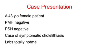
17. Case Presentation
A 57-year-old woman was admitted to our hospital due to epigastric abdominal pain. A physical
examination revealed no remarkable findings.
Laboratory studies showed an elevated white blood cell count (10400/μL), aspartate
aminotransferase (AST: 84 U/L), alanine aminotransferase (ALT: 39 U/L), alkaline phosphatase (ALP:
466 U/L), γ-glutamyl transpeptidase 205 U/L). Ultrasonography and computed tomography (CT)
revealed small gallstones in the gallbladder and some stones in the common bile duct. A large liver
cyst was also detected in S4. Thus, the patient was diagnosed with cholecysto-choledocholithiasis.
Laparoscopic cholecystectomy was planned after the complete removal of the gallstones in the
common bile duct following endoscopic sphincterotomy (EST).
However, a cystic duct which communicates with the intrahepatic bile duct of the posterior segmental
branch was suspected based on the gross X-ray images obtained after the extraction of the bile duct
stones. Therefore, the magnetic resonance cholangiopancreatography (MRCP) was performed.
MRCP showed strong suspicion of a single gallbladder with a double cystic duct.
18. Thus, to confirm this rare condition, endoscopic retrograde cholangiography (ERC) was additionally
performed for a second time. This time, a normal cystic duct was found to arise from the neck of the
gallbladder, from which it descended and joined the common bile duct.
In addition, the aberrant cystic duct arose from the cystic duct and communicated with the
intrahepatic bile duct of the posterior segmental branch.
Thus, we determined that the patient had a single gallbladder with a double cystic duct.
An endoscopic nasobiliary drainage (ENBD) tube was placed for intraoperative cholangiography in
order to protect the common bile and hepatic duct from injury during surgery. Laparoscopic
cholecystectomy was performed under general anesthesia.
During the procedure, the gallbladder was divided from the gallbladder bed in the fundus- to- hilar
direction, in order to explore the double bile ducts. The main and aberrant cystic ducts and the
common hepatic duct were identified by meticulous dissection and intraoperative cholangiography.
These cystic ducts were then ligated and precisely divided.
Laparoscopic cholecystectomy was finished and no drain was placed.
The excised specimen showed a duplicated cystic duct containing debris and chronic cholecystitis
19.
20. With more than half a million cholecystectomies performed annually, variations in
biliary anatomy present a significant challenge for laparoscopic surgeons.
Duplication of the cystic duct is extremely rare, with only 20 previous cases
reported in the literature.
Three types of duplicated cystic ducts have been previously described:
1) The ‘Y’ type, in which two cystic ducts meet to form a common channel.
2) the ‘H’ type, in which the accessory duct enters into the right, left or common
hepatic duct.
3) the trabecular type, in which the accessory duct enters the liver substance
directly.
While preoperative MRCP or endoscopic retrograde cholangiopancreatography
(ERCP) may identify abnormal anatomy, this is not guaranteed to prevent
encountering unexpected variations.
21.
22. Of the seven reported cases of cystic duct duplication where a preoperative ERCP was performed, only
three had the anomaly detected.
In most cases, the second cystic duct was an intraoperative finding.
This emphasizes the importance of constant vigilance during LC even when preoperative imaging yields
no abnormality.
In conclusion, the duplicated cystic duct is a highly uncommon variant of biliary anatomy, which may pose
a significant dilemma to the laparoscopic surgeon.
Constant vigilance for anatomical variance is essential to avoid adverse outcomes, with early involvement
of a hepatobiliary specialist if there is unexplained bile leakage.
Unremarkable preoperative imaging does not exclude the presence of abnormal anatomy.
However, the point at which the cystic duct joins the extrahepatic bile duct is variable, ranging from high at the level of the porta hepatis to low at the level of the ampulla.