Management of tibial plateau fracture
•Download as PPTX, PDF•
1 like•498 views

Update management of tibial plateau fracture

Recommended
More Related Content
What's hot
What's hot (20)
Similar to Management of tibial plateau fracture
Similar to Management of tibial plateau fracture (20)
More from Rizqi D Rosandi MD
Recently uploaded
Recently uploaded (20)
Management of tibial plateau fracture
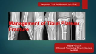
- 1. Rizqi D Rosandi Orthopaedi-Traumatology FK Univ. Brawijaya April 2020 Pengampu: Dr. dr. Edi Mustamsir, Sp. OT (K) Management of Tibial Plateau Fracture
- 2. Epidemiology (burden of disease/cost to society) • Tibial Plateau – Articular surface proximal tibia – +/- metaphyseal /diaphyseal extension • Account for 1.2% of all fractures • Lateral Plateau: 55-70% of fractures • Medial Plateau: 10-20% of fractures • Bicondylar Plateau: 10-30% of fractures
- 3. Epidemiology (burden of disease/cost to society) • Bimodal distribution – Young adults: high energy mechanism • Highest in 5th decade • Male > Female – Elderly: low energy mechanism • Osteoporotic bone • Female > Male • Significant functional impairment – Joint incongruity, malalignment, instability – Post-traumatic arthritis
- 4. Anatomy • Consist of medial and lateral plateau – Medial larger – Medial lower (concave) – Medial bone harder (thus less likely to fracture) – Lateral higher (convex) – Lateral cartilage thicker (3 vs 4 mm) MedialLateral
- 6. Mechanism of Injury • Valgus producing force – Lateral plateau • Varus producing force – Medial plateau • Axial compressive force – Bicondylar plateau • Combination – High energy – Bicondylar plateau
- 7. Mechanism of Injury • Low energy – Split depression – Increasing age – Poor bone quality • High energy – Pedestrian vs car (bumper) – Fall from height – Motor vehicle accident – Axial load (knee extended) – Bicondylar fracture – Associated injuries
- 8. Clinical presentation • History – High energy trauma in young – Low energy trauma in elderly • Assessment – Open or closed fracture – Compartment syndrome – Instability – Neurovascular – ATLS
- 9. Imaging • Radiographs – Knee AP/LAT – Oblique ( subtle plateau depression) – Plateau view ( 10 caudal tilt) • Knee CT – Articular involvement comminution – Schatzker IV V VI – Pre op planning • Knee MRI – Schatzker I II III – Assesment meniscus and ligament • Angiography
- 10. Personality of fracture • Soft tissue damage • Degree of dislocation • Degree of comminution • Degree of join involvement • Osteoporosis • Nerve / blood vessel injury
- 11. Personality of the fracture
- 12. Classification • Schatzker classification • AO/OTA • Hohl & Moore • Three column classification
- 16. The three column classification
- 17. Management • Non operative • Operative
- 18. Goals of treatment • Decompression and preservation of soft tissues • Reconstruction of joint surfaces • Reconstruction of normal mechanical axis • Early motion
- 19. Nonoperative treatment • No joint step > 2 mm • No axial instability • Severe osteoporosis • General and local contraindications
- 20. Nonoperative treatment • Traction may be of use in short term • Early active movements in a cast brace • Touch weight-bearing if patient’s condition allows • Weight-bearing to tolerance at 6 weeks • Nonoperative treatment does well in low-demand elderly patients
- 21. Emergency operative treatment • Vascular injury • Compartment syndrome • Open fractures • Gross dislocation • Floating knee • Polytrauma
- 22. Operative treatment—timing • Rarely as an emergency, unless: - Open fracture, dislocation, vascular injury … • Delayed surgery to allow soft tissue recovery and adequate investigations - (spanning external fixation may be required)
- 23. Timing of Surgery Low Energy: Fixed electively and early High Energy: Be patience
- 24. Delayed surgery • The use of a temporary spanning external fixator will allow • Optimal recovery of soft tissues while preserving length and axis
- 25. Decision-making • Surgical approaches can be open or arthroscopic
- 26. Approach • Straight anterior • Posteromedial • Lateral • Miniopen
- 27. Approach Anterolateral approach Posteromedial approach
- 28. Intraoperative procedure • • • • • Expose ligamentous and meniscal structures Reconstruct the joint surface usually with anatomical reduction and interfragmentary compression using lag screws Support the joint surface with bone or substitute Buttress with plate (conventional) Repair of the ligaments or menisci to achieve joint stability
- 29. ORIF • Fixation based on fracture type • Type I, II, III: Buttress plates with raft screws • Type IV: Medial plate (buttress) – Be cognizant of any impaction of lateral joint line • Type V, VI: – Important to understand plate function – Pattern dictates fixation – Single lateral base fixed angle implant – Dual plating (lateral and posteromedial)
- 30. ORIF
- 31. ORIF
- 32. Rehabilitation • Postoperative Care • Antibiotic x 24 hours • +/- drain • Knee brace – For comfort until able to do straight leg raise (SLR) – Associated ligamentous injuries • Elevate leg • NWB 10-12 weeks
- 33. Rehabilitation • Physical therapy • Early ROM • CPM • Strengthening – Isometric quad sets – Heel slides – SLR • Gait training – Crutches – D/c crutches when able to walk without limp and pain
- 34. Complications • Infection – Surgery timing is important – Careful soft tissue handling – Prolong operative time • Nonunion – Aseptic • Metadiaphyseal junction – Septic – Opened fracture Aseptic Nonunion Revised with ICBG
- 35. Complications • Contractures – Arthrofibrosis – Encourage early ROM and physical therapy – May require knee manipulation – Arthroscopic lysis of adhesion • Post Traumatic Osteoarthritis
- 36. Outcomes • 90% excellent or good results – Despite some incongruity • 10% fair or poor – > 10mm depression persisted • Conclusion – Instability (lateral or medial with knee extended) – Should be operative Lansinger et al. JBJS Am 1986
- 37. Outcomes • Risk factors for post-traumatic arthritis – Increase age – Removal of meniscus – Articular incongruity – Instability – Malalignment Honkonen JOT 1995
- 38. Summary • Anatomical reduction and rigid fixation of joint surface— absolute stability • Functional reduction and stable fixation of metaphysis— relative stability • Restoration of joint stability by appropriate soft-tissue reconstruction • Early active movement
- 39. Thank You Malang, 07 April 2020