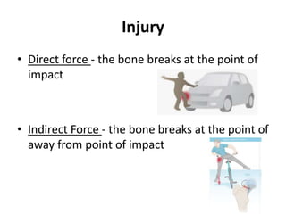
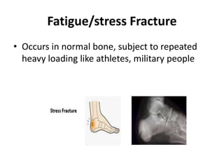
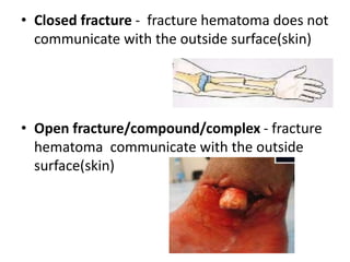
FRACTURE
Dr Ninad S Patil
MBBS,MD Pediatrics
Department of Pediatrics
NKP Salve & LMH ,Nagpur
Definition
 It is break/disruptions in the continuity of
bone
Fractures occurs when bone is subjected to
the stress greater than it can absorb
Causes
• Injury
• Repetitive stress
• Pathological fracture
Injury
• Direct force - the bone breaks at the point of
impact
• Indirect Force - the bone breaks at the point of
away from point of impact
Fatigue/stress Fracture
• Occurs in normal bone, subject to repeated
heavy loading like athletes, military people
Pathologic fracture
• Occurs in a bone that is made weak bye some
disease
• Causes
- inflammation eg osteomyelitis
- neoplastic- Ewing's sarcoma
- hereditary – osteogenesis imperfecta
- acquired- rickets ,osteomalacia
classification
• Type
• Communicating OR non-communication to
external environment
• Anatomical location
• Closed fracture - fracture hematoma does not
communicate with the outside surface(skin)
• Open fracture/compound/complex - fracture
hematoma communicate with the outside
surface(skin)
Anatomical classification
1) Impacted # - vertical force drives the distal
fragment of the # into the proximal fragment
2) Depressed # - type of skull fracture, segment
of the bone get depressed into the cranium
# type based upon “path of fracture line”
1) Transverse #
2) Oblique #
3) Longitudinal #
4) Spiral /torsion #
CLINICAL MANIFESTATION
• Pain
• Local swelling/tenderness/painful movement
• Discoloration of limb
• Deformity/shortening
• Loss of function of limb
Diagnosis of fracture
• History
• Physical examination- General and local
• X-ray- two view imaging
• CT scan- vertebral /joint /hairline #
• MRI scan- vertebral fracture compressing SC
Fracture Complication
STAGES OF FRACTURE HEALING
1) Inflammatory stage (A & B) –
A – stage of hematoma formation
B - Stage of granulation tissue (more fibrin to
hematoma & increased blood flow)
2) Reparative phase (A & B) –
A- stage of fibrocartilaginous callus
B- stage of bony callus (woven bone become calcified)
3) Remodeling Phase –excess material inside bone shaft
get replaced by more compact bone
Immediate
Damage to Vessel Hemorrhage/shock
Damage to Organ Brain/Liver/spleen etc
Damage to Nerve Power/sensation loss
Damage to Muscle compartment syndrome
Fat embolism
DIAGNOSIS
• History collection
• Physical examination
• X ray- 2 views
• CT scan
• MRI scan
PRINCIPLE OF MANAGEMENT
1st save life
2nd save limb
3rd save function of limb
SAVE THE LIFE
• Check for the Vitals and Injury to vital organs
act accordingly
A-airway
B-breathing
C-circulation
SAVE THE LIMB
• Early splinting
• Assess the fracture site and involved limb
• Look for the color and perfusion of limb
• Look for the injury to blood vessels and
Nerves
• Wound contamination
Splinting
• It is the most common procedure for
immobilizing an injury
• It stabilizes the limb and reduces the area
• Any material available can be used like towel,
paper etc
Any material can be used for splinting
Guideline for splinting
• Support the injured area
• Splint injury in the position that you find it
• Don’t try to realign the bones
• Check color, warmth and sensation
• Immobilize above and below the injury
SAVE THE LIMB FUNCTION
• Fracture reduction
• Wound/limb care
• Immobilization
• Early physiotherapy and rehabilitation
• Help to hasten recovery
• Look for complications
Reduction of fracture
(correction of displacement of fragments)
- Closed or Open reduction
CLOSED REDUCTION
Non-surgical procedure
plaster applied
OPEN REDUCTION
• Surgical correction of fractured bone alignment
• Internal Fixator used for stabilization
NURSING MANAGMENT
Closed Fracture
• Encourage patient to not to mobilize fracture
site
• Exercise to maintain the health of the
unaffected muscle for using assisted devices
like crutches /walker
• Patient education about – self care,
medication information, monitoring for
potential complication and need for
continuous supervision
Open fracture
• Injection TT
• Wound irrigation and debridement
• Heavily contaminated wounds – left
unsutured and dressed with sterile gauze to
permit swelling & wound drainage
• Administering IV antibiotics/painkiller
Nursing diagnosis
• Acute pain related breakdown of continuity of
the bone as evidenced by facial expression
and verbalization of patient
• Goal- patient should be pain free
• Interventions-
- asses the onset,duration,location,severity & intensity
of pain
- administer the analgesic as per prescription
- provide the comfort devices like sand bag for
immobilization of affected parts
Thank You

FRACTURE new.pptx

  • 1.
    FRACTURE Dr Ninad SPatil MBBS,MD Pediatrics Department of Pediatrics NKP Salve & LMH ,Nagpur
  • 2.
    Definition  It isbreak/disruptions in the continuity of bone Fractures occurs when bone is subjected to the stress greater than it can absorb
  • 3.
    Causes • Injury • Repetitivestress • Pathological fracture
  • 4.
    Injury • Direct force- the bone breaks at the point of impact • Indirect Force - the bone breaks at the point of away from point of impact
  • 5.
    Fatigue/stress Fracture • Occursin normal bone, subject to repeated heavy loading like athletes, military people
  • 6.
    Pathologic fracture • Occursin a bone that is made weak bye some disease • Causes - inflammation eg osteomyelitis - neoplastic- Ewing's sarcoma - hereditary – osteogenesis imperfecta - acquired- rickets ,osteomalacia
  • 7.
    classification • Type • CommunicatingOR non-communication to external environment • Anatomical location
  • 8.
    • Closed fracture- fracture hematoma does not communicate with the outside surface(skin) • Open fracture/compound/complex - fracture hematoma communicate with the outside surface(skin)
  • 9.
    Anatomical classification 1) Impacted# - vertical force drives the distal fragment of the # into the proximal fragment 2) Depressed # - type of skull fracture, segment of the bone get depressed into the cranium
  • 10.
    # type basedupon “path of fracture line” 1) Transverse # 2) Oblique # 3) Longitudinal # 4) Spiral /torsion #
  • 13.
    CLINICAL MANIFESTATION • Pain •Local swelling/tenderness/painful movement • Discoloration of limb • Deformity/shortening • Loss of function of limb
  • 14.
    Diagnosis of fracture •History • Physical examination- General and local • X-ray- two view imaging • CT scan- vertebral /joint /hairline # • MRI scan- vertebral fracture compressing SC
  • 15.
  • 16.
    STAGES OF FRACTUREHEALING 1) Inflammatory stage (A & B) – A – stage of hematoma formation B - Stage of granulation tissue (more fibrin to hematoma & increased blood flow) 2) Reparative phase (A & B) – A- stage of fibrocartilaginous callus B- stage of bony callus (woven bone become calcified) 3) Remodeling Phase –excess material inside bone shaft get replaced by more compact bone
  • 18.
    Immediate Damage to VesselHemorrhage/shock Damage to Organ Brain/Liver/spleen etc Damage to Nerve Power/sensation loss Damage to Muscle compartment syndrome Fat embolism
  • 19.
    DIAGNOSIS • History collection •Physical examination • X ray- 2 views • CT scan • MRI scan
  • 20.
    PRINCIPLE OF MANAGEMENT 1stsave life 2nd save limb 3rd save function of limb
  • 21.
    SAVE THE LIFE •Check for the Vitals and Injury to vital organs act accordingly A-airway B-breathing C-circulation
  • 22.
    SAVE THE LIMB •Early splinting • Assess the fracture site and involved limb • Look for the color and perfusion of limb • Look for the injury to blood vessels and Nerves • Wound contamination
  • 23.
    Splinting • It isthe most common procedure for immobilizing an injury • It stabilizes the limb and reduces the area • Any material available can be used like towel, paper etc
  • 24.
    Any material canbe used for splinting
  • 25.
    Guideline for splinting •Support the injured area • Splint injury in the position that you find it • Don’t try to realign the bones • Check color, warmth and sensation • Immobilize above and below the injury
  • 26.
    SAVE THE LIMBFUNCTION • Fracture reduction • Wound/limb care • Immobilization • Early physiotherapy and rehabilitation • Help to hasten recovery • Look for complications
  • 27.
    Reduction of fracture (correctionof displacement of fragments) - Closed or Open reduction CLOSED REDUCTION Non-surgical procedure plaster applied
  • 28.
    OPEN REDUCTION • Surgicalcorrection of fractured bone alignment • Internal Fixator used for stabilization
  • 29.
    NURSING MANAGMENT Closed Fracture •Encourage patient to not to mobilize fracture site • Exercise to maintain the health of the unaffected muscle for using assisted devices like crutches /walker • Patient education about – self care, medication information, monitoring for potential complication and need for continuous supervision
  • 30.
    Open fracture • InjectionTT • Wound irrigation and debridement • Heavily contaminated wounds – left unsutured and dressed with sterile gauze to permit swelling & wound drainage • Administering IV antibiotics/painkiller
  • 31.
    Nursing diagnosis • Acutepain related breakdown of continuity of the bone as evidenced by facial expression and verbalization of patient • Goal- patient should be pain free • Interventions- - asses the onset,duration,location,severity & intensity of pain - administer the analgesic as per prescription - provide the comfort devices like sand bag for immobilization of affected parts
  • 32.