SlideShare a Scribd company logo
1 of 44
Chapter-3
NCERT
By- Punya Mohanty
Class –XII
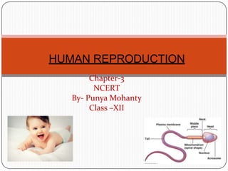
HUMAN REPRODUCTION
THE MALE REPRODUCTIVE SYSTEM.
 Located in the pelvis
region.
 Male reproductive system
includes
 A pair of testes.
 Accessory ducts.
 Accessory glands.
 External genitalia
Testes:
 Located outside the abdominal cavity within a pouch called scrotum.
 Scrotum provides low temperature required for spermatogenesis.
 Each testis is about 4 to 5 cm length and 2 to 3 cm width.
 Each testis has about 250 compartments called testicular lobules.
 Each lobule contains one to three seminiferous tubules.
 Seminiferous tubules lined by male germ cells and Sertoli cells.
 Male germ cell undergoes meiosis and produce sperm.
 Sertoli cells provide nutrition to the germ cell and the sperm.
 In between the seminiferous tubule there is interstitial cell or Leydig
cell.
 Leydig cells produce testicular hormones
called androgen (testosterone).
Accessory ducts:
 Includes rete testis, vasa efferentia, epididymis and vas
deferens.
 Seminiferous tubules open into vasa efferentia through rete
testis.
 The vasa efferentia leaves the testis and open into epididymis.
 The epididymis leads to vas deferens that ascends to the
abdomen through inguinal canal and loops over the urinary
bladder.
 Vas deferens receives a duct from seminal vesicle and opens
into the urethra as the ejaculatory duct.
 Urethra originates from the urinary bladder and extends
through the penis to its external opening calledurethral
meatus.
Accessory glands:
 Includes
 Paired seminal vesicle
 A prostate gland
 Paired bulbourethral gland.
 Secretion of these glands constitutes the seminal
plasma.
 Seminal plasma rich in fructose, calcium, and
certain enzyme.
 Secretion of bulbo-urethral glands helps in
lubrication of penis.
External genitalia:
 Penis is the external genitalia.
 It is made of special tissue that helps in erection
of the penis to facilitate insemination.
 The enlarged end of penis is called glans penis.
 Glans penis is covered by a loose fold of skin
called foreskin.
THE FEMALE REPRODUCTIVE
SYSTEM
 Located in the pelvic region
of the
female.
 The female reproductive
system includes:
 A pair of ovaries
 A pair of oviduct.
 Uterus
 Cervix
 Vagina
 External genitalia.
 A pair of mammary gland
Ovaries:
 It is the primary female sex organs that produce
the female gamete (ovum).
 It also produces several steroid hormones.
 The ovaries located in the lower abdomen.
 Each ovary is about 2-4 cm in length.
 Connected to the pelvic wall and uterus by
ligaments.
 Each ovary is covered by thin epithelium which
encloses the ovarian stroma
 The ovarian stroma has two zones
 A peripheral cortex.
 An inner medulla.
Oviduct:
 Oviducts, uterus and vagina constitute the female
accessory ducts.
 Each fallopian tube is about 10-12 cm long and extends
from the
periphery of each ovary to the uterus.
 Close to the ovary the oviduct has a funnel shaped
structure called infundibulum?
 The edges of the infundibulum possess finger-like
projections called fimbriae, which helps in collection
of the ovum after ovulation.
 The infundibulum leads to a wider part of the
oviduct called ampulla.
 The last part of the oviduct is called isthmus which
joined to uterus.
Uterus:
 It is single and is called womb.
 It is inverted pear shaped.
 Attached the pelvic wall by ligaments.
 The uterus opens into vagina through a narrow cervix.
 The lumen of cervix is called cervical canal.
 Cervical canal along with vagina form the birth canal.
 The wall of the uterus has three layers of tissues
 Perimetrium: external thin membranous.
 Myometrium: middle thick layer of smooth muscles
 Endometrium: inner glandular layer.
 Endometrium undergoes cyclical changes during
menstrual cycle.
 Myometrium exhibits strong contraction during delivery of
the baby.
External genitalia:
 It includes following structure:
 Mons Pubis: cushion of fatty covered by skin and
pubic hair.
 Labia majora: fleshy folds of tissue which extends
down from the mons pubis and surrounds the
vaginal opening.
 Labia minora: are paired folds of tissue under
the labia majora.
 Hymen: the opening of vagina is often covered
partially by a membrane called hymen.
 Clitoris: a tiny finger-like structure lies at the upper
junction of two labia minora above the urethral
opening.
Mammary glands:
 Mammary gland consists of glandular tissue and fat.
 Glandular tissue of each breast divided into 15-20
mammary
lobes.
 Mammary lobes contain cluster of cells called
alveoli.
 The cells of alveoli secrete milk, stored in the lumen
of alveoli.
 The alveoli open into mammary tubules.
 The tubules of each lobe join to form a mammary
duct.
 Several mammary ducts join to form a wider
mammary ampulla.
 Mammary ampulla connected to lactiferous duct,
GAMETOGENESIS: (formation of
gametes)
 Spermatogenesis:
 Formation of sperm from the germ cell in the testes is spermatogenesis.
 The process begins at puberty.
 Spermatogonia present in the lining of seminiferous tubules undergo
mitotic division to increase their number.
 Each spermatogonium is diploid (2n) which contain 46 chromosomes.
 Innermost layer of spermatogonial becomes larger called primary
spermatocyte.
 Primary spermatocyte undergoes meiosis-I to form two equal haploid (n)
secondary
spermatocytes (n).
 Each secondary spermatocyte undergoes meiosis-II to
form two equal, haploid spermatids.
 Each primary spermatocyte produces four spermatids.
 Spermatids transformed into spermatozoa (sperms) by
the process called spermiogenesis.
 The sperm head embedded in the Sertoli cell.
SPERMATOGENESIS PROCESS
Hormonal control of
spermatogenesis:
 This process is initiated at puberty due to
secretion of gonadotrophins releasing
hormone (GnRH)
 GnRH secreted form hypothalamus and
stimulate anterior
pituitary to secrete two gonadotrophins.
 Luteinizing hormone (LH) and
 Follicle stimulating Hormone (FSH)
 LH acts on Leydig cells and stimulates synthesis
of
androgens.
 Androgen stimulates spermatogenesis.
 FSH acts on Sertoli cells and stimulates
spermatogenesis in other ways.
Structure of sperm:
 Ultrastructure of sperm consists of a head, neck, a middle piece
and a tail.
 Whole body of sperm surrounded by plasma membrane.
 The sperm head contain an elongated haploid nucleus.
 Above the nucleus a cap like structure present called acrosome.
 The acrosome contains enzymes which help in fertilization of
ovum.
 The middle piece contains mitochondria, which provide
energy for movement of tail that facilitate sperm motility.
 Human male ejaculates 200-300 million sperms during coitus.
 60 percent must have normal shape and size and 40 percent of
them must show
vigorous motility.
 Sperm released from seminiferous tubules enters into accessory
ducts.
 On their way fluids from seminal vesicle and prostate gland added
which
SPERM
STRUCTURE
Oogenesis:
 Formation of a mature female gamete or ovum is calledoogenesis.
 Oogenesis starts during embryonic stage, 25th week of the fetal age.
 Germinal epithelium of ovary divided mitotically to produce millions of gamete
mother cell or oogonia.
 No oogonia formed or added after birth.
 Oogonia enters into meiosis-I and proceeds uptodiakinesis of Prophase-I and get
suspended, at this stage calledprimary Oocytes.
 Each primary oocyte surrounded by layers of granulose cells and then
called primary follicle.
 At puberty only 60,000 to 80,000 primary oocytes are left in each ovary.
 After puberty primary follicles get surrounded by more layers of granulosa cells and
a new theca to formsecondary follicles.
 The secondary follicle transformed into tertiary follicle, characterized by a fluid
filled cavity calledantrum.
 The theca layers organized into an inner theca interna and outer theca externa.
 During the growth of primary follicle into tertiary follicle during puberty, the
primary oocyte restarts its first meiotic division and completes it within tertiary
follicle resulting two unequal haploid cells.
 Large haploid cell is called secondary oocyte.
 A tiny cell called first polar body.
CONT
…. The secondary oocyte retains bulk of the nutrient rich cytoplasm of
primary oocyte.
 The tertiary follicle having secondary oocyte further changes
into Graafian follicle.
 The secondary oocyte surrounded by a new
membrane, zonapellucida.
 The secondary oocyte undergoes second meiotic division continued
upto metaphase-II and get suspended until entry of sperm.
 At this stage Graafian follicle releases secondary oocyte from the
ovary by the process called ovulation.
 On entry of a sperm into the secondary oocytes stimulates it to
complete meiosis-II and there is formation of a haploid ovum and
a second polar body (n).
Menstrual cycle:
 Reproductive cycle of female primates is
called menstrual cycle.
 The first menstruation begins at puberty is
called Menarche.
 Menstrual cycle repeated at an average interval of
28/29 days.
 One ovum is released in the middle of each
menstrual cycle.
Menstrual cycle has following phases:
 Menstrual phase:
 1st phase of menstrual cycle.
 Menstrual flow occurs.
 Lasts for 3-5 days.
 Breakdown of endometrial lining and blood vessel.
 Mucus and blood comes out through vagina.
 It occurs only when ovum released but no
fertilization.
 Lack of menstruation is the indication of pregnancy.
Follicular phase:
 Menstrual phase followed by follicular phase.
 Primary follicle becomes Graafian follicle.
 Regeneration and proliferation of uterine
endometrium.
 LH and FSH level increases gradually in follicular
phase.
 Level of estrogen increases as it is secreted from
growing follicle.
 It lasts for 5-13 days.
Ovulatory phase:
 FSH and LH attain peak level in this period (14th
day).
 This is called LH surge, which induces rupture of
Graafian follicle and release of ovum from the ovary
called ovulation.
Luteal phase:
 Remaining part of Graafian follicle transformed into corpus
luteum.
 Coupusluteum produces large amount of progesterone.
 Progesterone maintains the uterine endometrium, and
prepares it for implantation.
 Thickness of uterine endometrium increase in many folds, due
to proliferation.
 If there is fertilization, corpus luteum grows further and
pregnancy continued, menstrual cycle stopped.
 In the absence of fertilization corpus luteum degenerates.
 Disintegration of endometrium leading to menstruation.
 Menstrual cycle ceases around 50 years of age,
called menopause.
FERTILIZATION AND
IMPLANTATION:
 During copulation (coitus) semen is released by the
penis into the vagina is called insemination.
 The motile sperm swim rapidly, pass through cervix,
uterus and finally reach the junction of isthmus and
ampulla(ammpullary-isthmic junction).
 The ovum released from the ovary also transported to
ampullary isthmic junction where fertilization takes
place.
 Fertilization only takes place if both sperm and ovum
reach ampullary – isthmic junction simultaneously.
 The process of fusion of a sperm and ovum is
called fertilization.
 Acrosome of sperm secretes enzymes helps in
penetration into the ovum.
CONT
… Once a sperm comes contact with the zonapellucida of
ovum and induces the changes in the membrane that
blocks the entry of additional sperms.
 That ensures monospermy and prevents polyspermy.
 Only one sperm fertilize with one ovum.
 Entry of sperm into the ovum induces the ovum to
complete its second meiotic division of secondary
oocyte.
 Meiosis-II is also unequal cytokinesis resulting
production of one large ovum (ootid) and one
small second polar body.
 Haploid nucleus of sperm fused with the haploid
nucleus of ovum to form a diploid zygote.
 The zygote undergoes a series of mitotic cell divisions
called cleavage.
 The stages of development are:
Fertilized ovum (zygote)
2-cell stage
4-cell stage
8-cell stage
Morula
Blastula
 Gastrula
Sex determination:
 Sex of a baby has been decided during fertilization and in the
zygote.
 Sex is determined by the sex-chromosomes present in
gametes.
 Human female contain two XX chromosomes.
 Human male contain XY chromosomes.
 All the female gametes produced with only ǮXǯchromosome.
 Sperms produced by male, 50% with ǮXǯand 50 % with ǮYǯ
chromosome.
 After fertilization zygote either carries XX or XY
chromosomes.
 Zygote with XX chromosomes develop into female and with
XY chromosome develops into male
Cleavage:
 Repeated mitotic division of the zygote without growth resulting a
multicellular ball like embryo is calledcleavage.
 Cleavage starts soon after fertilization.
 Daughter cells produced during cleavage are called blastomeres.
 The product of cleavage is called Morula, which is 8 to 16 celled.
 The morula continues to divide and grow and transformed
into blastocyst.
 The blastomeres in blastocyst arranged into an outer layer
called trophoblast and an inner mass of cells attached to trophoblast
called inner cell mass.
 Trophoblast cells attached to the endometrium helps development of
placenta.
 Inner cell mass gets differentiated into the embryo.
 After attachment the uterine cells divide rapidly and cover the
blastocyst.
 Blastocyst completely embedded in the uterine endometrium. This is
called implantation.
Pregnancy and embryonic development:
 After implantation, finger like projections appears
on the trophoblast called chorionic villi.
 Chorionic villi surrounded by uterine tissue and
maternal blood.
 Temporary association between the fetal tissue
(chorionic villi) and maternal tissue (uterine
endometrium) is called placenta.
Function of placenta:
 The embryo connected to the placenta by umbilical
cord, which transports substances to and from the
embryo.
 Facilitate transport of oxygen and nutrient from mother
to embryo.
 Removes CO2 and waste material from the embryo.
 Acts as endocrine gland and produces several hormones
like:
 Human chorionic gonadotrophins (hCG)
 Human placental lactogen (hPL)
 Estrogen.
 Progesterone
 Relaxin produced from the ovary in the later stage of
pregnancy.
Embryonic development:
 After implantation the inner cell mass of blastocyst
differentiated into an outer layer
called ectoderm and an inner layer
called endoderm.
 Mesoderm differentiated in-between ectoderm and
endoderm.
 The inner cell mass thus called stem cells, having
potency to produce all types of cell, tissues and
organs by differentiation.
Organogenesis:
 Formation of different organs in the embryo is
called organogenesis.
 Human pregnancy lasts for 9 months.
 After one month of pregnancy heart is formed in the embryo.
 By the end of 2nd month the foetus develops limbs and digits.
 By the end of 12 weeks (first trimester) most of organ system is
formed (limbs and external genitalia are well developed).
 First movement of foetus and appearance of hairs observed in 5th
month.
 By the end of 24th week (2nd trimesters) the body is covered with
fine hairs, eye-lids separate, and eyelashes are formed.
 By the end of 9 months the foetus is fully developed and is ready for
delivery.
PARTURITION AND LACTATION:
 The period of pregnancy is called gestation period. (9 months).
 Ejection or expulsion or delivery of foetus is called parturition.
 Parturition is due to vigorous contraction of uterine Myometrium.
 The signal of parturition is originated from the fully developed
foetus and the placenta which induces mild contraction of uterus
called fetal ejection reflex.
 Fetal ejection reflex triggers the release of Oxytocin from pituitary.
 Oxytocin induces stronger contraction of uterine endometrium.
 Stimulatory reflex continues stronger contraction leads to
expulsion.
 After delivery the placenta is also expelled out of the uterus.
Lactation:
 The mammary gland of the female more
differentiated during pregnancy,
 Mammary gland starts producing milk towards the
end of the pregnancy.
 Process of milk production in mammary gland is
called lactation.
 Milk produced during initial days of lactation is
called colostrum.
 Colostrum contains several antibodies which
provide immunity to the new born baby.
Thanking
you

More Related Content

What's hot

Jeopardy reproductive system
Jeopardy reproductive systemJeopardy reproductive system
Jeopardy reproductive systema arg
 
Reproductive system of eartworms and insects
Reproductive system of eartworms and insectsReproductive system of eartworms and insects
Reproductive system of eartworms and insectsLoremie Jimenez
 
Introduction to General Embryology
Introduction to General EmbryologyIntroduction to General Embryology
Introduction to General EmbryologyDr. Sherif Fahmy
 
Extra embryonic membranes
Extra embryonic membranesExtra embryonic membranes
Extra embryonic membranesjyoti malik
 
Histology of Avian female reproductive system
Histology of Avian female reproductive systemHistology of Avian female reproductive system
Histology of Avian female reproductive systemDr. Waqas Nawaz
 
Female reproductive system
Female reproductive systemFemale reproductive system
Female reproductive systemUE
 
Reproductionsystem 131109102239-phpapp01
Reproductionsystem 131109102239-phpapp01Reproductionsystem 131109102239-phpapp01
Reproductionsystem 131109102239-phpapp01Chethan Durgan
 
Female Reproductive System
Female Reproductive System Female Reproductive System
Female Reproductive System Muhammadasif909
 
Human reproduction ppt
Human reproduction pptHuman reproduction ppt
Human reproduction pptnagamani42
 
Female reproductive anatomy
Female reproductive anatomyFemale reproductive anatomy
Female reproductive anatomyAhmed Ali
 
Embryology of genitourinary system
Embryology of genitourinary systemEmbryology of genitourinary system
Embryology of genitourinary systemAnuradha Sawant
 
Reproductive System
Reproductive SystemReproductive System
Reproductive Systemlogan_xavi93
 
Embryological development in insect
Embryological development in insectEmbryological development in insect
Embryological development in insectBhumika Kapoor
 
extra embryonic membranes in birds
extra embryonic membranes in birdsextra embryonic membranes in birds
extra embryonic membranes in birdsSKSampad
 

What's hot (20)

Reproduction
ReproductionReproduction
Reproduction
 
Female Reproductive system of Fowl, Egg formation and Egg structure
Female Reproductive system of Fowl, Egg formation and Egg structureFemale Reproductive system of Fowl, Egg formation and Egg structure
Female Reproductive system of Fowl, Egg formation and Egg structure
 
Jeopardy reproductive system
Jeopardy reproductive systemJeopardy reproductive system
Jeopardy reproductive system
 
OOGENESIS -GENETICS
OOGENESIS -GENETICSOOGENESIS -GENETICS
OOGENESIS -GENETICS
 
Reproductive system of eartworms and insects
Reproductive system of eartworms and insectsReproductive system of eartworms and insects
Reproductive system of eartworms and insects
 
Reproductive Systems
Reproductive SystemsReproductive Systems
Reproductive Systems
 
Introduction to General Embryology
Introduction to General EmbryologyIntroduction to General Embryology
Introduction to General Embryology
 
Human reproduction
Human reproductionHuman reproduction
Human reproduction
 
Extra embryonic membranes
Extra embryonic membranesExtra embryonic membranes
Extra embryonic membranes
 
Histology of Avian female reproductive system
Histology of Avian female reproductive systemHistology of Avian female reproductive system
Histology of Avian female reproductive system
 
Female reproductive system
Female reproductive systemFemale reproductive system
Female reproductive system
 
Embryology
Embryology Embryology
Embryology
 
Reproductionsystem 131109102239-phpapp01
Reproductionsystem 131109102239-phpapp01Reproductionsystem 131109102239-phpapp01
Reproductionsystem 131109102239-phpapp01
 
Female Reproductive System
Female Reproductive System Female Reproductive System
Female Reproductive System
 
Human reproduction ppt
Human reproduction pptHuman reproduction ppt
Human reproduction ppt
 
Female reproductive anatomy
Female reproductive anatomyFemale reproductive anatomy
Female reproductive anatomy
 
Embryology of genitourinary system
Embryology of genitourinary systemEmbryology of genitourinary system
Embryology of genitourinary system
 
Reproductive System
Reproductive SystemReproductive System
Reproductive System
 
Embryological development in insect
Embryological development in insectEmbryological development in insect
Embryological development in insect
 
extra embryonic membranes in birds
extra embryonic membranes in birdsextra embryonic membranes in birds
extra embryonic membranes in birds
 

Similar to Human Reproduction

Reproductive system
Reproductive systemReproductive system
Reproductive systemNisha Mhaske
 
HUMAN REPRODUCTION.pptx
HUMAN  REPRODUCTION.pptxHUMAN  REPRODUCTION.pptx
HUMAN REPRODUCTION.pptxbarunmahto001
 
Human Reproducton and biological change.pptx
Human Reproducton and biological change.pptxHuman Reproducton and biological change.pptx
Human Reproducton and biological change.pptxbholu803201
 
7. Male and Female reproductive system.pptx
7. Male and Female reproductive system.pptx7. Male and Female reproductive system.pptx
7. Male and Female reproductive system.pptxAbhiDabra
 
The reproductive system
The reproductive systemThe reproductive system
The reproductive systemhscteacher04
 
178-Anatomy-Reproductive-System.ppt
178-Anatomy-Reproductive-System.ppt178-Anatomy-Reproductive-System.ppt
178-Anatomy-Reproductive-System.pptsadiaahmad30
 
Female Reproductive system Slideshare
Female Reproductive system Slideshare Female Reproductive system Slideshare
Female Reproductive system Slideshare ankitkulpare
 
Reproductive sys a&p
Reproductive sys a&pReproductive sys a&p
Reproductive sys a&pJay Patel
 
2. Human reproduction.pptx
2. Human reproduction.pptx2. Human reproduction.pptx
2. Human reproduction.pptxSrivatsanSP1
 
Anatomy-Reproductive-System.ppt
Anatomy-Reproductive-System.pptAnatomy-Reproductive-System.ppt
Anatomy-Reproductive-System.pptpearl_joy18
 
Female reproductive system
Female reproductive systemFemale reproductive system
Female reproductive systemAbhay Rajpoot
 
The female Reproductive System
The female Reproductive SystemThe female Reproductive System
The female Reproductive SystemAlok Kumar
 
Reproductive system
Reproductive systemReproductive system
Reproductive systemGian Gonzaga
 
178-Anatomy-Reproductive-System.ppt
178-Anatomy-Reproductive-System.ppt178-Anatomy-Reproductive-System.ppt
178-Anatomy-Reproductive-System.pptManhainElementarySch
 
178-Anatomy-Reproductive-System.ppt
178-Anatomy-Reproductive-System.ppt178-Anatomy-Reproductive-System.ppt
178-Anatomy-Reproductive-System.pptMarktero2
 
178-Anatomy-Reproductive-System power point
178-Anatomy-Reproductive-System power point178-Anatomy-Reproductive-System power point
178-Anatomy-Reproductive-System power pointJRRolfNeuqelet
 

Similar to Human Reproduction (20)

human reproduction in males and females
 human reproduction in males and females  human reproduction in males and females
human reproduction in males and females
 
Reproductive system
Reproductive systemReproductive system
Reproductive system
 
Reproductive system
Reproductive  systemReproductive  system
Reproductive system
 
HUMAN REPRODUCTION.pptx
HUMAN  REPRODUCTION.pptxHUMAN  REPRODUCTION.pptx
HUMAN REPRODUCTION.pptx
 
Human Reproducton and biological change.pptx
Human Reproducton and biological change.pptxHuman Reproducton and biological change.pptx
Human Reproducton and biological change.pptx
 
7. Male and Female reproductive system.pptx
7. Male and Female reproductive system.pptx7. Male and Female reproductive system.pptx
7. Male and Female reproductive system.pptx
 
The reproductive system
The reproductive systemThe reproductive system
The reproductive system
 
Reproductive system
Reproductive systemReproductive system
Reproductive system
 
178-Anatomy-Reproductive-System.ppt
178-Anatomy-Reproductive-System.ppt178-Anatomy-Reproductive-System.ppt
178-Anatomy-Reproductive-System.ppt
 
Female Reproductive system Slideshare
Female Reproductive system Slideshare Female Reproductive system Slideshare
Female Reproductive system Slideshare
 
Reproductive sys a&p
Reproductive sys a&pReproductive sys a&p
Reproductive sys a&p
 
2. Human reproduction.pptx
2. Human reproduction.pptx2. Human reproduction.pptx
2. Human reproduction.pptx
 
Anatomy-Reproductive-System.ppt
Anatomy-Reproductive-System.pptAnatomy-Reproductive-System.ppt
Anatomy-Reproductive-System.ppt
 
Female reproductive system
Female reproductive systemFemale reproductive system
Female reproductive system
 
The female Reproductive System
The female Reproductive SystemThe female Reproductive System
The female Reproductive System
 
The reproductive system
The reproductive systemThe reproductive system
The reproductive system
 
Reproductive system
Reproductive systemReproductive system
Reproductive system
 
178-Anatomy-Reproductive-System.ppt
178-Anatomy-Reproductive-System.ppt178-Anatomy-Reproductive-System.ppt
178-Anatomy-Reproductive-System.ppt
 
178-Anatomy-Reproductive-System.ppt
178-Anatomy-Reproductive-System.ppt178-Anatomy-Reproductive-System.ppt
178-Anatomy-Reproductive-System.ppt
 
178-Anatomy-Reproductive-System power point
178-Anatomy-Reproductive-System power point178-Anatomy-Reproductive-System power point
178-Anatomy-Reproductive-System power point
 

More from Punya M

FAQs on Sexual Reproduction in Flowering Plants
FAQs on Sexual Reproduction in Flowering PlantsFAQs on Sexual Reproduction in Flowering Plants
FAQs on Sexual Reproduction in Flowering PlantsPunya M
 
Sexual Reproduction In Flowering Plants
Sexual Reproduction In Flowering PlantsSexual Reproduction In Flowering Plants
Sexual Reproduction In Flowering PlantsPunya M
 
Chapter 11 transport_in_plants
Chapter 11 transport_in_plantsChapter 11 transport_in_plants
Chapter 11 transport_in_plantsPunya M
 
Chapter 13 photosynthesis in higher plants
Chapter 13 photosynthesis in higher plantsChapter 13 photosynthesis in higher plants
Chapter 13 photosynthesis in higher plantsPunya M
 
Reprodution in organisms
Reprodution in organismsReprodution in organisms
Reprodution in organismsPunya M
 
16. environmental issues
16. environmental  issues16. environmental  issues
16. environmental issuesPunya M
 
Evolution xii
Evolution xiiEvolution xii
Evolution xiiPunya M
 
Nutrition in animals
Nutrition in animalsNutrition in animals
Nutrition in animalsPunya M
 
Nutrition in animals
Nutrition in animalsNutrition in animals
Nutrition in animalsPunya M
 
Chemotherapy
ChemotherapyChemotherapy
ChemotherapyPunya M
 

More from Punya M (11)

FAQs on Sexual Reproduction in Flowering Plants
FAQs on Sexual Reproduction in Flowering PlantsFAQs on Sexual Reproduction in Flowering Plants
FAQs on Sexual Reproduction in Flowering Plants
 
Sexual Reproduction In Flowering Plants
Sexual Reproduction In Flowering PlantsSexual Reproduction In Flowering Plants
Sexual Reproduction In Flowering Plants
 
Chapter 11 transport_in_plants
Chapter 11 transport_in_plantsChapter 11 transport_in_plants
Chapter 11 transport_in_plants
 
Chapter 13 photosynthesis in higher plants
Chapter 13 photosynthesis in higher plantsChapter 13 photosynthesis in higher plants
Chapter 13 photosynthesis in higher plants
 
Reprodution in organisms
Reprodution in organismsReprodution in organisms
Reprodution in organisms
 
16. environmental issues
16. environmental  issues16. environmental  issues
16. environmental issues
 
Evolution xii
Evolution xiiEvolution xii
Evolution xii
 
Cells
CellsCells
Cells
 
Nutrition in animals
Nutrition in animalsNutrition in animals
Nutrition in animals
 
Nutrition in animals
Nutrition in animalsNutrition in animals
Nutrition in animals
 
Chemotherapy
ChemotherapyChemotherapy
Chemotherapy
 

Recently uploaded

Full Stack Web Development Course for Beginners
Full Stack Web Development Course  for BeginnersFull Stack Web Development Course  for Beginners
Full Stack Web Development Course for BeginnersSabitha Banu
 
“Oh GOSH! Reflecting on Hackteria's Collaborative Practices in a Global Do-It...
“Oh GOSH! Reflecting on Hackteria's Collaborative Practices in a Global Do-It...“Oh GOSH! Reflecting on Hackteria's Collaborative Practices in a Global Do-It...
“Oh GOSH! Reflecting on Hackteria's Collaborative Practices in a Global Do-It...Marc Dusseiller Dusjagr
 
Presiding Officer Training module 2024 lok sabha elections
Presiding Officer Training module 2024 lok sabha electionsPresiding Officer Training module 2024 lok sabha elections
Presiding Officer Training module 2024 lok sabha electionsanshu789521
 
CARE OF CHILD IN INCUBATOR..........pptx
CARE OF CHILD IN INCUBATOR..........pptxCARE OF CHILD IN INCUBATOR..........pptx
CARE OF CHILD IN INCUBATOR..........pptxGaneshChakor2
 
Painted Grey Ware.pptx, PGW Culture of India
Painted Grey Ware.pptx, PGW Culture of IndiaPainted Grey Ware.pptx, PGW Culture of India
Painted Grey Ware.pptx, PGW Culture of IndiaVirag Sontakke
 
ECONOMIC CONTEXT - PAPER 1 Q3: NEWSPAPERS.pptx
ECONOMIC CONTEXT - PAPER 1 Q3: NEWSPAPERS.pptxECONOMIC CONTEXT - PAPER 1 Q3: NEWSPAPERS.pptx
ECONOMIC CONTEXT - PAPER 1 Q3: NEWSPAPERS.pptxiammrhaywood
 
Introduction to ArtificiaI Intelligence in Higher Education
Introduction to ArtificiaI Intelligence in Higher EducationIntroduction to ArtificiaI Intelligence in Higher Education
Introduction to ArtificiaI Intelligence in Higher Educationpboyjonauth
 
Framing an Appropriate Research Question 6b9b26d93da94caf993c038d9efcdedb.pdf
Framing an Appropriate Research Question 6b9b26d93da94caf993c038d9efcdedb.pdfFraming an Appropriate Research Question 6b9b26d93da94caf993c038d9efcdedb.pdf
Framing an Appropriate Research Question 6b9b26d93da94caf993c038d9efcdedb.pdfUjwalaBharambe
 
18-04-UA_REPORT_MEDIALITERAСY_INDEX-DM_23-1-final-eng.pdf
18-04-UA_REPORT_MEDIALITERAСY_INDEX-DM_23-1-final-eng.pdf18-04-UA_REPORT_MEDIALITERAСY_INDEX-DM_23-1-final-eng.pdf
18-04-UA_REPORT_MEDIALITERAСY_INDEX-DM_23-1-final-eng.pdfssuser54595a
 
History Class XII Ch. 3 Kinship, Caste and Class (1).pptx
History Class XII Ch. 3 Kinship, Caste and Class (1).pptxHistory Class XII Ch. 3 Kinship, Caste and Class (1).pptx
History Class XII Ch. 3 Kinship, Caste and Class (1).pptxsocialsciencegdgrohi
 
Crayon Activity Handout For the Crayon A
Crayon Activity Handout For the Crayon ACrayon Activity Handout For the Crayon A
Crayon Activity Handout For the Crayon AUnboundStockton
 
DATA STRUCTURE AND ALGORITHM for beginners
DATA STRUCTURE AND ALGORITHM for beginnersDATA STRUCTURE AND ALGORITHM for beginners
DATA STRUCTURE AND ALGORITHM for beginnersSabitha Banu
 
Pharmacognosy Flower 3. Compositae 2023.pdf
Pharmacognosy Flower 3. Compositae 2023.pdfPharmacognosy Flower 3. Compositae 2023.pdf
Pharmacognosy Flower 3. Compositae 2023.pdfMahmoud M. Sallam
 
Types of Journalistic Writing Grade 8.pptx
Types of Journalistic Writing Grade 8.pptxTypes of Journalistic Writing Grade 8.pptx
Types of Journalistic Writing Grade 8.pptxEyham Joco
 
Incoming and Outgoing Shipments in 1 STEP Using Odoo 17
Incoming and Outgoing Shipments in 1 STEP Using Odoo 17Incoming and Outgoing Shipments in 1 STEP Using Odoo 17
Incoming and Outgoing Shipments in 1 STEP Using Odoo 17Celine George
 
Historical philosophical, theoretical, and legal foundations of special and i...
Historical philosophical, theoretical, and legal foundations of special and i...Historical philosophical, theoretical, and legal foundations of special and i...
Historical philosophical, theoretical, and legal foundations of special and i...jaredbarbolino94
 

Recently uploaded (20)

Full Stack Web Development Course for Beginners
Full Stack Web Development Course  for BeginnersFull Stack Web Development Course  for Beginners
Full Stack Web Development Course for Beginners
 
“Oh GOSH! Reflecting on Hackteria's Collaborative Practices in a Global Do-It...
“Oh GOSH! Reflecting on Hackteria's Collaborative Practices in a Global Do-It...“Oh GOSH! Reflecting on Hackteria's Collaborative Practices in a Global Do-It...
“Oh GOSH! Reflecting on Hackteria's Collaborative Practices in a Global Do-It...
 
Presiding Officer Training module 2024 lok sabha elections
Presiding Officer Training module 2024 lok sabha electionsPresiding Officer Training module 2024 lok sabha elections
Presiding Officer Training module 2024 lok sabha elections
 
CARE OF CHILD IN INCUBATOR..........pptx
CARE OF CHILD IN INCUBATOR..........pptxCARE OF CHILD IN INCUBATOR..........pptx
CARE OF CHILD IN INCUBATOR..........pptx
 
OS-operating systems- ch04 (Threads) ...
OS-operating systems- ch04 (Threads) ...OS-operating systems- ch04 (Threads) ...
OS-operating systems- ch04 (Threads) ...
 
Painted Grey Ware.pptx, PGW Culture of India
Painted Grey Ware.pptx, PGW Culture of IndiaPainted Grey Ware.pptx, PGW Culture of India
Painted Grey Ware.pptx, PGW Culture of India
 
ECONOMIC CONTEXT - PAPER 1 Q3: NEWSPAPERS.pptx
ECONOMIC CONTEXT - PAPER 1 Q3: NEWSPAPERS.pptxECONOMIC CONTEXT - PAPER 1 Q3: NEWSPAPERS.pptx
ECONOMIC CONTEXT - PAPER 1 Q3: NEWSPAPERS.pptx
 
Introduction to ArtificiaI Intelligence in Higher Education
Introduction to ArtificiaI Intelligence in Higher EducationIntroduction to ArtificiaI Intelligence in Higher Education
Introduction to ArtificiaI Intelligence in Higher Education
 
Framing an Appropriate Research Question 6b9b26d93da94caf993c038d9efcdedb.pdf
Framing an Appropriate Research Question 6b9b26d93da94caf993c038d9efcdedb.pdfFraming an Appropriate Research Question 6b9b26d93da94caf993c038d9efcdedb.pdf
Framing an Appropriate Research Question 6b9b26d93da94caf993c038d9efcdedb.pdf
 
18-04-UA_REPORT_MEDIALITERAСY_INDEX-DM_23-1-final-eng.pdf
18-04-UA_REPORT_MEDIALITERAСY_INDEX-DM_23-1-final-eng.pdf18-04-UA_REPORT_MEDIALITERAСY_INDEX-DM_23-1-final-eng.pdf
18-04-UA_REPORT_MEDIALITERAСY_INDEX-DM_23-1-final-eng.pdf
 
Model Call Girl in Bikash Puri Delhi reach out to us at 🔝9953056974🔝
Model Call Girl in Bikash Puri  Delhi reach out to us at 🔝9953056974🔝Model Call Girl in Bikash Puri  Delhi reach out to us at 🔝9953056974🔝
Model Call Girl in Bikash Puri Delhi reach out to us at 🔝9953056974🔝
 
History Class XII Ch. 3 Kinship, Caste and Class (1).pptx
History Class XII Ch. 3 Kinship, Caste and Class (1).pptxHistory Class XII Ch. 3 Kinship, Caste and Class (1).pptx
History Class XII Ch. 3 Kinship, Caste and Class (1).pptx
 
Crayon Activity Handout For the Crayon A
Crayon Activity Handout For the Crayon ACrayon Activity Handout For the Crayon A
Crayon Activity Handout For the Crayon A
 
DATA STRUCTURE AND ALGORITHM for beginners
DATA STRUCTURE AND ALGORITHM for beginnersDATA STRUCTURE AND ALGORITHM for beginners
DATA STRUCTURE AND ALGORITHM for beginners
 
TataKelola dan KamSiber Kecerdasan Buatan v022.pdf
TataKelola dan KamSiber Kecerdasan Buatan v022.pdfTataKelola dan KamSiber Kecerdasan Buatan v022.pdf
TataKelola dan KamSiber Kecerdasan Buatan v022.pdf
 
Pharmacognosy Flower 3. Compositae 2023.pdf
Pharmacognosy Flower 3. Compositae 2023.pdfPharmacognosy Flower 3. Compositae 2023.pdf
Pharmacognosy Flower 3. Compositae 2023.pdf
 
Types of Journalistic Writing Grade 8.pptx
Types of Journalistic Writing Grade 8.pptxTypes of Journalistic Writing Grade 8.pptx
Types of Journalistic Writing Grade 8.pptx
 
Incoming and Outgoing Shipments in 1 STEP Using Odoo 17
Incoming and Outgoing Shipments in 1 STEP Using Odoo 17Incoming and Outgoing Shipments in 1 STEP Using Odoo 17
Incoming and Outgoing Shipments in 1 STEP Using Odoo 17
 
Historical philosophical, theoretical, and legal foundations of special and i...
Historical philosophical, theoretical, and legal foundations of special and i...Historical philosophical, theoretical, and legal foundations of special and i...
Historical philosophical, theoretical, and legal foundations of special and i...
 
Model Call Girl in Tilak Nagar Delhi reach out to us at 🔝9953056974🔝
Model Call Girl in Tilak Nagar Delhi reach out to us at 🔝9953056974🔝Model Call Girl in Tilak Nagar Delhi reach out to us at 🔝9953056974🔝
Model Call Girl in Tilak Nagar Delhi reach out to us at 🔝9953056974🔝
 

Human Reproduction

  • 1. Chapter-3 NCERT By- Punya Mohanty Class –XII HUMAN REPRODUCTION
  • 2. THE MALE REPRODUCTIVE SYSTEM.  Located in the pelvis region.  Male reproductive system includes  A pair of testes.  Accessory ducts.  Accessory glands.  External genitalia
  • 3. Testes:  Located outside the abdominal cavity within a pouch called scrotum.  Scrotum provides low temperature required for spermatogenesis.  Each testis is about 4 to 5 cm length and 2 to 3 cm width.  Each testis has about 250 compartments called testicular lobules.  Each lobule contains one to three seminiferous tubules.  Seminiferous tubules lined by male germ cells and Sertoli cells.  Male germ cell undergoes meiosis and produce sperm.  Sertoli cells provide nutrition to the germ cell and the sperm.  In between the seminiferous tubule there is interstitial cell or Leydig cell.  Leydig cells produce testicular hormones called androgen (testosterone).
  • 4. Accessory ducts:  Includes rete testis, vasa efferentia, epididymis and vas deferens.  Seminiferous tubules open into vasa efferentia through rete testis.  The vasa efferentia leaves the testis and open into epididymis.  The epididymis leads to vas deferens that ascends to the abdomen through inguinal canal and loops over the urinary bladder.  Vas deferens receives a duct from seminal vesicle and opens into the urethra as the ejaculatory duct.  Urethra originates from the urinary bladder and extends through the penis to its external opening calledurethral meatus.
  • 5. Accessory glands:  Includes  Paired seminal vesicle  A prostate gland  Paired bulbourethral gland.  Secretion of these glands constitutes the seminal plasma.  Seminal plasma rich in fructose, calcium, and certain enzyme.  Secretion of bulbo-urethral glands helps in lubrication of penis.
  • 6. External genitalia:  Penis is the external genitalia.  It is made of special tissue that helps in erection of the penis to facilitate insemination.  The enlarged end of penis is called glans penis.  Glans penis is covered by a loose fold of skin called foreskin.
  • 7. THE FEMALE REPRODUCTIVE SYSTEM  Located in the pelvic region of the female.  The female reproductive system includes:  A pair of ovaries  A pair of oviduct.  Uterus  Cervix  Vagina  External genitalia.  A pair of mammary gland
  • 8. Ovaries:  It is the primary female sex organs that produce the female gamete (ovum).  It also produces several steroid hormones.  The ovaries located in the lower abdomen.  Each ovary is about 2-4 cm in length.  Connected to the pelvic wall and uterus by ligaments.  Each ovary is covered by thin epithelium which encloses the ovarian stroma  The ovarian stroma has two zones  A peripheral cortex.  An inner medulla.
  • 9. Oviduct:  Oviducts, uterus and vagina constitute the female accessory ducts.  Each fallopian tube is about 10-12 cm long and extends from the periphery of each ovary to the uterus.  Close to the ovary the oviduct has a funnel shaped structure called infundibulum?  The edges of the infundibulum possess finger-like projections called fimbriae, which helps in collection of the ovum after ovulation.  The infundibulum leads to a wider part of the oviduct called ampulla.  The last part of the oviduct is called isthmus which joined to uterus.
  • 10. Uterus:  It is single and is called womb.  It is inverted pear shaped.  Attached the pelvic wall by ligaments.  The uterus opens into vagina through a narrow cervix.  The lumen of cervix is called cervical canal.  Cervical canal along with vagina form the birth canal.  The wall of the uterus has three layers of tissues  Perimetrium: external thin membranous.  Myometrium: middle thick layer of smooth muscles  Endometrium: inner glandular layer.  Endometrium undergoes cyclical changes during menstrual cycle.  Myometrium exhibits strong contraction during delivery of the baby.
  • 11. External genitalia:  It includes following structure:  Mons Pubis: cushion of fatty covered by skin and pubic hair.  Labia majora: fleshy folds of tissue which extends down from the mons pubis and surrounds the vaginal opening.  Labia minora: are paired folds of tissue under the labia majora.  Hymen: the opening of vagina is often covered partially by a membrane called hymen.  Clitoris: a tiny finger-like structure lies at the upper junction of two labia minora above the urethral opening.
  • 12. Mammary glands:  Mammary gland consists of glandular tissue and fat.  Glandular tissue of each breast divided into 15-20 mammary lobes.  Mammary lobes contain cluster of cells called alveoli.  The cells of alveoli secrete milk, stored in the lumen of alveoli.  The alveoli open into mammary tubules.  The tubules of each lobe join to form a mammary duct.  Several mammary ducts join to form a wider mammary ampulla.  Mammary ampulla connected to lactiferous duct,
  • 13. GAMETOGENESIS: (formation of gametes)  Spermatogenesis:  Formation of sperm from the germ cell in the testes is spermatogenesis.  The process begins at puberty.  Spermatogonia present in the lining of seminiferous tubules undergo mitotic division to increase their number.  Each spermatogonium is diploid (2n) which contain 46 chromosomes.  Innermost layer of spermatogonial becomes larger called primary spermatocyte.  Primary spermatocyte undergoes meiosis-I to form two equal haploid (n) secondary spermatocytes (n).  Each secondary spermatocyte undergoes meiosis-II to form two equal, haploid spermatids.  Each primary spermatocyte produces four spermatids.  Spermatids transformed into spermatozoa (sperms) by the process called spermiogenesis.  The sperm head embedded in the Sertoli cell.
  • 15. Hormonal control of spermatogenesis:  This process is initiated at puberty due to secretion of gonadotrophins releasing hormone (GnRH)  GnRH secreted form hypothalamus and stimulate anterior pituitary to secrete two gonadotrophins.  Luteinizing hormone (LH) and  Follicle stimulating Hormone (FSH)  LH acts on Leydig cells and stimulates synthesis of androgens.  Androgen stimulates spermatogenesis.  FSH acts on Sertoli cells and stimulates spermatogenesis in other ways.
  • 16. Structure of sperm:  Ultrastructure of sperm consists of a head, neck, a middle piece and a tail.  Whole body of sperm surrounded by plasma membrane.  The sperm head contain an elongated haploid nucleus.  Above the nucleus a cap like structure present called acrosome.  The acrosome contains enzymes which help in fertilization of ovum.  The middle piece contains mitochondria, which provide energy for movement of tail that facilitate sperm motility.  Human male ejaculates 200-300 million sperms during coitus.  60 percent must have normal shape and size and 40 percent of them must show vigorous motility.  Sperm released from seminiferous tubules enters into accessory ducts.  On their way fluids from seminal vesicle and prostate gland added which
  • 18. Oogenesis:  Formation of a mature female gamete or ovum is calledoogenesis.  Oogenesis starts during embryonic stage, 25th week of the fetal age.  Germinal epithelium of ovary divided mitotically to produce millions of gamete mother cell or oogonia.  No oogonia formed or added after birth.  Oogonia enters into meiosis-I and proceeds uptodiakinesis of Prophase-I and get suspended, at this stage calledprimary Oocytes.  Each primary oocyte surrounded by layers of granulose cells and then called primary follicle.  At puberty only 60,000 to 80,000 primary oocytes are left in each ovary.  After puberty primary follicles get surrounded by more layers of granulosa cells and a new theca to formsecondary follicles.  The secondary follicle transformed into tertiary follicle, characterized by a fluid filled cavity calledantrum.  The theca layers organized into an inner theca interna and outer theca externa.  During the growth of primary follicle into tertiary follicle during puberty, the primary oocyte restarts its first meiotic division and completes it within tertiary follicle resulting two unequal haploid cells.  Large haploid cell is called secondary oocyte.  A tiny cell called first polar body.
  • 19. CONT …. The secondary oocyte retains bulk of the nutrient rich cytoplasm of primary oocyte.  The tertiary follicle having secondary oocyte further changes into Graafian follicle.  The secondary oocyte surrounded by a new membrane, zonapellucida.  The secondary oocyte undergoes second meiotic division continued upto metaphase-II and get suspended until entry of sperm.  At this stage Graafian follicle releases secondary oocyte from the ovary by the process called ovulation.  On entry of a sperm into the secondary oocytes stimulates it to complete meiosis-II and there is formation of a haploid ovum and a second polar body (n).
  • 20. Menstrual cycle:  Reproductive cycle of female primates is called menstrual cycle.  The first menstruation begins at puberty is called Menarche.  Menstrual cycle repeated at an average interval of 28/29 days.  One ovum is released in the middle of each menstrual cycle.
  • 21. Menstrual cycle has following phases:  Menstrual phase:  1st phase of menstrual cycle.  Menstrual flow occurs.  Lasts for 3-5 days.  Breakdown of endometrial lining and blood vessel.  Mucus and blood comes out through vagina.  It occurs only when ovum released but no fertilization.  Lack of menstruation is the indication of pregnancy.
  • 22. Follicular phase:  Menstrual phase followed by follicular phase.  Primary follicle becomes Graafian follicle.  Regeneration and proliferation of uterine endometrium.  LH and FSH level increases gradually in follicular phase.  Level of estrogen increases as it is secreted from growing follicle.  It lasts for 5-13 days.
  • 23. Ovulatory phase:  FSH and LH attain peak level in this period (14th day).  This is called LH surge, which induces rupture of Graafian follicle and release of ovum from the ovary called ovulation.
  • 24. Luteal phase:  Remaining part of Graafian follicle transformed into corpus luteum.  Coupusluteum produces large amount of progesterone.  Progesterone maintains the uterine endometrium, and prepares it for implantation.  Thickness of uterine endometrium increase in many folds, due to proliferation.  If there is fertilization, corpus luteum grows further and pregnancy continued, menstrual cycle stopped.  In the absence of fertilization corpus luteum degenerates.  Disintegration of endometrium leading to menstruation.  Menstrual cycle ceases around 50 years of age, called menopause.
  • 25.
  • 26. FERTILIZATION AND IMPLANTATION:  During copulation (coitus) semen is released by the penis into the vagina is called insemination.  The motile sperm swim rapidly, pass through cervix, uterus and finally reach the junction of isthmus and ampulla(ammpullary-isthmic junction).  The ovum released from the ovary also transported to ampullary isthmic junction where fertilization takes place.  Fertilization only takes place if both sperm and ovum reach ampullary – isthmic junction simultaneously.  The process of fusion of a sperm and ovum is called fertilization.  Acrosome of sperm secretes enzymes helps in penetration into the ovum.
  • 27. CONT … Once a sperm comes contact with the zonapellucida of ovum and induces the changes in the membrane that blocks the entry of additional sperms.  That ensures monospermy and prevents polyspermy.  Only one sperm fertilize with one ovum.  Entry of sperm into the ovum induces the ovum to complete its second meiotic division of secondary oocyte.  Meiosis-II is also unequal cytokinesis resulting production of one large ovum (ootid) and one small second polar body.  Haploid nucleus of sperm fused with the haploid nucleus of ovum to form a diploid zygote.
  • 28.
  • 29.
  • 30.  The zygote undergoes a series of mitotic cell divisions called cleavage.  The stages of development are: Fertilized ovum (zygote) 2-cell stage 4-cell stage 8-cell stage Morula Blastula  Gastrula
  • 31.
  • 32.
  • 33. Sex determination:  Sex of a baby has been decided during fertilization and in the zygote.  Sex is determined by the sex-chromosomes present in gametes.  Human female contain two XX chromosomes.  Human male contain XY chromosomes.  All the female gametes produced with only ǮXǯchromosome.  Sperms produced by male, 50% with ǮXǯand 50 % with ǮYǯ chromosome.  After fertilization zygote either carries XX or XY chromosomes.  Zygote with XX chromosomes develop into female and with XY chromosome develops into male
  • 34. Cleavage:  Repeated mitotic division of the zygote without growth resulting a multicellular ball like embryo is calledcleavage.  Cleavage starts soon after fertilization.  Daughter cells produced during cleavage are called blastomeres.  The product of cleavage is called Morula, which is 8 to 16 celled.  The morula continues to divide and grow and transformed into blastocyst.  The blastomeres in blastocyst arranged into an outer layer called trophoblast and an inner mass of cells attached to trophoblast called inner cell mass.  Trophoblast cells attached to the endometrium helps development of placenta.  Inner cell mass gets differentiated into the embryo.  After attachment the uterine cells divide rapidly and cover the blastocyst.  Blastocyst completely embedded in the uterine endometrium. This is called implantation.
  • 35. Pregnancy and embryonic development:  After implantation, finger like projections appears on the trophoblast called chorionic villi.  Chorionic villi surrounded by uterine tissue and maternal blood.  Temporary association between the fetal tissue (chorionic villi) and maternal tissue (uterine endometrium) is called placenta.
  • 36. Function of placenta:  The embryo connected to the placenta by umbilical cord, which transports substances to and from the embryo.  Facilitate transport of oxygen and nutrient from mother to embryo.  Removes CO2 and waste material from the embryo.  Acts as endocrine gland and produces several hormones like:  Human chorionic gonadotrophins (hCG)  Human placental lactogen (hPL)  Estrogen.  Progesterone  Relaxin produced from the ovary in the later stage of pregnancy.
  • 37.
  • 38. Embryonic development:  After implantation the inner cell mass of blastocyst differentiated into an outer layer called ectoderm and an inner layer called endoderm.  Mesoderm differentiated in-between ectoderm and endoderm.  The inner cell mass thus called stem cells, having potency to produce all types of cell, tissues and organs by differentiation.
  • 39. Organogenesis:  Formation of different organs in the embryo is called organogenesis.  Human pregnancy lasts for 9 months.  After one month of pregnancy heart is formed in the embryo.  By the end of 2nd month the foetus develops limbs and digits.  By the end of 12 weeks (first trimester) most of organ system is formed (limbs and external genitalia are well developed).  First movement of foetus and appearance of hairs observed in 5th month.  By the end of 24th week (2nd trimesters) the body is covered with fine hairs, eye-lids separate, and eyelashes are formed.  By the end of 9 months the foetus is fully developed and is ready for delivery.
  • 40.
  • 41.
  • 42. PARTURITION AND LACTATION:  The period of pregnancy is called gestation period. (9 months).  Ejection or expulsion or delivery of foetus is called parturition.  Parturition is due to vigorous contraction of uterine Myometrium.  The signal of parturition is originated from the fully developed foetus and the placenta which induces mild contraction of uterus called fetal ejection reflex.  Fetal ejection reflex triggers the release of Oxytocin from pituitary.  Oxytocin induces stronger contraction of uterine endometrium.  Stimulatory reflex continues stronger contraction leads to expulsion.  After delivery the placenta is also expelled out of the uterus.
  • 43. Lactation:  The mammary gland of the female more differentiated during pregnancy,  Mammary gland starts producing milk towards the end of the pregnancy.  Process of milk production in mammary gland is called lactation.  Milk produced during initial days of lactation is called colostrum.  Colostrum contains several antibodies which provide immunity to the new born baby.