
Recommended
More Related Content
What's hot
What's hot (20)
Similar to CHAPTER-3. Human reproduction
Similar to CHAPTER-3. Human reproduction (20)
Recently uploaded
Recently uploaded (20)
CHAPTER-3. Human reproduction
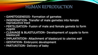
- 1. HUMAN REPRODUCTION • GAMETOGENESIS- Formation of gametes • INSEMINATION- Transfer of male gametes into female reproductive tract • FERTILISATION- Fusion of male and female gamete to form zygote • CLEAVAGE & BLASTULATION- Development of zygote to form blastocyst • IMPLANTATION- Attachment of blastocyst to uterine wall • GESTATION- Embryonic development • PARTURITION- Delivery of baby
- 2. MALE REPRODUCTIVE SYSTEM It includes: 1. Primary sex organs – a pair of testes 2. Accessory ducts 3. Glands 4. External genitalia
- 3. •Testes • Paired Structures; Located outside the abdomen in Scrotum • FUNCTION OF SCROTUM:- Maintain optimal temperature • Each testis divided into compartments called Testicular Lobules. • Each testicular lobe contain 1-3 highly coiled Seminiferous tubules.
- 4. • Seminiferous tubule:- Main site of Sperm formation • Each seminiferous tubules is lined by : 1. Male germ cells- Form sperms 2. Sertoli cells- Provide Nutrition • Outside region of seminiferous tubules contain Interstitial spaces which have 1. Blood vessels 2. Leydig cells or Interstitial cells- Secrete Androgens
- 5. ACCESSORY DUCTS • Seminiferous tubules Retetestis Vasaefferentia Epididymis Vas deferens • Vas deferens + duct of seminal vesicle Ejaculatoryduct whichopen into urethra.
- 6. MALE ACCESSORY GLANDS • Pair of Seminal Vesicles • A Prostate Gland • A Bulbourethral Gland • Secretions of seminal vesicles, prostate gland and Bulbourethral gland – Seminal Plasma • Sperms alongwith Seminal plasma is calledSemen.
- 7. MALE EXTERNAL GENITALIA •Male external genitalia Penis •Made up of Special Erectile tissue •There is common passage for both Urine and Sperms •The urethra runs through Penis. •The function of Penis is to facilitate insemination
- 8. FEMALE REPRODUCTIVE SYSTEM • All these parts are integrated structurally and functionally to support 1. the processes of ovulation 2. fertilisation 3. Pregnancy 4. Birth 5. child care. 1. Primary sex organs- a pair of OVARIES. 2. Secondary sex organs- • OVIDUCTS • UTERUS • CERVIX VAGINA. 3. External genitalia. 4. Mammary glands.
- 9. OVARIES • Location:- LOWER ABDOMEN • Thin EPITHELIUM covers it • Ovarian Stroma inside. • Ovarian stroma divided into:- 1. Outer CORTEX 2. Inner MEDULLA. • Functions:- 1. Female gamete formation 2. Releasing Female sex hormones (ESTROGEN & PROGESTERONE)
- 10. SECONDARY SEX ORGANS 1. OVIDUCTS:-Has three parts • InfundibulumwithFIMBRIAE • Ampulla • Isthmus 2. UTERUS:-InvertedPear • Perimetrium • Myomoetrium • Endometrium 3. CERVIX+ VAGINA:- Birth Canal
- 11. EXTERNAL GENITALIA • Mons pubis:- Outermost cushion of fatty tissue covered with Skin and pubic hairs. • Labia majora and Labia minora:- Folds of tissue surrounding vaginal opening. • Hymen:- Thin membrane that partially covers vaginal opening • Clitoris:- A tiny finger-like structure located between folds of labia minora above the urethral opening.
- 12. MAMMARY GLANDS • Paired structures with glandular tissue and fattytissue. • Glandular tissue 15-20 Mammarylobes. • Eachmammarylobe contains cluster of cells called Alveoli. • Alveoli is the mainsite of milksecretion and storage (lumen of alveoli) • Alveoli Mammarytubules Mammary duct Mammaryampulla Lactiferous duct.
- 13. GAMETOGENESIS • SPERMATOGENESIS • The Formation of Sperms from immature male germ cell (Spermatogonia) in testes. • It initiates on the onset of pubery. • OOGENESIS • The Formation of Ovumfrom immature female germ cell (oogonia) in ovaries. • It initiates during embryonic development
- 14. SPERMATOGENESIS • MULTIPLICATIONPHASE :- Male Germ Cells called Spermatogonia (2n) undergoes Mitotic division to increase their number and Form Primary Spermatocytes(2n). • GROWTH PHASE:-Primary spermatocytes enlarges in size and prepares to undergo maturation division.
- 15. • MATURATION PHASE:- The Primary spermatocyte (2n) undergoes Meiosis-1 to form Two Secondary spermatocytes (n) which undergoes Meiosis-2 to form Four Spermatids (n). • SPERMIOGENESIS- Differentiation Phase- The Spermatids transform into Spermatozoa (Sperms). The mature sperms heads got embedded into SERTOLI cells. • SPERMIATION:- Release of mature sperms from Seminiferous Tubules.
- 18. STRUCTURE OF SPERMATOZOA • Motile and flagellated structure • Have four parts: 1. Head – Comprises of Acrosome and Nucleus 2. Neck- contain two centrioles 3. Middle piece- contain Mitochondria 4. Tail- Longest structure.
- 19. OOGENESIS • Starts during Embryonic Development • Comprises of three phase: 1. Multiplicative phase:- Female germ cells called Oogonia undergo Mitosis. 2. Growth phase: Oogonia increase in size to form Primary oocytes. 3. Multiplicative phase:- Primary oocyte undergo Meiosis
- 20. STRUCTURE OF OVUM• It is pherical, non-motile gamete with nutrition rich cytoplasm. • Enclosed in egg envelopes 1. Vitelline membrane 2. Zona Pellucida 3. Corona radiata
- 21. MENSTRUAL CYCLE • The cyclic changes occuring in reproductive system of females of primate mammals is called Menstrual cycle. • MENARCHE: The first Menstruation beginning at puberty. • MENOPAUSE:Ceasing of Menstrual cycle in the age of 50s • It is periodically repeated on an average of 28/29 days.
- 23. FERTILISATION • Insemination • Movement of Sperm and Seondary oocyte to Ampullary-ishtmic junction. • Enzymes of acrosome dissolve egg envelopes • Completion of Meiosis-2 of secondary oocyte • Prevention of polyspermy • Fusion of nuclei of sperm and ovum.
- 24. SEX- DETERMINATION IN HUMANS • Human beings have 46 chromosomes. • Out of which one pair is sex chromosome. • Male have XY • Female have XX • Each gamete carries only one sex chromosome
- 25. CLEAVAGE AND BLASTULATION • Zygote starts dividing by rapid maitotic division called Cleavage. • Daughter cells are called Blastomeres. • Embryo with 8-16 celled stage is called Morula. • Morula divide further and transform into Blastula (Blastocyst in humans). • The process of formation of Blastula is called Blastulation. • Blastocyst has: 1. Outer layer called Trophoblast. 2. Inner cell Mass.
- 26. • Trophoblast layer attach to endometrium. Thus, blastocyst become embedded in uterine wall. The process is called Implantation and it leads to pregnancy.
- 27. POST EMBRYONIC DEVELOPMENT • Trophoblast develop finger-like projections called cchorionic villi. • Chorionic villi and uterine tissue together form Placenta. • Placenta is connected to embryo through an umbilical cord which help in transport of substances to and from the foetus.
- 28. • Placenta also acts as an endocrine tissue and produces several hormones like 1. humanchorionic gonadotropin(hCG) 2. humanplacental lactogen (hPL) 3. Estrogens 4. progestogens, etc. Relaxin released by ovary- later in pregnancy.
- 29. Te inner cell mass (embryo) differentiates into three layers:- Ectoderm, Mesoderm & Endoderm. • Foetal development • 1 month- heart is formed • 2 months- limbs and digits are formed • (First trimester) 3 months- major organ systems are formed • 5 months- appearance of hairs and the first movements of the foetus • (Second trimester) 6 months- body is covered with fine hair, eyelids separate, eyelashes are formed. • 9 months- foetus is fully developed
- 30. PARTURITION • Induced by complex Neuro- endocrine Mechanism. • First signal for paturition arises from fully developed foetus and placenta- Foetal EjectionReflex.
- 31. LACTATION • First milk produced- COLOSTRUM. • Rich in antibodies especially IgA. • Only source of immunity to newborn