
Recommended
More Related Content
What's hot
What's hot (20)
Similar to cardiomyopathy seminar.pptx
Similar to cardiomyopathy seminar.pptx (20)
Recently uploaded
Recently uploaded (20)
cardiomyopathy seminar.pptx
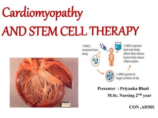
- 1. Presenter : Priyanka Bhati M.Sc. Nursing 2ND year CON ,AIIMS Cardiomyopathy AND STEM CELL THERAPY
- 2. OBJECTIVES: • At the end of the class, the students will be able to, • Define cardiomyopathy • List the different types of cardiomyopathy • Describe dilated cardiomyopathy and its treatment. • Explain hypertrophic cardiomyopathy and its management • Explain restrictive cardiomyopathy. • Formulate nursing diagnosis for patients with cardiomyopathy.
- 3. INTRODUCTION •Cardiomyopathy is an irreversible primary disease of the heart muscle. •Cardiomyopathy affects the myocardial layer of the heart, but it can also affect endocardial, subendocardial and pericardial layers.
- 4. Cardiomyopathy is defined as a heart muscle disease, characterized by ventricle dilation, myocyte and wall thickening (hypertrophy), interstitial fibrosis, decreased contractility and conduction disturbances. DEFINITION:
- 6. INCIDENCE • Affect any age group, clinical manifestations most commonly appear in the third or fourth decade. • Incidence in males is twice that of females • In blacks it is 2.4 times that of the white • Africo- American people most affected • The incidence of dilated cardiomyopathy DCM discovered at autopsy is estimated to be 4.5 cases per 100,000 populations per year, whereas the clinical incidence is 2.45 cases per 100,000 populations per year. • DCM occurs in age group of 20-60 yrs.
- 7. Types of cardiomyopathy based on etiology: Primary cardiomyopathies are those conditions in which the etiology of the heart disease is unknown. Portion of the heart is involved and other cardiac structures are unaffected. Secondary cardiomyopathies, the cause of the myocardial disease is known and is secondary to another disease process.
- 10. CAUSES • Unknown • Ischemic: = 50% atherosclerosis, Kawasaki disease, anomalous origin of left coronary artery. • Idiopathic = 45% • Hereditary: = 25 – 35% autosomal dominant, autosomal recessive, X-linked, mitochondrial. • Acute and chronic myocarditis: coxsackievirus, HIV adenovirus, • Autoimmune diseases • Chronic tachycardia
- 11. CAUSES • Drugs: alcohol, sympathomimetics, anthracyclines, amphetamines ,cocaine and cancer drugs doxorubicin and daunorubicin. • End-stage hypertrophic cardiomyopathy • Endocrine: growth hormone deficiency, hyperthyroidism, hypothyroidism, hypocalcemia, diabetes mellitus, pheochromocytoma • Inborn errors of metabolism • Muscular dystrophies • Nutritional deficiency: selenium, carnitine, thiamine • Peripartum • Structural heart disease • Systemic hypertension • Toxins: cobalt, lead
- 12. Heart becomes weak and the chambers get large. Heart cannot pump enough blood out to the body Decreased cardiac output
- 14. CLINICAL MANIFESTATION -signs • May be asymptomatic • The onset is insidious, but sometimes symptoms of heart failure occur suddenly • Dyspnea ,Nocturnal dry cough • Palpitation • Chest pain ,pulmonary edema • Dizziness and syncope • CVA , Hemiplegia, Paraplegia • Systemic embolism • Infants and younger children have respiratory symptoms and failure to thrive.
- 15. Cont.. • Pulse : low amplitude and pulse alternans are common . • BP : Low BP , Narrow pulse pressure • Cardiomegaly( board and displaced point of max .impulse , right ventricular heave) • JVP : Elevated Precordium: – Apex shifted, forceful and ill sustained – S2 may be palpable due to PH – May be parasternal lift – On auscultation:S1 and S2 soft, S2 loud from PH , Pansystolic murmur at mitral and tricuspid area,S3 and S4 are common
- 16. Diagnostic evaluation Assessment Finding Auscultation Chest X-ray ECG{RULE OUT} Echo { MAIN} Point of maximal intensity away from the normal position. Heart sound reveal S3 Cardiomegaly with signs of pulmonary venous hypertension. Non-specific ST segment and T wave abnormalities / arrhythmias Dilated chambers with normal/decreased wall thickness, decreased ejection fraction (<30%)
- 17. Diagnostic evaluation Assessment Finding Left ventriculogram Cardiac catheterization Laboratory investigations Endomyocardial biopsy{ NOT DEFINITIVE} Thin walled ventricles that are dilated. Coronary vasodilatation is impaired by increased LV filling. Cardiac troponin I is done to detect recent MI LFT, BUN and creatinine. To identify the aetiology.
- 18. INVESTIGATIONS(HEART FAILURE EVALUATION) • Laboratory Studies – Blood count, urinalysis, electrolytes, renal function, glucose, LFTs (class I; level C) • Thyroid stimulating hormone (class I; level C) • Fe/TIBC, ferritin (class IIa, level C) • Urinary screening for hemochromatosis (class IIa; level C) • HIV testing (class IIb; level C) • Electrocardiogram (class I; level C • Chest x-ray (class I; level C) • Echocardiogram/Doppler or radioventriculogram (class I;level C)
- 19. ECG changes: • Sinus tachycardia, Bradycardia • Low voltage ECG • LVH or Rt or Lt Atrial hypertrophy • Atrial or Ventricular arrythmia • Pseudo infraction pattern(Deep but narrow ‘q’ suggestive of antero-septal MI) • Shallow invasion of ‘T’ wave • LBBB, RBBB ,LAHB, LPHB, Bifascicular or Trifascicular block • AV block( 1’or 2’ or 3’degree)
- 21. X-ray • Cardiomegaly ,simulating pericardial effusion or multi valvular disease or Ischemic cardiomyopathy. • Pulmonary edema with Kerley’ B line. • Pleural effusion may be present . • Pulmonary infraction is not uncommon . • Usually no LA enlargement so single right border. No calcified valve and normal aorta.
- 22. Pulmonary edema with Kerley’ B line
- 23. MRI • black blood images: enlarged cardiac chambers and thin myocardial walls. • Cine images: show LV hypokinesia, increased volumes, (end- diastolic volumes that constitute a dilated CMP: > 140 mL for the LV and > 150 mL for the RV. • Phase-contrast sequences: impaired diastolic function. transvalvular flow may be characterized by a restrictive pattern . • Late gadolinium-enhancement.
- 26. MANAGEMENT Goal •Enhance myocardial contractility •Decrease afterload.
- 27. sw
- 28. Supportive medical treatment: • Treatment of HF: – Rest – Salt and water restriction – Diuretics (eg. Frusemide) • Digoxin • Vasodilators (eg.ACE inhibitor , Nitrates, Prazocin ) - Potassium supplements -Spironolactone • Vitamin: B1 -Levocarnitine
- 29. Supportive medical treatment: • Anti arrythmic drugs: – 90% patient has complex ventricular arrythmia – • IF AF: Digoxin • If VT or VF :Amiodorone. • Long term anticoagulant therapy: – To prevent systemic and pulmonary embolism . • Consider adding beta blocker if symptoms persist Intravenous Dobutamine / Dopamine.
- 30. • Implantable cardiac defibrillators. • Biventricular pacemakers.(CRT) OTHER MANAGEMEMT
- 31. HEART TRANSPLANT •Heterotopic Procedure Orthotopic procedure
- 32. EMERGING THERAPIES • Agents to eradicate persistent viral infections and immunomodulatory agents. • Stem cells for cardiac regeneration and gene therapy approaches are in clinical trials.
- 33. Prevention • Modification of contributory factor: • Avoid alcohol • Avoid pregnancy – Genetic counselling – Limit exercise – Weight reduction – Control of blood pressure – Prompt control of Infection – Correction of anemia and malnutrition
- 34. Complication of DCM • Heart failure • Arrythmias : AF ,SVT,PVCs ,VT VF • AV blocks:1’ 2’ and CHB • BBB : RBBB ,LBBB, LAHB ,LPHB • Systolic or pulmonary embolism • Syncope • Sudden cardiac death
- 35. PROGNOSTIC FEATURES • VENTRICULOGRAPHIC FINDINGS • Degree of impairment in LVEF – Extent of left ventricular enlargement – Coexistent right ventricular dysfunction – Ventricular mass/volume ratio – Global wall motion abnormalities – Left ventricular sphericity . • CLINICAL FINDINGS • Favorable prognosis: NYHA < IV, younger age, female sex • Poor prognosis: Syncope, persistent S3 gallop, rightsided heart failure, AV or bundle branch block, hyponatremia, troponin elevation, increased BNP, maximum oxygen uptake < 12 ml/kg/min.
- 37. DEFINITION • HCM is a disorder of cardiac muscle characterized by myocardial hypertrophy. • It most commonly affects the intraventricular septum and disorganization of the cardiac myocytes and myofibrils. • It is the most common cause of sudden cardiac death in young competitive athletes.
- 38. Common causes •Genetic(autosomal dominant) or familial. •Hypertension. •Ischemia( coronary artery disease). •Aortic stenosis. •Idiopathic.
- 39. PATHOPHYSIOLOGY • Involves 4 interrelated process • LVOT • Diastolic dysfunction • Myocardial ischemia • Mitral regurgitation Pathophysiology Left ventricular hypertrophy (thick ventricular wall) ventricular chamber size hold less blood CO pressure in the ventricles and lungs changes in the cardiac muscles interfere with the heart's electrical signals, leading to arrhythmias sudden cardiac arrest
- 40. PATHOPHYSIOLOGY Hypertrophy and asymmetric septal wall Changes in left ventricular cavity shape Misaligned papillary muscles. systolic anterior motion of the mitral valve during systole
- 41. Inward movement of the mitral valve leaflet due to increased pressure in the LVOT( Venturi phenomenon) Mitral regurgitation, Cresendo- decresendo murmur , Blockage of blood flow, during increased HR , The blood flow to heart is compromised This condition is also known as idiopathic hypertrophic subaortic stenosis(IHSS
- 42. LV Outflow Obstruction in HCM • Long-standing LV outflow obstruction is a major determinant for heart failure symptoms and death in HCM patients Subaortic outflow obstruction is caused by systolic anterior motion (SAM) of the mitral valve – leaflets move toward the septum.
- 43. LV Outflow Obstruction in HCM • Explanations for the SAM of the mitral valve • 1. Mitral valve is pulled against the septum by contraction of the papillary muscles, which occurs because of the valve's abnormal location and septal hypertrophy altering the orientation of the papillary muscles. • 2. Mitral valve is pushed against the septum because of its abnormal position in the outflow tract. • 3. Mitral valve is drawn toward the septum because of the lower pressure that occurs as blood is ejected at high velocity through a narrowed outflow tract (Venturi effect).
- 45. Physiological Consequences of Obstruction • Elevated intraventricular pressures. • Prolongation of ventricular relaxation . • Increased myocardial wall stress . • Increased oxygen demand. • Decrease in forward cardiac output .
- 47. CLINICAL MANIFESTATIONS • SUDDEN DEATH • Rapid, pounding heartbeat. • Chest tightness or pressure. • Fatigue. • Fluid retention resulting in swollen feet or ankles or unexplained weight gain. • In children and adolescents, pre-syncope and syncope identify patients at increased risk of sudden death. Often diagnosis is made during screening. Sometimes, the first sign is sudden death.
- 48. Diagnostic studies •History and physical examination •Palpation and auscultation of the chest.( Split S2, harsh systolic ejection murmur worsening with Valsalva ( crescendo decrescendo murmur) •S4 heard •ECG findings- (ST segment changes, not specific, SVT ,VT) may be normal . •Echocardiogram( MAINSTAY OF DIAGNOSIS) •Chest X ray. •Biopsy –myofibril disarray
- 49. Physical Examination • Holosystolic murmur at the apex and axilla of mitral regurgitation is heard in patients with systolic anterior motion of the mitral valve and significant LV outflow gradients • Diastolic decrescendo murmur of aortic regurgitation is heard in 10% of patients, although mild aortic regurgitation can be detected by Doppler echocardiography in 33% of patients
- 50. Electrocardiogram in HCM • LVH with nonspecific ST/T wave abnormalities . • Left or right axis deviation, Conduction abnormalities . • Abnormal and prominent Q wave in the anterior precordial and lateral limb leads A fib with preexcitation implies poor prognosis. • Findings on Holter monitoring include APC’s VPC’s, sinus pauses, wandering atrial pacemaker, atrial tachycardia, AF/flutter and nonsustained ventricular tachycardia.
- 52. ECHO 2-D echocardiography is diagnostic for HCM. Abnormal systolic anterior leaflet motion of the mitral valve . LV hypertrophy. Left atrial enlargement . Diastolic dysfunction. Small ventricular chamber size . Septal hypertrophy with septal to free wall ratio greater than 1.4:1 (absolute septal wall thickness >15mm) . SAM of anterior and rarely posterior mitral valve leaflet and mitral regurgitation . Decreased mid aortic flow . Partial systolic closure of the aortic valve in mid systole.
- 53. Cardiac MRI in HCM • Useful when echocardiography is questionable, particularly with apical hypertrophy . • Cines loops typically show obstruction and velocity mapping is useful in the assessment of peak velocities . • SAM of the mitral valve is clearly seen on cardiac MRI. • Improvement in obstruction after septal ablation or myomectomy can be demonstrated, as can the location and size of the associated infarction, which are useful for planning repeat procedures. • Cardiac MRI tagging identifies abnormal patterns of strain, shear, and torsion in cases of HCM, demonstrating significant dysfunction in hypertrophic areas of the ventricle.
- 54. Cardiac MRI in HCM • Gadolinium contrast cardiac MRI - differentiating HCM from other causes of cardiac hypertrophy and other types of cardiomyopathy such as, amyloidosis, athletic heart, and Fabry’s disease • Late gadolinium enhancement occurring in HCM represents myocardial fibrosis • The greater the degree of late gadolinium enhancement, the more likely that the particular HCM patient has 2 or more risk factors for sudden death. • More likely the patient has or will develop progression of ventricular dilation toward heart failure, thereby indicating a poorer prognosis
- 55. Cardiac Catheterization • Diagnostic cardiac catheterization is useful to determine the degree of LVOT obstruction, cardiac hemodynamics, the diastolic characteristics of the left ventricle, LV anatomy and coronary anatomy • Reserved for situations when invasive modalities of therapy, such as a pacemaker or surgery, are being considered • Therapeutic cardiac catheterization interventions, include transcatheter septal alcohol ablation • The arterial pressure tracing found on cardiac catheterization may demonstrate a "spike and dome" configuration
- 56. Cardiac Catheterization • Approximately one fourth of patients demonstrate pulmonary hypertension - usually mild • Results in characteristic change recorded on arterial pressure tracing - exhibits a pulse pressure that fails to increase as expected or actually decreases (the so-called Brocken broughBraunwald phenomenon) • One of the more reliable signs of dynamic obstruction of the LVOT, intensity of murmur also increased
- 57. GOAL • The main goals of management are alleviate symptoms, prevent complications and decrease risk of sudden death • Improve ventricular filling. • Reduce ventricular contractility • Relieve left ventricular outflow obstruction. TO DECREASE CONTRACTILITY TO IMPROVE FILLING
- 58. MANAGEMENT OF HCM • Medical therapy • Device therapy • Surgical septal myomectomy • Alcohol septal ablation
- 59. MANAGEMENT OF HCM Beta-blockers( Mainstay) Increase ventricular diastolic filling/relaxation (decreases HR) Decrease myocardial oxygen consumption Have not been shown to reduce the incidence of SCD Verapamil - Augments ventricular diastolic filling/relaxation Amiodarone Disopyramide - Used in combination with beta-blocker Negative inotrope – Diuretics DIGOXIN IS CONTRAINDICATED IN HYPERTROPHIC CARDIOMYOPATHY.
- 60. Dual-Chamber Pacing • Proposed benefit: • Pacing the RV apex will decrease the outflow tract gradient by decreasing projection of basal septum into LVOT • Several RCTs have found that the improvement in subjective measures provided by dual-chamber pacing is likely a placebo effect • Objective measures such as exercise capacity and oxygen consumption are not improved
- 61. SURGICAL MANAGEMENT OF HCM: Mitral valve replacement: Septal myectomy:
- 62. SYMPTOMATIC MANAGEMENT Percutaneous Transluminal Septal Myocardial Ablation(PTSMA)
- 63. Alcohol Septal Ablation • Successful short-term outcomes • LVOT gradient reduced from a mean of 60-70 mmHg to <20 mmHg • Symptomatic improvements, increased exercise tolerance • Long term data not available yet • Complications • Large MI • Complete heart block • No randomized efficacy trials yet for alcohol septal ablation vs. surgical myectomy
- 64. Sudden Cardiac Death in HCM Most frequent in young adults<30-35 yrs old Primary VF/VT Tend to die during or just following vigorous physical activity Often is 1st clinical manifestation of disease HCM is most common cause of SCD among atheletes.
- 66. RESTRICTIVE CARDIOMYOPATHY • It is a disease of the heart muscle that impairs diastolic filling and stretch. • Systolic function remains unaffected. • Least common of the cardiomyopathic conditions. • The heart chambers are unable to fill with blood because the heart muscle is stiff.
- 67. RESTRICTIVE CARDIOMYOPATHY • The average age at diagnosis is 5 to 6 years. • RCM appears to affect girls somewhat more often than boys. • There is a family history of cardiomyopathy in approximately 30% of cases. • In most cases the cause of the disease is unknown (idiopathic), although a genetic cause is suspected in most cases of pediatric RCM
- 68. CAUSES •Endomyocardial fibrosis •Sarcoidosis •amyloidosis •Lofflers syndrome •Neoplastic tumor •Ventricular thrombus •Fibrosis of different etiology •Radiation to the thorax
- 69. PATHOPHYSIOLOGY Increase in stiffness of the ventricular walls Impaired diastolic filling of the ventricle(pulmonary congestion, decreased CO) Reducing preload and end-diastolic volume Heart failure As the disease progresses Systolic dysfunction
- 70. Disease course • The most common symptoms at first may appear to be lung related. Children with RCM frequently have a history of “repeated lung infections” or “asthma.” • In these cases, referral to a cardiologist eventually occurs when a large heart is seen on chest x-ray. The second most common reason for referral is an abnormal physical finding during a doctor’s examination. • Children who have ascites hepatomegaly and edema are often sent to see a gastroenterologist first.
- 71. Disease course • Referral to a cardiologist is made when additional cardiac signs or symptoms occur, a chest x-ray is found to be abnormal or no specific gastrointestinal cause is found for the edema or enlarged liver. • In approximately 10% of cases, fainting is the first symptom causing concern. • Unfortunately, sudden death has been the initial presentation in some patients.
- 73. ASSESSMENT FINDINGS •Most common symptoms are dyspnea and edema. - Ascites may be evident. •Auscultation: - AV valve regurgitation. •Echo: - Biatrial enlargement due to rigid ventricular walls is seen.(balloning of the RA and LA) - PA pressure is greater than 50mm of Hg in patients with RCM. - Ejection fraction is 25-50%
- 74. COMPLICATIONS OF CARDIOMYOPATHY • Congestive cardiac failure. • Arrhythmias. • Thrombus and emboli. • Stroke. • Valvular disorders. • Cardiac arrest. • Infective endocarditis for patients HOCM.
- 75. STEM CELL THERAPY • Stem cell therapy has emerged as a promising strategy for the treatment of dead myocardium, directly or indirectly, and seems to offer functional benefits to patients. • The ideal candidate donor cell for myocardial reconstitution is a stem-like cell that can be easily obtained, has a robust proliferation capacity and a low risk of tumor formation and immune rejection, differentiates into functionally normal cardiomyocytes, and is suitable for minimally invasive clinical transplantation.
- 77. Types of Stem Cells for Cardiac Cell Therapy • Skeletal Myoblasts Autologous transplantation of SKMs • Bone Marrow-Derived Stem Cells • Bone Marrow-Derived Mesenchymal Stem Cells • Adipose-Derived Mesenchymal Stem Cells • Cardiac Stem Cells • Cardiosphere-Derived Cells • Embryonic Stem Cells and Induced Pluripotent Stem Cells
- 78. • The variable and moderate benefits associated with stem cell treatments were initially ascribed to inefficient cell delivery, with only 10% (or less) of the cells retained in the heart after 24 hours regardless of the cell type or delivery route. • The cells are usually washed out through the coronary venous system or mechanically ejected from the injection site . • Repeated administrations of BMCs, MSCs, and CDCs were demonstrated to boost therapeutic benefits.
- 79. Route • The following are three routes for delivering stem cells for cardiac therapy: • systemic intravenous infusion, Uptake only 0.04% • intracoronary infusion, balloon catheters to infuse stem cells , uptake 1.3% to 2.6% • and intramyocardial injection(open-chest surgery) Uptake of 6.2%
- 80. STEM CELL THERAPY • Effects of stem cell therapy on dilated cardiomyopathy • Rong Jiao, MD, Yuan Liu, MD, Wen-Jian Yang, MD, Xiao-Yan Zhu, MD, Jin Li, MD, and Qi-Zhu Tang, 2014 Abstract Objectives: • To perform a meta-analysis of clinical trials and investigate the effect of stem cell therapy on dilated cardiomyopathy. Methods: • A systematic literature search was carried out between May 2012 and July 2013 in PubMed, Medline, Cochrane Library, and Excerpta Medica Database (EMBASE). The study took place in the Department of Cardiology, Renmin Hospital of Wuhan University, Wuhan, China. The weighted mean difference (WMD) was calculated for left ventricular ejection fraction (LVEF), left ventricular end-diastolic diameter (LVEDD), mortality and heart transplantation, and the 6-minute walk test (6-MWT) distance using the RevMan 5.0 software.
- 81. CONT… Results: • Seven trials with 599 participants evaluated the association between the stem cell therapy and control groups. Compared with the control group, stem cell therapy group improved the LVEF (WMD: 3.98%, 95% confidence interval [CI]: 0.55 - 7.41%, p=0.02) and the 6-MWT distance (WMD: 132.12 m, 95% CI: 88.15-176.09 m, p<0.00001), and reduced mortality and heart transplantation (odds ratio [OR]: 0.48, 95% CI: 0.29-0.80, p=0.005). However, the LVEDD showed no significant difference between the 2 groups (WMD: -1.53 mm, 95% CI: -1.15-0.10 mm, p=0.10). Conclusion: • This meta-analysis demonstrated that stem cell therapy improves cardiac function and reduces mortality in dilated cardiomyopathy patients, which suggested that stem cell therapy may represent a new therapy option for dilated cardiomyopathy.
- 84. NURSING ASSESSMENT • Take proper History • Assess sign and symptoms • Assess the psychosocial factors and stressors among pt Physical assessment oVitals oPulse pressure oWeight –gain/loss oPoint of maximum impulse oCheck for s3 and s4 oPulmonary assessment for crackles oJugular venous distention oEdema and severity
- 85. •Assess BP, heart rate & respiratory rate. •Assess for change in mental status. •Monitor intake and output regularly. •Assess peripheral pulse for strength and quality. •Encourage physical and psychological rest. •Administer prescribed medications and evaluate responses Decreased cardiac output related to structural disorders caused by cardiomyopathy
- 86. •Monitor intake and output every 4 hourly. •Weigh patients daily. •Assess for the presence of peripheral edema •Assess for jugular vein distension, hepatomegaly and abdominal pain •Follow low sodium diet and fluid restriction •Auscultate breath sounds 2 hourly for the presence of crackles and monitor for sputum production •Administer diuretics as ordered and evaluate effectiveness Excess fluid volume related to decreased contractility of heart
- 87. •Assess respiratory rate, rhythm and saturation •Auscultate breath sounds every 2 hourly •Encourage client to turn, cough and deep breath. Use incentive spirometer 2 hourly. •Provide fowler’s position to facilitate breathing and observe for PND. •Administer oxygen as ordered. •Obtain ABG if ordered •Administer diuretics as ordered Impaired gas exchange related to pulmonary congestion caused by myocardial failure
- 88. . •Assess for the manifestations of angina •Assess respirations, BP, & HR with each episode of chest pain •Obtain a 12 lead ECG each time chest pain recurs •Administer nitrates, monitor response, notify if pain does not abate in 15 – 20 min •Provide care in a calm environment that minimizes anxiety •Instruct patient to avoid physical exertion if pain occurs •Stay with patient until discomfort is relieved. Angina related to decreased coronary perfusion
- 89. Summary •Definition •Types •Causes •Clinical classification •Dilated cardiomyopathy •Hypertrophic cardiomyopathy •Restrictive cardiomyopathy •Nursing management.
- 90. Conclusion Although cardiomyopathy affects the myocardium of the heart, the management of each cardiomyopathy is different. Hence, early diagnosis should be done to prevent complications.
- 91. References •Lewis, “medical surgical nursing, assessment and management of clinical problems, 6th edition, mosby publications, page no:852- 857. •Susan woods, “cardiac nursing” 5th edition, lippincott publications, page no:782-786. •https://www.hindawi.com/journals/acp/2017/94 04057/ •https://www.ncbi.nlm.nih.gov/pmc/articles/PMC 4362159/#targetText=Conclusion%3A,therapy%2 0option%20for%20dilated%20cardiomyopathy.
- 92. THANK YOU
Editor's Notes
- Idiopathic ischemic Autoimmune mediated inflammation Metabolic disorders Muscular dystrophy Myocarditis Pregnancy(PAH) Valvular disorder Cardiotoxic agents Genetic/familial Hypertension Viral infections MOSTLY IDIOPATHIC MYOCARDITIS ( COCKAXIE VIRUS), CHAGAS DISEASE( protozoal disease) dystrophy.HAEMOCHROMATOSIS AS THE HEART DILATES THE VALVES MOVE APART AND REGURGITATION OCCOURS MR HEARD AS A HOLOSYSTOLIC REGURGITATION DIABETIES ,GAUCHERRS DISEASE ,ALCOHOL INTAKE METABOLIC DISEASE Cobalt is another cause Glycogen storage diseThe liver cannot control the use of glycogen and glucose because certain enzymes are missing that control the change of sugar (glucose) into its storage form (glycogen) or release of glucose ...ase (GSD, also glycogenosis and dextrinosis) is the result of defects in the processing of glycogen synthesis or breakdown within muscles, liver, and other cell types
- THIAMINE, (WET BERI BERI,) ALCOHOL STRONGLY ASSOCIATED WITH DILATED CARDIOMYOPATHY ITS METABOLITES HAS A DIRECT AFFECT ON THE HEART MUSCLE GENETIC CAUSE DUCCHE MUSCULAR DYSTROPHY DMD-associated dilated cardiomyopathy is a form of heart disease that is caused by mutations in the DMD gene. In addition to heart disease, these conditions are characterized by progressive weakness and wasting of muscles used for movement (skeletal muscles). People with DMD-associated dilated cardiomyopathy typically do not have any skeletal muscle weakness or wasting, although they may have subtle changes in their skeletal muscle cells that are detectable through laboratory testing. Based on these skeletal muscle changes, DMD-associated dilated cardiomyopathy is sometimes classified as subclinical Becker muscular
- Irregular heart rate with an abnormal S3 and/or S4 Tachycardia or bradycardia Pulmonary crackles Edema Weak peripheral pulses
- Holosystolic murmur due to regurgitation
- Cardiac Ventriculography is a medical imaging test DONE DURING CARDIAC CATHETERIZATION used to determine a patient's cardiac function in the right, or more typically, left ventricle. Cardiac ventriculography involves injecting contrast media into the heart's ventricle(s) to measure the volume of blood pumped. Cardiac ventriculography can be performed with a radionuclide in radionuclide ventriculography or with an iodine-based contrast incardiac chamber catheterization. The 3 major measurements obtained by cardiac ventriculography are: Ejection Fraction Stroke Volume Cardiac Output These three measurements share a commonality of ratios between End Systolic Volume and End Diastolic Volume and all lend mathematical structure to the common medical term Systole.
- Kerley lines are a sign seen on chest radiographs with interstitial pulmonary edema. They are thin linear pulmonary opacities caused by fluid or cellular infiltration into the interstitium of the lungs. They are named after Irish neurologist and radiologist Peter Kerley
- Phase contrast imaging is an MRI technique that can be used to visualise moving fluid. It is typically used for MR venography as a non-IV-contrast requiring technique. Spins that are moving in the same direction as a magnetic field gradient develop a phase shift that is proportional to the velocity of the spins. This is the basis of phase-contrast angiography. In the simplest phase-contrast pulse sequence, bipolar gradients (two gradients with equal magnitude but opposite direction) are used to encode the velocity of the spins. Stationary spins undergo no net change in phase after the two gradients are applied. Moving spins will experience a different magnitude of the second gradient compared to the first, because of its different spatial position. This results in a net phase shift. This information can be used directly to determine the velocity of the spins. Alternatively, the image can be subtracted from one acquired without the velocity encoding gradients to obtain an angiogram.
- Gradient echo (GE), a.k.a. gradient recalled echo (GRE), has fast imaging speeds, but is more susceptible to metal induced artifacts. Gradient echo is the workhorse of cardiac imaging because of its speed and versatility. Gradient echo imaging is employed in the assessment of ventricular function, blood velocity and flow measurements, assessment of valvular disease, myocardial perfusion, delayed enhanced imaging, and magnetic resonance angiography.
- Indication for biopsy Acute dilated cardiomyopathy with refractory heart failure symptoms Rapidly progressive ventricular dysfunction in an unexplained cardiomyopathy of recent onset New onset cardiomyopathy with recurrent ventricular tachycardia or high grade heart block Heart failure in the setting of fever, rash, and peripheral eosinophilia Dilated cardiomyopathy in setting of systemic diseases known to affect the myocardium (systemic lupus erythematosus, polymyositis, sarcoidosis)
- VT is a pulse rate of more than 100 beats per minute, with at least 3 irregular heartbeats in a row.
- Orthotopic procedure[edit] The orthotopic procedure begins with a median sternotomy, opening the chest and exposing the mediastinum. The pericardium is opened, the great vessels are dissected and the patient is attached to cardiopulmonary bypass. The donor's heart is injected with potassium chloride (KCl). Potassium chloride stops the heart beating before the heart is removed from the donor's body and packed in ice. Ice can usually keep the heart usable for four[9] to six hours depending on preservation and starting condition. The failing heart is removed by transecting the great vessels and a portion of the left atrium. The patient's pulmonary veins are not transected; rather a circular portion of the left atrium containing the pulmonary veins is left in place. The donor heart is trimmed to fit onto the patient's remaining left atrium and the great vessels are sutured in place. The new heart is restarted, the patient is weaned from cardiopulmonary bypass and the chest cavity is closed. The orthotopic procedure was developed by Shumway and Lower at Stanford-Lane Hospital in San Francisco in 1958.[5] Heterotopic procedure[edit] In the heterotopic procedure, the patient's own heart is not removed. The new heart is positioned so that the chambers and blood vessels of both hearts can be connected to form what is effectively a 'double heart'. The procedure can give the patient's original heart a chance to recover, and if the donor's heart fails (e.g., through rejection), it can later be removed, leaving the patient's original heart. Heterotopic procedures are used only in cases where the donor heart is not strong enough to function by itself (because either the patient's body is considerably larger than the donor's, the donor's heart is itself weak, or the patient suffers from pulmonary hypertension). This procedure is used when the recipient's body is too large for the donor's heart, or when the patient has pulmonary high blood pressure (that is, high blood pressure in the vessels that transport blood from the heart to the lungs). The advantage of this is that the procedure can give the patient's original heart time to recover, and even if the donor's heart fails, the original heart can continue beating. However, complications like organ rejection, infection and side effects of immunosuppressive medications can still occur.
- Oxygen uptake is VO2. It's the oxygen consumption or uptake per kilogram of body weight. It's the most relevant measure of the cardiorespiratory system. It's the highest rate at which oxygen can be consumed is called VO2Max, distributed and used by the body during exercise.
- Familial hypertrophic cardiomyopathy Theβ -myosin heavy chain (MyHC), cardiac troponin T (cTnT) and myosin binding protein-C (MyBP-C) are the most common genes accounting for approximately 2/3 of all HCM cases Mostly autosomal dominant disease caused by missence mutation Different aa formed mostly of the sarcomere The B myosin trop T, myocyte binding protein C etc involved OF THE SARCOMERE
- Approximately 25% of patients with HCM have left ventricular outflow tract obstruction Crescendo murmurs progressively increase in intensity. Decrescendo murmurs progressively decrease in intensity. With crescendo—decrescendo murmurs (diamond or kite-shaped murmurs), a progressive increase in intensity is followed by a progressive decrease in intensity.
- USUALLY LV AFFECTED VENTURI EFFECT THE INTERVENTRICULAR HYPERTROPHIC MUSCLE OBSTRUCTING THE LVOutflowTO CRESENDO DECRESENDO MURMUR S4 heard Muscle need oxygen thus sudden death due to arrrythmia occours Mostly autosomal dominant disease caused by missence mutation Different aa formed mostly of the sarcomere The B myosin trop T, myocyte binding protein C etc involved OF THE SARCOMERE MYOCYTE ARRAY FREIDREICHS ATAXIA DIGOXIN CONTRAINDICATED
- VALSAVA IS A SCRRENING TEST DURING VALSALVA THERE IS DECREASE IN THE BLOOD FLOW INTO THE HEART AND THUS AND THUS THE SIZE OF THE VENTICLE DECREASES THUS THERE IS MORE TURBULENT BLOOD FLOW THROUGH THE AORTIC VALVE AND THUS CAUSES THE SYSTOLIC EJECTION MURMUR
- The interventricular septum increased from a median of 8.3 mm in the age group 20-29 to 11.2 mm in the group 60-70, whereas the posterior left ventricular wallincreased from 7.5 mm to 9.8 mm. Morphologic diagnosis is based on the presence of myocardial thickness of >15mm in adults or a septum to posterior wall ratio of >1.5. Septal wall thickness (WT) >30 mm is one of several high risk features that are used to guide the decision for prophylactic implantable cardioverter-defibrillator (ICD) placement.
- Fabry disease is a rare genetic disease a deficiency of the enzyme alpha-galactosidase A (a-Gal A) that causes a buildup of a type of fat called globotriaosylceramide (Gb3, or GL-3) in the body.
- Pharmacological management Beta-blockers.( Mainstay) Calcium channel blockers.(disopyramide) Anti-coagulants for patients with chronic atrial fibrillation. Dual chamber pacing for patients with outflow tract gradient. Diuretics, digitalis, vasodilators, dopamine are contra-indicated for HOCM.
- No correlation has been found between pacing and reduction of LVOT gradient
- SEPTAL MYECTOMY MEDIAN STERNOTOMY INCISION NEEDED , LOWER AORTA CUT AORTIC VALVE SEEN , AND THROUGH IT IS SEEN THE INTERVENTRICULAR SEPTUM AND UNDER DIRECT VISUALIZATION IT IS CUT
- Alcohol septal ablation is performed in the cardiac catheterization laboratory, and should only be performed by interventional cardiologists with specific training in the procedure. (Current guidelines suggest at least 20 successful procedures to demonstrate competence.) As such, it is only available in a few institutions. The technique is similar to coronary angioplasty, and utilizes similar equipment. Using wires and balloons to localize the septal artery feeding the diseased muscle under both fluoroscopic (X-ray) and echocardiographic (ultrasound) guidance, a small amount of pure alcohol is infused into the artery to produce a small heart attack. Patients typically experience mild chest discomfort during the procedure, which takes approximately 60–90 minutes to complete. Analgesics and mild sedatives are administered as needed. Patients typically are maintained in the hospital for three to four days to monitor for any complications, including the need for a permanent pacemaker in 5–10%.
- LVOT obstruction in HCM was defined as a resting LVOT gradient of ≥30 mm Hg, with severe obstruction defined as ≥50 mm Hg
- THE SIZE IS NORMAL BUT NO STREATCH Small QRS LOW AMPLITUDE QRS
- AMYLOIDOSIS PROTEINS WHICH ARE MISFOLDED ONCE MISFOLDED DEPOSIT IN THE BODY FAMILIAL AMYLOID CARDIOMYOPATHY – TRANSTHYRETIN (MUTANTED) NORMALLY TRANSPORTS THYROXINE AND RETINOL SENILE CARDIAC AMYLOIDOSIS-(ELDERLY Pathologic specimens in Loeffler endocarditis show eosinophilic myocarditis, a tendency toward endomyocardial fibrosis and clinical manifestations of thromboembolism, and acute heart failure. Eosinophilic states that may occur in association with Loeffler endocarditis include hypereosinophilic syndrome, eosinophilic leukemia, carcinoma, lymphoma, drug reactions or parasites, as reported in multiple case series. SARCOIDOSIS –COLLECTION OF IMMUNE CELLS ENDOCARDIAL FIBROELASTOSIS- Fibrosis of endocardium and subendocardium mostly in children due to infection Haemochromatosis Radiation causes reactive oxygen species which cause inflamation then fibrosis
- When the first sign of the disease is an abnormal heart sound, or signs of heart failure are recognized, then earlier referral to a cardiologist occurs.
- Review Article Cell Therapies in Cardiomyopathy: Current Status of Clinical Trials Ming Hao,1,2 Richard Wang,1,2 and Wen Wang1,2 2017