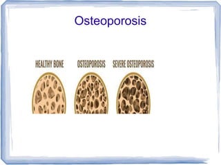
2. Definition
Porous bone – holes become bigger, bone
becomes fragile and easier to break
Progressive systemic skeletal disease,
characterized by low bone mass and micro-
architectural deterioration of bone tissue with
consequent increase in bone fragility and
susceptibility to fracture
Different from osteomalacia – deficient
mineralization, normal matrix
Porosis – deficient matrix and normal
mineralization
3. Porosis – decrease in bony density with
increase in porosity.
Insufficiency fracture – bone fails with normal
weight bearing, (due to physiological stresses)
Fragility fracture – fall from standing height or
less
Primary osteoporosis
Postmenopausal
50-70, Colle's # and vertebral compression
more common, high turnover
Senile – >70, hip fractures more common,
low turnover
5. Pathology
Normally, young patients have more blastic
activity
Plateaus for the majority of life (blastic =
clastic)
Older – more osteoclastic activity
Overactive remodelling – deeper resorption
cavities
Continuous remodelling of microfractures
Patients with history of one fragility fracture
are upto 10 times more likely to have future
fractures
6. WHO
Bone mineral density = T score < -2.5 SD
Guidelines to screen
Women > 65, males >70
Young postmenopausal (post hysterectomy)
or with risk factors like long term
corticosteroid
Post menopausal with history of fracture
Diagnosis
FRAX (fracture risk assessment)
Risk factors + BMD
Age, sex, height, weight, family history,
previous fracture, steroid, tobacco, alcohol,
7. 10 year fracture risk – hip, and other.
Screening
Ca, Vit D3, PTH, S. Creat
Can also screen for ddx
Infection – ESR, CRP
Secondaries
Multiple myeloma - SPEP
8. X-ray
Empty bone appearance – resorption of
trabeculae
Ground glass appearance
Fish mouth appearance
Spine, end plate weakening and intra discal
expansion
12. DEXA – dual energy Xray absorptiometry
2 xray beams – difference is calculated to
eliminate the amount of radiation absorbed
by the soft tissue
BMD – bone mineral content (g) / area
(cm^2)
Bone density indicates bone strength (60-
80%)
Early predictor of fracture risk
Permits diagnosis even before first fracture
Mean and standard deviation
T score – is considered for a young healthy
person of same gender
Z score – gender + age group
13. Commonly measured in spine and hips (L2,
L3, L4 vertebrae are good as they don't have
other bones nearby)
T Scores:
Normal = 0 to -1
Osteopenia = -1 to -2.5
Osteoporosis = -2.5 or less
14.
15. Treatment
Risk factors – tobacco, alcohol
Nutrition + supplement
Ca – 1200 mg
Vit D – 1500 IU
Mg, Si, Vit K, boron
Exercise
Strength
BMD increase
Prevent fall
Post fracture rehab
16. Medical Management
T < -2.5 without risk factors, or < -1 with
history of fracture or other risk factors
Fracture risk of 3% hip and 20% other.
Goal
Lower fracture risk
Safe, affordable, well tolerable
17. Anti resorptive
HRT – estrogen/progestin
SERM – tamoxifen (10 mg), raloxifen (60
mg/day)
Bisphosphate
Calcitonin (nasal spray – 200 IU)
Denosumab – RANKL monoclonal antibody
60 mg/6 month, upto 10 years. Nephro safe
Bone forming
Teriparitide
New trabeculae formation
Osteoblast lifespan
More bone formation occurs
Daily 20 ug sub-cut “pulse” doses are anabolic
18. Bisphosphonates
Inhibit osteoclastic activity, thereby allowing for
more mineralization
Non nitrogenous (tilduronate, clodronate,
etidronate) – interferes with ATP in osteoclast,
leading to apoptosis
Nitrogenous (pami, neri, olpad, alendronate,
ibandronate, risedronate, zolendronate)
Disrupts enzyme Farnesyl diphosphate
synthase, which disrupts the protein
synthesis in cell membrane and cytoskeleton
Complication – gastritis, AVN mandible
Patient should be able to sit for at least 30 min
19. Dose
Alendronate – 70 mg per week, upto 4 years
Risedronate – 35 mg per week
Zolendronic acid – 5 mg per year
Outcome
BMD improvement
Bone turnover markers
Formation
bone specific Alk Phos
Osteocalcin
Propeptide of Type 1 Collagen
Resorption
N telopeptide of Type 1 collagen
C telopeptide
Urine and free deoxypyridinoline
Tartrate resistant acid phos (Trap5b)
20. Atypical fracture
3-5 years of treatment
Remodelling less, therefore microfractures and
stress fractures healing impaired, poor quality
Atypical femur fracture (subtrochanteric)
Stress fracture of tensile surface due to
repeated wt bearing
On X-ray – thick cortex, transverse or short
oblique, locally dense cortex, thigh pain
before the fracture occurs, bisphophonate
line
Fracture – lateral to medial, non
comminuted, minimal trauma, delayed
healing
NOT NOF, IT, periprosthetic, Mets, Fibrous
24. Spine
Osteoporotic vertebral compression fractures
Transitional +/- radicular pain
Management
Fresh #
Late presentation with neurodeficit
Deformity
Evaluation
Sitting vs supine Xrays
CT/MRI/DEXA
Blood work
25. Kummel's sign
Collapse and cleavage within the body with a
fluid cleft – doubtful union
26. Management
Minimal bed rest, prefer bracing + mobilization
Medical management
Surgical Modalities:
Cement Augmentation
Subacute (6 weeks) vertebral #
Vertebroplasty
Kyphoplasty if there is kyphosis
Fenestrated screws
Posterior decompression with fixation
Neurodeficit and instability, with cord
compression
27. Anterior – not preferred
Hartshill – very old bone where screws won't
hold even with cement
Neurodeficit with kyphosis – PSO
Deformity correction
Mesh + graft can be used
Challenges – reduce the failure rate
Pedicle screws
Hydroxyappetite coated
Cement augmented
Bicortical
Laminar hooks, sublaminar wiring