Formation of low mass protostars and their circumstellar disks
Print slidess
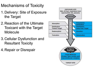
1. Mechanisms of Toxicity
1.Delivery: Site of Exposure to
the Target
2.Reaction of the Ultimate
Toxicant with the Target
Molecule
3.Cellular Dysfunction and
Resultant Toxicity
4.Repair or Disrepair
2. Chemical Factors that Cause Cellular Dysfunction
Chemicals that cause DNA adducts
Chemicals that cause protein adducts
Chemicals that cause oxidative stress
Chemicals that specifically interact with protein targets
Chemicals that inhibit cellular respiration
3. Chemical Factors that Cause Cellular Dysfunction
• Chemicals that cause DNA adducts
can lead to DNA mutations which can activate cell
death pathways; if mutations activate oncogenes or
inactivate tumor suppressors, it can lead to
uncontrolled cell proliferation and cancer (e.g.
benzopyrene)
4. • Chemicals that cause protein adducts
can lead to protein dysfunction which can activate cell
death pathways; protein adducts can also lead to
autoimmunity; if protein adducts activate oncogenes
or inactivate tumor suppressors, it can lead to
uncontrolled cell proliferation and cancer (e.g.
diclofenac glucuronidation metabolite)
5. • Chemicals that cause oxidative stress
can oxidize DNA or proteins leading to DNA
mutations or protein dysfunction and all of the
above. (e.g. benzene, CCl4)
Chemical Factors that Cause Cellular Dysfunction
7. • Chemicals that specifically interact with protein
targets
• chemicals that activate or inactivate ion channels
can cause widespread cellular dysfunction and cause
cell death and many physiological symptoms—Na+,
Ca2+, K+ levels are extremely important in
neurotransmission, muscle contraction, and nearly
every cellular function (e.g. tetrodotoxin closes
voltage-gated Na+ channels)
8. • Chemicals that inhibit cellular respiration—
inhibitors of proteins or enzymes involved in oxygen
consumption, fuel utilization, and ATP production will cause
energy depletion and cell death (e.g. cyanide inhibits
cytochrome c oxidase)
Chemical Factors that Cause Cellular Dysfunction
9. • Chemicals that inhibit the production of cellular
building blocks, e.g. nucleotides, lipids, amino acids (e.g.
amanita from Deathcap mushrooms)
• Chemicals that inhibit enzymatic processes of
bioactive metabolites that alter ion channels and metabolism
(e.g. sarin inhibits acetylcholinesterase and elevates
acetylcholine levels to active signaling pathways and ion
channels)
• All of the above can also cause inflammation which can
lead to cellular dysfunction
12. Two Forms of Cell Death
Necrosis: unprogrammed cell death (dangerous)
A. Passive form of cell death induced by accidental
damage of tissue and does not involve activation of any
specific cellular program.
B. Early loss of plasma membrane integrity and swelling of
the cell body followed by bursting of cell.
C. Mitochondria and various cellular processes contain
substances that can be damaging to surrounding cells and
are released upon bursting and cause inflammation.
D. Cells necrotize in response to tissue damage [injury by
chemicals and viruses, infection, cancer, inflammation,
ischemia (death due to blockage of blood to tissue)].
13. Mechanisms of Necrosis
• Cells must synthesize endogenous molecules,
assemble macromolecular complexes, membranes,
and cell organelles, maintain intracellular
environment, and produce energy for operation.
• Agents that disrupt these functions (especially
energy-producing function of the mitochondria
and protein synthesis) will cause cell death.
14.
15. I. ATP Depletion
ATP plays a central role in cellular maintenance both as a chemical
for biosynthesis and as the major source of energy.
1. ATP drives ion transporters such as Na+/K+-ATPase (plasma
membrane), Ca2+ -ATPase (endoplasmic reticulum and plasma
membrane) to maintain cellular ion gradients. (most important for
necrosis!)
2. Used in biosynthetic reactions (phosphorylation and adenylation)
3. Used for signal transduction regulation (e.g. phosphorylation of
receptor tyrosine kinase and kinase pathways)
4. Incorporated into DNA
5. Muscle contraction (actin/myosin interaction) and neurotransmission
6. Polymerization of cytoskeleton (actin and tubule polymerization)
7. Cell division
8. Maintenance of cell morphology
16. Direct Consequences of ATP Depletion
ATP Depletion
compromised
ion pumps (eg Na/K ATPase and Ca2+-ATPases)
loss of ionic and volume
regulatory controls
cell swelling
(water influx)
(rise in intracellular Na+)
cell lysis
necrosis
Ca2+/Na+ levels rise intracellularly
and leads to opening of voltage-gated channels
that depolarize membranes leading to further
Ca2+ and Na+ influx into the cell
17. Agents That Impair ATP Synthesis
1. Inhibitors of electron transport
1. Cyanide inhibits cytochrome oxidase
2. Rotenone inhibits complex I—insecticide that may be an
environmental cause of Parkinson’s Disease
3. Paraquat inhibits complex I—herbicide, but also causes lung
hemorrhaging in humans
2. Inhibitors of oxygen delivery
1. Ischemic agents such as ergot alkaloids, cocaine
2. Carbon monoxide—displaces oxygen from hemoglobin
3. Inhibitors of ADP phosphorylation – DDT
4. Chemicals causing mitochondrial DNA damage - antivirals,
chronic ethanol
18. II. Sustained Rise of Intracellular Ca2+
Ca2+ is involved in :
1. signal transduction regulation and exocytosis
2. muscle contraction (actin/myosin interaction)
3. cytoskeletal polymerization
4. neurotransmission and synaptic plasticity
5. enzyme induction (i.e. citrate and -ketoglutarate
dehydrogenases from the TCA cycle)
6. Transporters (Ca2+/ATPase, Na/Ca2+ exchanger, etc.)
19. Intracellular Ca2+ levels are highly regulated
•The 10,000-fold difference between extracellular and
cytosolic Ca2+
concentration is maintained by: impermeability of plasma
membrane
to Ca2+ and by transport mechanisms that remove Ca2+ from
cytoplasm
(0.1 M inside versus 1000 M outside).
• Ca2+ sources are from outside cell or Ca2+ stores in ER or
mitochondria (as calcium phosphate).
20. Excitotoxicity: Consequence of Increased Intracellular Ca2+
1. Depletion of energy reserves—decreased mitochondrial
ATP production and increased loss of ATP by activation
of Ca+2-ATPase.
2. Dysfunction of microfilaments—impaired cell motility,
disruption in cell morphology, cellular functions
3. Activation of hydrolytic enzymes—disintegration of
membranes, proteins, DNA, etc.
4. Generation of ROS/RNS—disintegration of
membranes, proteins, DNA, etc.
21. Reactive Oxygen and Nitrogen Species Generation
A. Direct generation of ROS/RNS
a. Xenobiotic bioactivation (i.e. carbon tetrachloride, benzene)
b. Redox cycling (paraquat, MPP+)
c. Transition metals (Fe2+, Cu2+)
d. Inhibition of mitochondrial electron transport (many
phytochemicals)
22. B. Indirect generation of ROS/RNS
Increased Ca2+ can cause ROS/RNS
i. Activates dehydrogenases in citric acid cycle and
increases electron output (NADH and
FADH2)leads to an increase in O2
.- (superoxide) by
the e- transport chain.
ii. Ca2+ -activated proteases proteolytically convert
xanthine dehydrogenase to xanthine oxidase, the
by-products of which are O2
-. and H2O2.
iii. Neurons and endothelial cells constitutively express
NOS that is activated by Ca2+ increase .NO
production which reacts with O2
.- to produce highly
reactive ONOO- (peroxynitrite).
23. Consequences of ROS/RNS
1. ROS can directly oxidize and affect protein function and can mutate DNA
leading to cellular dysfunction
2. ROS/RNS oxidatively inactivate Ca2+ /ATPases and elevate Ca2+
3. ROS and RNS also drain ATP reserves:
NO. is a reversible inhibitor of cytochrome oxidase
ROS can disrupt mitochondrial membranes and dissipate the
electrochemical gradient needed for ATP synthase.
4. Lipid peroxidation, cell swelling, and cell rupture
24. decreased ATP
decreased Ca2/ATPase
decreased mitochondrial
potential
increased mitochondrial e-
transport
induction of NOS, XO
decreased ATP synthase
increased e- transport
(increased NADH)
decreased
Ca2+/ATPase
inactivation of e-
transport complexes
DNA injury
decreased NADPH/
NADH
increased ROS
increased RNS
increased Ca2+
lipid peroxidation
membrane destruction
and/or cell swelling
cell lysis
microfilamental dissociation
membrane blebbing
cell destruction
Cell osmolarity disruption
(transporter disruption)
cell swelling
cell destruction