
Recommended
More Related Content
Similar to Overview of Cell Injury and Cell Death.pptx
Similar to Overview of Cell Injury and Cell Death.pptx (20)
Recently uploaded
Recently uploaded (20)
Overview of Cell Injury and Cell Death.pptx
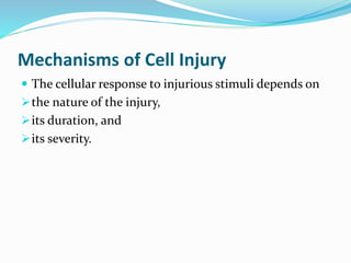
- 1. Mechanisms of Cell Injury The cellular response to injurious stimuli depends on the nature of the injury, its duration, and its severity.
- 2. The consequences of cell injury depend on the type, state, and adaptability of the injured cell.
- 3. Cell injury results from different biochemical mechanisms acting on several essential cellular components. The cellular components that are most frequently damaged by injurious stimuli include mitochondria, cell membranes, the machinery of protein synthesis and packaging, and DNA.
- 5. Mitochondrial Damage Mitochondria can be damaged by increases of cytosolic Ca2+, reactive oxygen species and oxygen deprivation In addition, mutations in mitochondrial genes are the cause of some inherited diseases. So they are sensitive to virtually all types of injurious stimuli, including hypoxia and toxins.
- 6. There are three major consequences of mitochondrial damage: 1. Mitochondrial damage often results in the formation of a high-conductance channel in the mitochondrial membrane, called the mitochondrial permeability transition pore. The opening of this conductance channel leads to the loss of mitochondrial membrane potential, resulting in failure of oxidative phosphorylation and progressive depletion of ATP, culminating in necrosis of the cell.
- 7. 2. Abnormal oxidative phosphorylation leads to the formation of reactive oxygen species, which have many deleterious effects.
- 8. 3. The mitochondria sequester between their outer and inner membranes several proteins that are capable of activating apoptotic pathways; these include cytochrome c and proteins that indirectly activate apoptosis inducing enzymes called caspases. Increased permeability of the outer mitochondrial membrane may result in leakage of these proteins into the cytosol and death by apoptosis.
- 9. Influx of Calcium and Loss of Calcium Homeostasis Cytosolic free calcium is normally maintained at very low concentrations (~0.1 µmol) compared with extracellular levels of 1.3 mmol, and most intracellular calcium is sequestered in mitochondria and the ER. Ischemia and certain toxins cause an increase in cytosolic calcium concentration, initially because of release of Ca2+ from intracellular stores, and later due to increased influx across the plasma membrane.
- 10. Increased intracellular Ca2+ causes cell injury by several mechanisms. 1. The accumulation of Ca2+ in mitochondria results in opening of the mitochondrial permeability transition pore and failure of ATP generation. 2. Increased cytosolic Ca2+ activates a number of enzymes.
- 11. These enzymes include a) phospholipases (which cause membrane damage), b) proteases (which break down both membrane and cytoskeletal proteins), c) endonucleases (which are responsible for DNA and chromatin fragmentation), and d) ATPases (thereby hastening ATP depletion).
- 12. 3. Increased intracellular Ca2+ levels also result in the induction of apoptosis, by direct activation of caspases and by increasing mitochondrial permeability.
- 13. Accumulation of Oxygen-Derived Free Radicals (Oxidative Stress) Cell injury induced by free radicals, particularly reactive oxygen species, is an important mechanism of cell damage in many pathologic conditions, such as chemical and radiation injury, ischemia-reperfusion injury (induced by restoration of blood flow in ischemic tissue), cellular aging, and microbial killing by phagocytes.
- 14. Free radicals are chemical species that have a single unpaired electron in an outer orbit. Unpaired electrons are highly reactive and “attack” and modify proteins, lipids, carbohydrates, nucleic acids—many of which are key components of cell membranes and nuclei.
- 15. Reactive oxygen species (ROS) are a type of oxygen derived free radical produced normally in cells during mitochondrial respiration and energy generation, but are degraded and removed by cellular defense systems. Increased production or decreased scavenging of ROS may lead to an excess of these free radicals, a condition called oxidative stress. Oxidative stress has been implicated in a wide variety of pathologic processes, including cell injury, cancer, aging, and some degenerative diseases such as Alzheimer disease.
- 16. Free radicals may be generated within cells in several ways: 1. The reduction-oxidation reactions that occur during normal metabolic processes. As a part of normal respiration, small amounts of partially reduced intermediates are produced in which different numbers of electrons have been transferred from O2; these include superoxide anion (O2 • − , one electron), hydrogen peroxide (H2O2, two electrons), and hydroxyl ions (˙OH, three electrons).
- 17. 2. Absorption of radiant energy (e.g., ultraviolet light, x- rays). For example, ionizing radiation can hydrolyze water into ˙OH and hydrogen (H) free radicals. 3. Rapid bursts of ROS are produced in activated leukocytes during inflammation. 4. Enzymatic metabolism of exogenous chemicals or drugs can generate free radicals that are not ROS but have similar effects (e.g., CCl4 can generate ˙CCl3).
- 18. 5. Transition metals such as iron and copper donate or accept free electrons during intracellular reactions and catalyze free radical formation, as in the Fenton reaction (H2O2 + Fe2 → Fe3 + ˙ OH + OH− ). Because most of the intracellular free iron is in the ferric (Fe3+ ) state, it must be reduced to the ferrous (Fe2+ ) form to participate in the Fenton reaction. This reduction can be enhanced by O2 • − , and thus sources of iron and O2 • − may co-operate in oxidative cell damage.
- 19. 6. Nitric oxide (NO), an important chemical mediator generated by endothelial cells, macrophages, neurons, and other cell types, can act as a free radical and can also be converted to highly reactive peroxynitrite anion (ONOO− ) as well as NO2 and NO3 − .
- 20. Removal of Free Radicals. Free radicals are inherently unstable and generally decay spontaneously; e.g. O2 • − is unstable and decays (dismutates) spontaneously to O2 and H2O2 in the presence of water. In addition, cells have developed multiple nonenzymatic and enzymatic mechanisms to remove free radicals and thereby minimize injury.
- 21. Antioxidants either block free radical formation or inactivate (e.g., scavenge) free radicals. Examples are the lipid-soluble vitamins E and A as well as ascorbic acid and glutathione in the cytosol.
- 22. Free iron and copper can catalyze the formation of ROS. Under normal circumstances, the reactivity of these metals is minimized by their binding to storage and transport proteins (e.g., transferrin, ferritin, lactoferrin, and ceruloplasmin) This prevents these metals from participating in reactions that generate ROS.
- 23. A series of enzymes acts as free radical-scavenging systems and breaks down H2O2 and O2 • − . 1. Catalase, present in peroxisomes, decomposes H2O2 (2H2O2 → O2 + 2H2O). 2. Superoxidase dismutases (SODs) are found in many cell types and convert O2 • − to H2O2 (2O2 • − + 2H → H2O2 + O2). This group of enzymes includes both manganese SOD, which is localized in mitochondria, and copper-zinc- SOD, which is found in the cytosol.
- 24. 3. Glutathione peroxidase also protects against injury by catalyzing free radical breakdown (H2O2 + 2GSH → GSSG + 2H2O, or 2˙OH + 2GSH → GSSG + 2H2O). The intracellular ratio of oxidized glutathione (GSSG) to reduced glutathione (GSH) is a reflection of the oxidative state of the cell and is an important indicator of the cell’s ability to detoxify ROS.
- 25. Pathologic Effects of Free Radicals. 1. Lipid peroxidation in membranes. In the presence of O2, free radicals may cause peroxidation of lipids within plasma and organellar membranes. The lipid-free radical interactions yield peroxides, which are themselves unstable and reactive, and an autocatalytic chain reaction ensues that can result in extensive membrane damage.
- 26. 2. Oxidative modification of proteins. Free radicals promote oxidation of amino acid side chains, formation of covalent protein-protein cross- links (e.g., disulfide bonds), and oxidation of the protein backbone. Oxidative modification of proteins may damage the active sites of enzymes, disrupt the conformation of structural proteins, and enhance proteasomal degradation of unfolded or misfolded proteins.
- 27. 3. Lesions in DNA. Free radicals are capable of causing single- and double-strand breaks in DNA, cross-linking of DNA strands, and formation of adducts. Oxidative DNA damage has been implicated in cell aging and in malignant transformation of cells.
- 28. Defects in Membrane Permeability Early loss of selective membrane permeability, leading ultimately to overt membrane damage, is a consistent feature of most forms of cell injury (except apoptosis). Membrane damage may affect the functions and integrity of all cellular membranes.
- 29. Mechanisms of Membrane Damage. 1. In ischemic cells, membrane defects may be the result of ATP depletion and calcium-mediated activation of phospholipases. 2. The plasma membrane can also be damaged directly by various bacterial toxins, viral proteins, lytic complement components, and a variety of physical and chemical agents.
- 30. 3. Several biochemical mechanisms may contribute to membrane damage. Reactive oxygen species. Oxygen free radicals cause injury to cell membranes by lipid peroxidation. Decreased phospholipid synthesis. The decreased phospholipid synthesis may affect all cellular membranes, including the mitochondria themselves.
- 31. Increased phospholipid breakdown. Phospholipid breakdown leads to the accumulation of lipid breakdown products, including unesterified free fatty acids, acyl carnitine, and lysophospholipids, which have a detergent effect on membranes. They may also either insert into the lipid bilayer of the membrane or exchange with membrane phospholipids, potentially causing changes in permeability and electrophysiologic alterations.
- 32. Cytoskeletal abnormalities. Cytoskeletal filaments serve as anchors connecting the plasma membrane to the cell interior. Activation of proteases by increased cytosolic calcium may cause damage to elements of the cytoskeleton. In the presence of cell swelling, this damage results, particularly in myocardial cells, in detachment of the cell membrane from the cytoskeleton, rendering it susceptible to stretching and rupture.
- 33. Consequences of Membrane Damage. 1. Mitochondrial membrane damage results in opening of the mitochondrial permeability transition pore, leading to decreased ATP generation and release of proteins that trigger apoptotic death. 2. Plasma membrane damage results in loss of osmotic balance and influx of fluids and ions, as well as loss of cellular contents. The cells may also leak metabolites that are vital for the reconstitution of ATP, thus further depleting energy stores.
- 34. 3. Injury to lysosomal membranes results in leakage of their enzymes into the cytoplasm and activation of the acid hydrolases in the acidic intracellular pH of the injured cell. Lysosomes contain RNases, DNases, proteases, phosphatases, and glucosidases. Activation of these enzymes leads to enzymatic digestion of proteins, RNA, DNA, and glycogen, and the cells die by necrosis.
- 35. Damage to DNA and Proteins Cells have mechanisms that repair damage to DNA, but if DNA damage is too severe to be corrected (e.g., after exposure to DNA damaging drugs, radiation, or oxidative stress), the cell initiates a suicide program that results in death by apoptosis. A similar reaction is triggered by improperly folded proteins, which may be the result of inherited mutations or acquired triggers such as free radicals.
- 40. Selected Examples of Cell Injury and Necrosis Ischemic and Hypoxic Injury Ischemia is the most common type of cell injury in clinical medicine and it results from hypoxia induced by reduced blood flow, most commonly due to a mechanical arterial obstruction. It can also be caused by reduced venous drainage. In contrast to hypoxia, during which energy production by anaerobic glycolysis can continue, ischemia compromises the delivery of substrates for glycolysis.
- 41. So in ischemic tissues, anaerobic energy generation also stops after glycolytic substrates are exhausted, or glycolysis is inhibited by the accumulation of metabolites that would otherwise be washed out by flowing blood. For this reason, ischemia tends to cause more rapid and severe cell and tissue injury than does hypoxia in the absence of ischemia.
- 42. Mechanisms of Ischemic Cell Injury Reduction in ATP levels is the fundamental cause of hypoxic cell injury. ATP is produced in two ways. The major pathway is oxidative phosphorylation of adenosine diphosphate by the electron transfer system of mitochondria. The second is the glycolytic pathway, which can generate ATP in the absence of oxygen using glucose derived either from body fluids or from the hydrolysis of glycogen.
- 43. ATP is required for virtually all synthetic and degradative processes within the cell such as membrane transport, protein synthesis, lipogenesis, and the deacylation-reacylation reactions necessary for phospholipid turnover.
- 44. Depletion of ATP to 5%-10% of normal levels has widespread effects on many critical cellular systems: 1. The activity of the plasma membrane energy- dependent sodium pump (ouabain-sensitive Na+ , K+ -ATPase) is reduced. Failure of this active transport system causes sodium to enter and accumulate inside cells and potassium to diffuse out. The net gain of solute is accompanied by isosmotic gain of water, causing cell swelling, and dilation of the ER.
- 45. 2. Cellular energy metabolism is altered. If the supply of oxygen to cells is reduced, as in ischemia, oxidative phosphorylation ceases, resulting in a decrease in cellular ATP and increase in adenosine monophosphate. These changes stimulate phosphofructokinase and phosphorylase activities, leading to an increased rate of anaerobic glycolysis (maintain the cell’s energy sources by generating ATP through metabolism of glucose derived from glycogen).
- 46. As a consequence glycogen stores are rapidly depleted. Anaerobic glycolysis results in the accumulation of lactic acid and inorganic phosphates from the hydrolysis of phosphate esters. This reduces the intracellular pH, resulting in decreased activity of many cellular enzymes.
- 47. 3. Failure of the Ca2+ pump leads to influx of Ca2+ , with damaging effects on numerous cellular components. 4. With prolonged or worsening depletion of ATP, structural disruption of the protein synthetic apparatus occurs, manifested as detachment of ribosomes from the rough ER and dissociation of polysomes, with a consequent reduction in protein synthesis.
- 48. The cytoskeleton disperses, resulting in the loss of ultrastructural features such as microvilli and the formation of “blebs” at the cell surface. “Myelin figures,” derived from degenerating cellular membranes, may be seen within the cytoplasm (in autophagic vacuoles) or extracellularly. If oxygen is restored, all of these disturbances are reversible.
- 49. 5. In cells deprived of oxygen or glucose, proteins may become misfolded, and accumulation of misfolded proteins in the endoplasmic reticulum (ER) triggers a cellular reaction called the unfolded protein response that may culminate in cell injury and even death.
- 51. If ischemia persists, irreversible injury and necrosis ensue. Irreversible injury is associated morphologically with severe swelling of mitochondria, extensive damage to plasma membranes and swelling of lysosomes. Large, flocculent, amorphous densities develop in the mitochondrial matrix. Ultimately, there is irreversible damage to mitochondrial and lysosomal membranes, and the cell undergoes necrosis.
- 52. Massive influx of calcium into the cell then occurs, particularly if the ischemic zone is reperfused. Death is mainly by necrosis, but apoptosis also contributes; the apoptotic pathway is probably activated by release of pro-apoptotic molecules from leaky mitochondria.
- 53. The cell’s components are progressively degraded, and there is widespread leakage of cellular enzymes into the extracellular space and, conversely, entry of extracellular macromolecules from the interstitial space into the dying cells.
- 54. Finally, the dead cells may become replaced by large masses composed of phospholipids in the form of myelin figures. These are then either phagocytosed by leukocytes or degraded further into fatty acids. Calcification of such fatty acid residues may occur, with the formation of calcium soaps.
- 55. Mammalian cells have developed protective responses to deal with hypoxic stress. The best-defined of these is induction of a transcription factor called hypoxia-inducible factor-1, which promotes new blood vessel formation, stimulates cell survival pathways, and enhances anaerobic glycolysis.
- 56. There are still no reliable therapeutic approaches for reducing the injurious consequences of ischemia in clinical situations. The strategy that is perhaps the most useful in ischemic (and traumatic) brain and spinal cord injury is the transient induction of hypothermia (reducing the core body temperature to 92°F).
- 57. This treatment reduces the metabolic demands of the stressed cells, decreases cell swelling, suppresses the formation of free radicals, and inhibits the host inflammatory response. All of these may contribute to decreased cell and tissue injury.
- 58. The sequential morphologic changes in cell injury progressing to cell death
- 59. Ischemia-Reperfusion Injury Restoration of blood flow to ischemic tissues can promote recovery of cells if they are reversibly injured, but can also paradoxically exacerbate the injury and cause cell death. As a consequence, reperfused tissues may sustain loss of cells in addition to the cells that are irreversibly damaged at the end of ischemia.
- 60. This process, called ischemia-reperfusion injury, is clinically important because it contributes to tissue damage during myocardial and cerebral infarction and following therapies to restore blood flow.
- 61. How does reperfusion injury occur? Oxidative stress. New damage may be initiated during reoxygenation by increased generation of reactive oxygen and nitrogen species. These free radicals may be produced in reperfused tissue as a result of incomplete reduction of oxygen by damaged mitochondria, or because of the action of oxidases in leukocytes, endothelial cells, or parenchymal cells. Cellular antioxidant defense mechanisms may be compromised by ischemia, favoring the accumulation of free radicals.
- 62. Intracellular calcium overload. Intracellular and mitochondrial calcium overload begins during acute ischemia; it is exacerbated during reperfusion due to influx of calcium resulting from cell membrane damage and ROS mediated injury to sarcoplasmic reticulum. Calcium overload favors opening of the mitochondrial permeability transition pore with resultant depletion of ATP. This in turn causes further cell injury.
- 63. Inflammation. Ischemic injury is associated with inflammation as a result of “dangers signals” released from dead cells, cytokines secreted by resident immune cells such as macrophages, and increased expression of adhesion molecules by hypoxic parenchymal and endothelial cells, all of which act to recruit circulating neutrophils to reperfused tissue. The inflammation causes additional tissue injury.
- 64. Activation of the complement system may contribute to ischemia-reperfusion injury.
- 65. Chemical (Toxic) Injury Chemical injury remains a frequent problem in clinical medicine and is a major limitation to drug therapy. Because many drugs are metabolized in the liver, this organ is a frequent target of drug toxicity. Direct toxicity. In mercuric chloride poisoning, mercury binds to the sulfhydryl groups of cell membrane proteins, causing increased membrane permeability and inhibition of ion transport.
- 66. In such instances, the greatest damage is usually to the cells that use, absorb, excrete, or concentrate the chemicals—in the case of mercuric chloride, the cells of the gastrointestinal tract and kidney. Cyanide poisons mitochondrial cytochrome oxidase and thus inhibits oxidative phosphorylation. Many antineoplastic chemotherapeutic agents and antibiotics also induce cell damage by direct cytotoxic effects.
- 67. Conversion to toxic metabolites. Most toxic chemicals are not biologically active in their native form but must be converted to reactive toxic metabolites, which then act on target molecules. This modification is usually accomplished by the cytochrome P-450 mixed-function oxidases in the smooth ER of the liver and other organs. The toxic metabolites cause membrane damage and cell injury mainly by formation of free radicals and subsequent lipid peroxidation; direct covalent binding to membrane proteins and lipids may also contribute.
- 68. CCl4, which was once widely used in the dry cleaning industry, is converted by cytochrome P-450 to the highly reactive free radical ˙CCl3, which causes lipid peroxidation and damages many cellular structures. Acetaminophen, an analgesic drug, is also converted to a toxic product during detoxification in the liver, leading to cell injury.