SlideShare a Scribd company logo
1 of 226
Download to read offline
HEAD AND
NECK
ANATOMY
1
Regions of the head
2
Bone
Surface
Markings
• Depressions and openings:
– Fissure: narrow
opening between
adjacent parts of
bones for nerves and
vessels
– Foramen: hole,
opening
– Fossa: shallow
depression
– Sulcus: groove
– Meatus: tubelike
passageway
• Processes that form joints
– Condyle: large
rounded prominence
– Facet: smooth flat
surface
– Head: rounded
articular projection
• Processes for tendon and
ligament attachment:
– Crest: prominent
border or ridge
– Epicondyle:
prominence above a
condyle
– Linea: line, less
prominent than a
crest
– Trochanter: large
projection of bone
found only on the
femur
– Tubercle: small
rough process
– Tuberosity: large,
rounded
3
4
Gross Anatomy
Osteology of the Skull: Cranium
Skull
5
• (1) Frontal Bone
• (2) Temporal Bones
• (2) Parietal Bones
• (1) Occipital Bone
• (1) Sphenoid Bone
• (1) Ethmoid Bone
Cranium: Consists of 8 bones
• (2) Maxilla
• (2) Zygomatic bones
• (2) Lacrimal bones
• (2) Palatine bones
• (2) Nasal bones
• (2) Inferior nasal conchae
• (1) Vomer
• (1) Mandible
Face: Consists of 14 bones
(Studied in lecture 2)
6
Bones of the Cranium
Frontal Bone
7
Frontal
Bone
8
The large bone that makes up the forehead and
supplies the upper edge and roof of the orbit
(eye socket).
The frontal bone articulates (comes together)
with a number of other bones including the
parietal, nasal, ethmoid, maxillary, and
zygomatic bones.
Landmarks:
• Squama: flat portion that forms the forehead
• Supraorbital margin: ridge under the eyebrow, forming
the upper part of the orbit (eye socket)
• Supraorbital foramen: small hole within supraorbital
margin for blood vessels and nerves
• Frontal sinuses: hollow spaces behind the squama, act as
sound chambers to give the voice resonance.
• Frontal Bone
(Anterior View) (Blue
Colored Bone)
9
10
Frontal Bone (Lateral View) (Blue Colored Bone)
11
Temporal Bones
12
Temporal Bones
• A large irregular bone situated at the base and side of the skull. The temporal
bone is connected with the mandible (the jaw bone) via the
temporomandibular (TM) joint.
• The temporal bone is formed of three parts (squamous, tympanic and
petrous) that are distinct at birth but then fuse. The petrous portion of the
temporal bone contains the structures of the inner ear.
• Landmarks:
– Squama: flat portion of the temporal bone forming the anterior and superior part
of the temple
– Zygomatic process: process forming part of the cheek
– Petrous portion: internal, forming part of the floor of the cranium. Contains the
ear canal and internal ear structures.
– Mandibular fossa: socket between squama and petrous portion, articulates with
the condyle of the mandible (TMJ)
– External auditory meatus: opening to the ear canal
– Mastoid process: bony prominence behind the external auditory meatus
– Styloid process: looks like an elephant’s tusk located between the mastoid
process and the jaw. Acts as a point of attachment for muscles and ligaments.
13
Temporal Bone (Lateral View) (Purple Colored Bone)
14
Parietal Bones
• The main bone on the side of
the skull.
• The word "parietal" comes
from the Latin "parietalis"
meaning "belonging to the
wall."
• It articulates (joins) with the
other parietal bone in the
midline (top of the head),
with the frontal bone in front
of it, with the occipital bone
behind it, and with the
sphenoid and temporal bones
lower down on the side of
the skull.
15
Parietal Bone (Lateral View) (Red Colored Bone)
16
Occipital
17
Occipital
• From the Latin, meaning the part of the head opposite the front.
• The bone that forms the rear and the rear bottom of the skull.
• The occipital bone articulates (joins) with the parietal and
temporal bones of the skull, the sphenoid bone in front of it, and
the first cervical vertebra (the atlas) beneath it
• Landmarks:
– Foramen magnum: large hole, allowing passage of the
spinal cord
– External occipital protuberance (EOP): prominent
projection on back of occipital
– Nuchal lines: a superior and inferior line running laterally
from the midline, serve as a point of muscle attachment
18
Occipital Bone (Posterior View) (Orange Colored Bone)
19
Occipital Bone (Lateral View) (Orange Bone)
20
Sphenoid Bone
21
Sphenoid Bone
• A prominent, irregular, wedge-shaped bone at the base of the
skull. The sphenoid bone has been called the "keystone" of the
cranial floor since it is in contact with all of the other cranial
bones.
• The Greek physician Galan wrote that the sphenoid bone was "like
a wedge thrust between the skull and the superior maxilla."
• Landmarks
– Greater wings: large lateral projections of bone that help to
form the lateral border of the skull
– Lesser wings: smaller lateral projections of bone above the
greater wings
– Pterygoid processes: two long downward projections from the
greater wings that act as a point of muscle attachment.
– Sella turcica: known as the Turkish Saddle which cradles the
pituitary gland.
22
Sphenoid Bone (Lateral View) (Green Colored Bone)
23
Sphenoid Bone (Floor of Cranium) (Green Colored Bone)
24
Ethmoid Bone
• An irregularly shaped, spongy bone that provides the floor of the front part of the
skull and the roof of the nasal cavity.
• The ethmoid consists of two masses of thin plates enclosing air cells and looks
like a sieve.
• Landmarks:
– Lateral masses: form most of the wall between the nasal cavity and the
orbits
– Perpendicular plate: forms the superior portion of the nasal septum
– Cribriform plate: forms the roof of the nasal cavity
– Olfactory foramina: small holes within the cribriform plate for passage of
the first cranial nerve (for smell)
– Crista Galli: upward extension of bone above the cribriform plate, acts as an
anchoring point for one of the coverings of the brain.
– Nasal concha (turbinates): two scroll-shaped projections with a mucus
membrane on either side of the nasal septum. Function to cause air
turbulence and trap inhaled particles.
25
26
Bones of the Face
27
Maxilla
28
Maxilla
• The largest bones of the face, except for the mandible and form,
by their union, the whole of the upper jaw.
• They hold the upper teeth, and connect on the left and right to
the zygomatic bones (cheek bones).
• Each assists in forming the boundaries of three cavities, namely,
the roof of the mouth, the floor and lateral wall of the nose, and
the floor of the orbit.
• Landmarks:
– Infra Orbital foramen: hole below the orbit, for blood
vessels and nerves
– Alveolar process: arch of the maxilla containing the upper
teeth
– Palatine process: horizontal projection of the maxilla
forming the anterior Âľ of the hard palate.
29
Maxilla (Anterior View) (Yellow Colored Bones)
30
Zygomatic Bones
Commonly referred to as
the cheekbone.
It is situated at the upper
and lateral part of the
face: it forms the
prominence of the cheek
and part of the lateral wall
and floor of the orbit.
It articulates with the
zygomatic arch of the
temporal bone.
31
Zygomatic Bones (Anterior View) (Pink Colored Bones)
32
Lacrimal Bones
Smallest and most
fragile bone of the
face, is situated at
the front part of the
Medial of the
orbit.
Contains the lacrimal
sac and the naso-
lacrimal duct.
Lacrimal bone
33
Lacrimal Bones (Anterior View) (Blue Colored Bones below frontal bone)
34
Palatine bones
It contributes to the
walls of three
cavities: the floor
and lateral wall of
the nasal cavity,
the roof of the
mouth, and the
floor of the orbit
35
Nasal Bones
Varying in size and
form in different
individuals
They are placed side by
side at the middle and
upper part of the face
and form, by their
junction, "the bridge" of
the nose
36
Nasal Bones (Anterior View) (Bridge of the nose, below frontal bone)
37
Inferior Nasal Conchae
Extends horizontally
along the lateral wall
of the nasal cavity
and consists of a
lamina of spongy
bone, curled upon
itself like a scroll.
Inferior Nasal Conchae
38
Inferior Nasal Conchae (Anterior View) (Inside nasal cavity on lateral walls)
39
Vomer
One of the unpaired
facial bones of the
skull.
Located in
the midsagittal line,
and touches the
sphenoid, the
ethmoid, the left and
right palatine bones,
and the left and right
maxillary bones.
40
Vomer (Anterior View) (Center wall in nasal cavity)
41
Mandible
Largest and strongest
bone of the face.
Forms the lower jaw and
holds the lower teeth in
place.
The mandible consists of
a curved, horizontal
portion, the body, and two
perpendicular portions,
the rami, which unite with
the ends of the body nearly
at right angles.
42
Mandible
• Largest and strongest bone of the face.
• Forms the lower jaw and holds the lower teeth in place.
• Landmarks:
– Body: curved horizontal portion of the mandible
– Rami: two upward projections of bone that are perpendicular to the
body of the mandible.
– Angle of the mandible: angle formed where the body meets the
ramus
– Condylar process: a condyle on the posterior portion of the ramus
that articulates with the mandibular fossa of the temporal bone.
– Coronoid process: a sharp projection of bone on the anterior portion
of the ramus that acts as a point of muscle attachment.
– Alveolar process: arch of bone containing the lower teeth
– Mental foramen: small hole on the side of the body for blood vessels
and nerves.
43
Mandible (Anterior View) (Lower Jaw)
44
Mandible (Lateral View) (Lower Jaw)
45
Palpation of the Cranium
Occipital: Prone; place hands on the back of the head between partner’s ears. Slide your
fingers superiorly to the External Occipital Protuberance (EOP) two to three inches.
Then slide fingers laterally to the mastoid process behind the ears.
Superior Nuchal Lines: Prone or supine; locate the EOP and then slide your fingers
laterally moving your finger pads up and down feeling for the edge of the superior
nuchal line.
Parietal Bone: Prone or supine; place both hands on the top of the cranium. Palpate the
sagittal suture between the parietals. From the suture, palpate the parietal bones down
towards the ears
Temporal Bone: Supine; locate the mastoid process by placing your fingers behind the ear
lobe. The zygomatic arch can be palpated by placing your fingers anterior to the
external auditory meatus. Palpate anteriorly along the arch with your finger and
thumb. The flat squamous portion can be palpated superior to the mastoids and
external auditory meatus. The styloid process can be palpated between the mandible
and the mastoid process (palpate very gently)
46
Frontal bone: Supine; palpate the region of the forehead from the eyebrows up toward the
coronal sutures
Mandible: Supine: place your fingers inferior to the bottom teeth and palpate the body of
the mandible. Move inferiorly and palpate the base of the mandible from the chin to
the angle of the mandible. Then curl your fingertips underneath the edge to palpate the
submandibular fossa. To palpate the angle of the mandible slide posterior alone the
base of the mandible. The angle is located between the body and the ramus. To palpate
the mandibular condyle place your finger anterior to the ear canal and below the
zygomatic arch. Ask your partner to open his/her mouth fully, the condyle will protrude
laterally and become more palpable.
Nasal bones: Supine; locate the bridge of the nose
Zygomatic bone: Supine, return to the zygomatic arch of the temporal bone and continue
to move anteriorly until you reach the zygomatic (cheek) bone.
Maxilla: Supine; palpate inferior to the zygomatic bone down to the mouth. The maxilla
forms the center of the face. The alveolar processes can also be palpated where the
teeth insert into the maxilla.
HYIOD
BONE
HYIOD BONE
• the hyoid is suspended between the
mandible and the larynx
• it does not articulate with any other bone
• is suspended from the styloid process of
the temporal bone by two stylohyoid
ligaments.
48
HYIOD BONE CONTD.
• It is shaped like a horseshoe
• consists of a central body with
two lateral projections
• functions as a primary support for the
tongue and floor of the mouth
49
AUDITORY
OSSICLES
INCUS
51
STAPES
52
stapes
• The stapes or stirrup is a bone in the middle
ear of humans and other animals which is
involved in the conduction of sound vibrations
to the inner ear.
• This bone is connected to the oval window by
its annular ligament, which allows the
footplate to transmit sound energy through the
oval window into the inner ear.
• The stapes is the smallest and lightest bone in
the human body.
53
MALLEOUS
54
55
TEMPOROMANDIBULAR
JOINT
56
TMJ
Composed of 3 bony parts
Glenoid fossa
Articular eminence
Condyle process
57
tmj
58
TMJ CONTD
59
• It is the only movable joint in the skull
• It have 2 type of movement
• Gliding action and hinge action
• Hinge action allows for opening and closing
to the jaw
• Gliding allows the side to side movement,
protrusion and retrusion
MALE VS FEMALE SKULL
60
61
FUSION OF BONES
(POSTNATAL)
62
63
Pterion
The pterion is the region where the frontal, parietal,
temporal, and sphenoid bones join.
• The pterion is known as the weakest part of the
skull. The anterior division of the middle meningeal
artery runs underneath the pterion.
• A traumatic blow to the pterion may rupture the
middle meningeal artery causing an epidural
hematoma.
• The pterion may also be fractured indirectly by
blows to the top or back of the head that place
sufficient force on the skull to fracture the pterion.
64
PTERION
65
LAMBDA
• The lambda is the meeting point of the
sagittal suture and the lambdoid suture. This
is also the point of the occipital angle.
• In the fetus, the lambda is membranous, and
is called the posterior fontanelle.
66
LAMBDA
67
BREGMA
• The bregma is the anatomical point on the
skull at which the coronal suture is
intersected perpendicularly by the sagittal
suture.
• The bregma is known as the anterior
fontanelle during infancy. The anterior
fontanelle is membranous and closes in the
first 18-36 months of life.
68
bregma
69
MYOLOGY
OF THE
HEAD AND
NECK
70
• Muscles of facial expression
• Muscles of mastication
• Muscles of the floor of the mouth
• Muscles of the soft palate
• Muscles of the tongue (extrinsic)
• Muscles of the tongue (intrinsic)
• Muscles of the neck
• Muscles of the pharynx
71
Muscles of
facial
expression
72
buccinator
73
buccinator
• Origin: alveolar processes
of the maxilla and mandible
• Insertion: orbicularis oris
• Function: compresses the
cheeks against the teeth and
is used in acts such as
blowing. It assists muscle of
chewing
in newborns it is used to
suckle.
74
75
nasalis
76
Nasalis
• Origin: maxilla
• Insertion: nasal bone
• Function: flares the nostrils,
compresses the bridge of the
nose & depresses the tip of
the nose
77
78
Orbicularis
oculi
79
Orbicularis
oculi
80
Origin: nasal part of the frontal
bone, maxilla and lacrimal bone
Insertion: skin of the orbital
region
Function: voluntary closing of
eyelids (winking and forced
squeezing) and pumping of tears
81
frontalis
82
frontalis
• Origin: Galea aponeurotica
(epicranial aponeurosis)
• Insertion: orbicularis oculi
• Function: elevating
eyebrows
83
mentalis
84
mentalis
• Origin: Anterior mandible
• Insertion: Chin
• Function: Elevates and
wrinkles the chin , protrudes
the lower lip
85
risorius
86
RISORIUS
• Origin: Parotid fascia
• Insertion: Modiolus
• Function: draws back the
angles of the mouth
(sometimes referred to as
the smiling muscles)
87
Zygomaticus
major&
minor
88
Zygomaticus
major
• Origin: anterior of the
zygomatic
• Insertion: modiolus of the
mouth
• Function: draws angles of
the mouth upwards laterally
89
Zygomaticus
minor
• Origin: zygomatic bone
• Insertion: skin of the upper
lip
• Function: elevates three
upper lip
90
Orbicularis
oris
Orbicularis
oris
• Origin: maxilla and
mandible
• Insertion: skin around the
lips
• Function: puckers the lips (
sometimes referred to as the
kissing muscle)
92
93
Depressor anguli oris
94
Depressor
anguli oris
• Origin: tuberosity of the
mandible
• Insertion: modiolus of the
mouth
• Function: depresses the
angles of the mouth
95
Depressor
labii
inferioris
Depressor
labii
inferioris
• Origin: oblique line of the
mandible, between the
symphysis and the mental
foramen
• Insertion: orbicularis oris
fibers
• Function: depresses the
lower lip
97
Levator
labii
superioris
Levator
labii
superioris
• Origin: medial infraorbital
margin
• Insertion: skin of the upper
lip
• Function: elevates the upper
lip
99
Levator
labii
superioris
aleaque
nasi
Levator
labii
superioris
aleaque
nasi
• Origin: nasal bone
• Insertion: nostril and upper
lip
• Function: dilates the
nostrils, elevates the upper
lip & wing of the nose
101
Depressor
supercilli
Depressor
supercilli
• Origin: medial orbital rim
• Insertion: medial aspects of
the bony orbits
• Function: depressing the
eyebrows
103
Corrugator
supercilli
Corrugator
supercilli
• Origin: supraorbital ridge
• Insertion: skin of the
forehead near the eyebrows
• Function: wrinkles the
foreheads and draws the
eyebrows medially
105
Muscles of
mastication
Temporalis
/ temporal
muscle
107
temporalis
• Origin: temporal lines of the
parietal bone and the
superior temporal surface of
the sphenoid bone
• Insertion: coronoid process
of the mandible and the
retromolar fossa
• Function: elevation and
retraction of the mandible
108
masseter
109
masseter
• Origin: zygomatic arch and
maxillary process of the
zygomatic
• Insertion: coronoid process,
angle and lateral surfaces of
ramus
• Function: elevation
(closing) and protrusion of
the mandible
110
Internal/
medial
pterygoid
111
Medial
pterygoid
• Origin: deep head-medial
side of the lateral pterygoid
plate
superficial head-
process of the palatine bone
and maxillary tuberosity
• Insertion: angle of the
mandible
• Function: elevates the
mandible and closes the jaw
and side to side movements
112
External/lateral
pterygoid
113
Lateral
pterygoid
• Origin: superior head- sphenoid
bone
inferior head- lateral
pterygoid plate
• Insertion: superior head- anterior
side of the
condyle
inferior head- pterygoid
fossa
• Function: depresses and
protrudes the jaw
side to side movement
of the mandible
114
Muscles
of the
floor of
the
mouth
digastric
116
digastric
• Origin: anterior body-
mandible
posterior body- mastoid of
the temporal
bone
• insertion: hyoid bone
• Function: opens the jaw
when the masseter and
temporalis are relaxed
117
mylohyoid
118
mylohyoid
• Origin: mylohyoid line of
the mandible
• Insertion: body of the hyoid
bone
• Function: raises the floor of
the oral cavity
elevates the tongue
depresses the mandible
119
geniohyoid
120
geniohyoid
• Origin: genial tubercle
• Insertion: hyoid bone
• Function: draws the tongue
and the hyoid bone forward
121
stylohyoid
stylohyoid
• Origin: styloid process of
temporal bone
• Insertion: hyoid process
• Function: Elevates the
hyoid bone during
swallowing
123
Muscles of
the soft
palate
124
Palatoglossus/anterior
faucal pillar
12
5
palatoglossus
• Origin: arises from the soft
palate
• Forms the anterior arch on
each side of the throat
• Insertion: tongue
• Function: arching the
tongue against the soft
palate
depressing the soft palate
towards the tongue
126
Palatopharyngeal/posterior
faucal pillar
12
7
palatopharyngeal
• Origin: soft palate, posterior border of the
thyroid cartilage
• Insertion: upper borders of the thyroid
cartilage
• Function: helps to shut the nasopharynx
• Forms the posterior arch of the throat
128
Isthumus fauces
129
Isthumus fauces
• The fauces is a part of the oropharynx
Between these two arches on the lateral
walls of the oropharynx is the tonsillar fossa
which is the location of the palatine tonsil.
The arches are also known together as the
palatine arches.
130
tonsilitis
131
tonsilitis
• Tonsillitis is inflammation of the tonsils in
the upper part of the throat. It can be acute
or chronic.
• Acute tonsillitis typically has a rapid onset.
Symptoms may include sore throat, fever,
enlargement of the tonsils, trouble
swallowing, and enlarged lymph nodes
around the neck.
• Can be caused by viral or bacterial
infections
132
TONSILLAR HYPERTROPHY
• Tonsillar hypertrophy is the enlargement of
the tonsils, but without the history of
inflammation. Obstructive tonsillar
hypertrophy is currently the most common
reason for tonsillectomy. These patients
symptoms of loud snoring, irregular
breathing, nocturnal choking and coughing,
frequent awakenings& sleep apnea.
133
TONSILLAR HYPERTROPHY
134
Extrinsic muscles of the tongue
135
styloglossus
styloglossus
• Origin: styloid process of
the temporal bone
• Insertion: the sides of the
tongue
• Function: retraction and
elevation the tongue
137
genioglossus
genioglossus
• Origin: superior part of the
symphysis menti
( genial tubercle)
• Insertion: under surface of
the tongue
• Function: protrudes and
depresses the tongue
139
hyoglossus
hyoglossus
• Origin: hyoid bone
• Insertion: side of the tongue
• Function: depresses and
retracts the tongue
141
Intrinsic muscles of the tongue
142
Transverse/horizontal
muscle & Vertical
muscle
Transverse muscle
• Origin: median fibrous septum
• Insertion: sides of the tongue
• Function: makes the tongue narrow and
elongated
144
Vertical
muscle
• Origin: Submucosal fibrous
layer of the dorsum of the
tongue
• Insertion: inferior surface
borders of the tongue
• Function: flattens and
broadens the tongue
145
Superior
longitudinal
& inferior
longitudinal
Superior
longitudinal
muscle
• Origin: median fibrous
septum
• Insertion: edges of the
tongue
• Function: retracts the
tongue making the tongue
short and thick
147
Inferior
longitudinal
muscle
• Origin: root of the tongue
• Insertion: apex of the
tongue
• Function: retracts the
tongue making it short and
thick
148
Muscles of the neck
149
sternocleidomastoid
sternocleidomastoid
• Origin: sternum and clavicle
• Insertion: mastoid process
• Function: divide the neck
into anterior and posterior
triangles important in
extraoral examination
151
trapezius
trapezius
• Origin: occipital bone
• Insertion: clavicle and
scapula
• Function: shrugging of the
shoulders, rotation,
retraction and elevation of
the scapula
153
platysma
platysma
• Origin: clavicle
• Insertion: base of the
mandible, orbicularis oris,
angle of the mouth, skin of
the cheeks and lips
• Function: draws the angles
of the mouth inferiorly
155
CRANIAL NERVES
156
157
Cranial nerves
158
NERVE TYPE FUNCTION
I. OLFACTORY SENSORY SENSE OF SMELL
II. OPTIC SENSORY SENSE OF SIGHT
III. OCULOMOTOR MOTOR EYE MUSCLES
IV. TROCHEAR MOTOR EYE MUSCLES
V. TRIGEMINAL MOTOR& SENSORY MUSCLES OF MASTICATION,
SENSATION IN THE
MOUTH.TEETH, TONGUE & FACE
VI. ABDUCENS MOTOR EYE MUSCLES
VII. FACIAL MOTOR & SENSORY FACIAL EXPRESSION MUSCLES,
SALIVARY GLANDS AND SENSE
OF TASTE
VIII.
VESTIBULOCONCLEAR
SENSORY SENSE OF BALANCE
IX.
GLOSSOPHARYNGEAL
MOTOR & SENSORY PAROTID GLAND
SENSATION AROUND THE EARS
X. VAGUS MOTOR & SENSORY SOFT PALATE, PHARYNIX &
LARYNIX MUSCLES
SENSE OF TASTE, SENSATION
AROUND EARS
XI. ACESSORY MOTOR MUSCLES OF THE NECK &
159
Trigeminal
nerve
160
Trigeminal
branches
16
1
• V1 Ophthalmic nerve
• V2 Maxillary nerve
• V3 Mandibular nerve
162
TRIGEMINAL
OPHTHALMIC MAXILARY MANDIBULAR
V1 (ophthalmic nerve)
• The ophthalmic nerve is purely a sensory
nerve
• Supplying sensory innervation to certain
parts of the eye, the lacrimal gland & some
paranasal sinuses
• Also supplies the upper eyelid, dorsum of
nose and anterior part of the scalp
163
164
V2 (maxillary)
• Has 5 branches that innervates the oral
cavity
1. Anterior Superior Alveolar nerve
2. Middle Superior Alveolar Nerve
3. Posterior Superior Alveolar Nerve
4. Greater palatine Nerve
5. Nasopalatine Nerve
165
166
167
168
MAXILLARY
NASOPALATINE
MUCOPERIOSTEUM
OG ANTERIOR TEETH
GREATER PALATINE
MUCOPERIOSTEUM
OF ANTERIOR TEETH
ANTERIOR SUPERIOR
ALVEOLAR NERVE
INCISORS AND
CANINES
MAXILLARY
SINUSES
MIDDLE SUPERIOR
ALVEOLAR NERVE
1ST & 2ND PREMOLARS
MESIOBUCCAL ROOT
OF 1ST MOLAR
POSTERIOR
SUPERIOR
ALVEOLAR NERVE
2 ROOTS OF 1ST
MOLAR, 2ND AND 3RD
MOLARS
V3 (mandibular)
• Has 3 branches that innervates the oral
cavity
1. Inferior alveolar nerve( branches out into)
• mylohyoid nerve
• incisive nerve
• mental nerve
2. Lingual nerve
3. Buccal nerve
169
170
171
Mandibular
Inferior
alveolar nerve
mental
incisive
mylohyoid
lingual Buccal
Trigeminal neuralgia
• This chronic pain condition affects the trigeminal nerve,
which carries sensation from your face to your brain.
Symptoms may include:
• Severe shooting pain that may feel like an electric shock
• Pain or attacks activated by touching the face, biting,
talking or brushing
• Pain areas include the ear, eyes, forehead, jaw, or mouth
and face
• Over sensitivity, sensitivity to pain, or uncomfortable
tingling and burning
• Can be only one attack of pain, some may experience sharp
pain every hour or every few seconds
172
Blood
circulation
arteries
CAROTID ARTERY
• Rises from the aorta
• Branches into
1. internal carotid- supplies the brain and
eyes
2. external carotid- oral cavity. Sinuses ,
nose and tongue
174
175
External carotid branches
• Facial artery- supplies muscles of the face,
nasal septum, tonsils and posterior part of
the tongue
• Lingual artery- supplies the tongue, soft
palate and tonsils
• Maxillary artery – oral cavity and the teeth
176
177
Maxillary branches
• It divides into 3 branches
1. Pterygoid – supplies muscles of
mastication and the upper jaw
2. Pterygopalatine- gingiva, maxillary
sinuses. Palate & nasal septum
3. Mandibular- supplies the lower jaw, chin
and lower lip
178
179
Pterygoid artery branches
• Greater palatine artery- supplies the hard
palate & nasal septum
• infraorbital artery-
• Anterior superior alveolar artery
• Anterior and middle superior alveolar artery
• Posterior superior alveolar artery
180
Mandibular artery branches
• Divides into 5 branches
1. Inferior alveolar artery
2. Lingual artery
3. Mylohyoid artery
4. Incisive artery
5. Mental artery
181
182
Veins
• Maxillary vein corresponds with the
maxillary artery
• The retromandibular vein is formed by the
union of the maxillary vein and the
temporal vein.
• The retromandibular has 2 branches
1. The anterior branch which joins the facial
vein
183
Veins contid
2. Posterior vein joins the auricular vein to
make the external jugular vein
• The facial vein joins the anterior division of
the retromandibular vein to form the
common facial vein
• The external jugular vein empties into the
subclavian vein
184
Veins contid
• The common facial vein and the deep facial
vein enter the internal jugular which
empties into the superior vena cava
• The superior vena cava returns blood from
the upper body to the right atrium of the
heart
185
186
187
188
Lymph
nodes
DEEP AND SUPERFICIAL
NODES
• The superficial cervical lymph nodes are
lymph nodes that lie near the surface of the
neck.
• The deep cervical lymph nodes are a group
of cervical lymph nodes found near the
internal jugular vein in the neck.
190
Cervical lymph nodes
191
• Cervical lymph node swelling can be a reliable
indicator of infection or other inflammation in the
area
• Generally, swollen cervical lymph nodes
(lymphadenopathy) are nonthreatening. Many
things can cause cervical lymph node swelling,
including:
• bronchitis
• common cold
• ear infection
• scalp infection
• strep throat
• Tonsillitis
• Oral infections
192
Axillary
lymph
nodes
• also known as adenopathy
• Unilateral swelling is often (but not always) a
symptom of an infection or disease on that side of the
body.
• Bilateral swelling tends to point to systemic illness—
that is, an illness affecting the entire body.
• Common causes of axillary lymphadenopathy include;
• Localized infection and short-term inflammation(e.g.,
a tattoo on arm)
• HIV
• Breast cancer
• Lymphoma
• Vaccinations
194
Abdominal/
inguinal
lymph
nodes
• More often than not, swollen inguinal lymph nodes are
caused by infections or injury affecting the lower body.
This can include the:
• groin
• genitals
• urinary tract
• leg
• Foot
• In rare cases, swollen lymph nodes in the groin could be
due to cancer. Some of these types of cancer include:
• melanoma
• testicular cancer
• ovarian cancer
196
Salivary
glands
Importance of saliva
• Lubricates and cleanses the oral cavity
• Aids in digestion through enzymatic process
• Helps to soften and mix food in mechanical
digestion
• Maintains the integrity of tooth surfaces
through chemical protection
• Prevents infections and promotes healing in
the oral cavity through antibacterial
protection
198
Types of saliva
• Serous saliva (protein fluid)
• Predominantly produced by the parotid
gland
• Mucous saliva (carbohydrate fluid)
• Produced by the sublingual and
submandibular gland (these two are
seromucous)
199
Saliva
• Serous saliva is watery, thin and contains
enzymes, antibodies and inorganic ions
involved in digestion and immune defense
• Mucous saliva is this and viscous involved
in lubrication
200
Minor salivary
glands
• Located through out the mouth
• Produces 5% of saliva in the mouth
• The von Ebner gland Is located on
the posterior surfaces of the tongue
alongside the circumvallate papillae
Parotid
gland
202
Parotid
gland
• Found of both sides of the
mouth in front of the ear
• connects to the stensen’s duct
which empties saliva into the
mouth
• Produces 25% of saliva
• Produces serous saliva, rich in
enzymes
• A small projection called the
parotid papillae is the opening
to the parotid duct can be felt
on the buccal vestibule
203
Sublingual
gland
Sublingual
• Located under the tongue, it
is the smallest of the major
glands and has the most
dispersal
• Produces 10% of saliva
• Associated with the
Bartholin’s duct
• Has small ducts known as
Rivinus ducts
205
RIVINUS
DUCTS
• Small ducts
associated with the
sublingual gland
20
6
Submandibular
gland
207
Submandibular gland
• Located below the mandible
• Produces 65% of saliva
• Releases saliva through the Wharton’s duct
208
Salivary glands diseases
• Sialolithiasis : occurs when stones made of
calcium form in the salivary glands. These
stones can block the glands, and that can
partially or completely stop the flow of
saliva.
• Sialadenitis (or sialoadenitis) is an infection
involving a salivary gland. It often results
from stones blocking the gland. Staph or
strep bacteria can cause this infection. Older
adults and infants are most likely to develop
this condition.
209
Sialolithiasis
(salivary
stones)
210
Sialadenitis
(swollen
salivary
glands)
Sjogren syndrome
• the immune system primarily attacks the
glands that produce tears (the lacrimal
glands) and saliva (the salivary glands),
impairing the glands' ability to secrete these
fluids.
212
213
Cancerous and noncancerous
tumors
• Salivary gland tumors, also known as
mucous gland adenomas or neoplasms, are
tumors that form in the tissues of salivary
glands.
214
Parotid
gland tumor
• Could be benign or malignant
215
Paranasal
sinuses
21
6
Paranasal sinuses
• Paranasal sinuses are a group of four paired
air-filled spaces that surround the nasal
cavity.
• The paranasal sinuses have several
functions of which reducing the weight of
the head is the most important. Other
functions include air humidification and
aiding in voice resonance .
217
Maxillary
sinuses
21
8
MAXILLARY SINUSES
• They are the largest of the paranasal sinuses
• They are the first of the paranasal sinuses to
form
• Account for both transverse and vertical
growth of the face
• At birth, its 5-8 cubic cm in volume and
continues to increase in size up to the age of
18years
219
Frontal
sinuses
22
0
FRONTAL SINUSES
• Frontal sinuses are absent at birth but are
developed and well functioning by ag 6-8
years
• Maximum growth is achieved after puberty
• 5% of people have absent frontal sinuses
• Assists in immune defense and air filtration
performed by the nose
221
Ethmoid
sinuses
22
2
ETHMOID SINUSES
• Present at birth and is complete by 5 years
• Acute ethmoiditis in childhood and
ethmoidal carcinoma may spread superiorly
causing meningitis and cerebrospinal fluid
leakage
223
Sphenoid
sinuses
22
4
Sphenoid sinuses
• They are not present at birth, they slowly
develop with the growth of the skull. Just
after puberty the sinuses finish
development.
225
Paranasal sinuses disease
• characterized by decreased aeration,
mucosal thickening, soft tissue masses (e.g.,
mucus retention cyst, polyp, mucocele,
tumor), air-fluid levels, and
demineralization or bone destruction.
• Inflammation in the posterior ethmoid and
sphenoid sinus causes optic nerve
dysfunction
226

More Related Content

What's hot

Dural venous sinuses
Dural venous sinusesDural venous sinuses
Dural venous sinusesmgmcri1234
 
blood supply nerve supply of face
blood supply nerve supply of faceblood supply nerve supply of face
blood supply nerve supply of facespmc bikaner
 
Muscles of mastication & TMJ Dr.N.Mugunthan
Muscles of mastication & TMJ Dr.N.MugunthanMuscles of mastication & TMJ Dr.N.Mugunthan
Muscles of mastication & TMJ Dr.N.Mugunthanmgmcri1234
 
Infra temporal fossa
Infra temporal fossaInfra temporal fossa
Infra temporal fossamgmcri1234
 
Norma basalis (skull base).pps
Norma basalis (skull base).ppsNorma basalis (skull base).pps
Norma basalis (skull base).ppsKifayat Khan
 
Parotid gland
Parotid glandParotid gland
Parotid glandnadia omer
 
Suboccipital triangle
Suboccipital triangleSuboccipital triangle
Suboccipital trianglemgmcri1234
 
DEEP CERVICAL FASCIA.pptx
DEEP CERVICAL FASCIA.pptxDEEP CERVICAL FASCIA.pptx
DEEP CERVICAL FASCIA.pptxDr. sana yaseen
 
Suboccipital triangle
Suboccipital triangleSuboccipital triangle
Suboccipital triangleIdris Siddiqui
 
Submandibular salivary gland
Submandibular salivary glandSubmandibular salivary gland
Submandibular salivary glandChitransha03
 
Parasympathetic ganglia of Head and Neck
 Parasympathetic ganglia of Head and Neck Parasympathetic ganglia of Head and Neck
Parasympathetic ganglia of Head and NeckCIMS
 
Parotid gland & Facial nerve
Parotid gland & Facial nerveParotid gland & Facial nerve
Parotid gland & Facial nerveMehul Tandel
 
Osteology of the Skull
Osteology of the SkullOsteology of the Skull
Osteology of the SkullHeatherSeghi
 

What's hot (20)

Anatomy of parotid gland
Anatomy of parotid glandAnatomy of parotid gland
Anatomy of parotid gland
 
Muscles of Pharynx
Muscles of PharynxMuscles of Pharynx
Muscles of Pharynx
 
Fourth ventricle
Fourth ventricleFourth ventricle
Fourth ventricle
 
Dural venous sinuses
Dural venous sinusesDural venous sinuses
Dural venous sinuses
 
blood supply nerve supply of face
blood supply nerve supply of faceblood supply nerve supply of face
blood supply nerve supply of face
 
Muscles of mastication & TMJ Dr.N.Mugunthan
Muscles of mastication & TMJ Dr.N.MugunthanMuscles of mastication & TMJ Dr.N.Mugunthan
Muscles of mastication & TMJ Dr.N.Mugunthan
 
Scalp
Scalp Scalp
Scalp
 
Infra temporal fossa
Infra temporal fossaInfra temporal fossa
Infra temporal fossa
 
Cranial fossa-SCALP
Cranial fossa-SCALPCranial fossa-SCALP
Cranial fossa-SCALP
 
Norma basalis (skull base).pps
Norma basalis (skull base).ppsNorma basalis (skull base).pps
Norma basalis (skull base).pps
 
Parotid gland
Parotid glandParotid gland
Parotid gland
 
Overview Cranial Foramina
Overview Cranial ForaminaOverview Cranial Foramina
Overview Cranial Foramina
 
Suboccipital triangle
Suboccipital triangleSuboccipital triangle
Suboccipital triangle
 
DEEP CERVICAL FASCIA.pptx
DEEP CERVICAL FASCIA.pptxDEEP CERVICAL FASCIA.pptx
DEEP CERVICAL FASCIA.pptx
 
Suboccipital triangle
Suboccipital triangleSuboccipital triangle
Suboccipital triangle
 
Submandibular salivary gland
Submandibular salivary glandSubmandibular salivary gland
Submandibular salivary gland
 
Parasympathetic ganglia of Head and Neck
 Parasympathetic ganglia of Head and Neck Parasympathetic ganglia of Head and Neck
Parasympathetic ganglia of Head and Neck
 
Parotid gland & Facial nerve
Parotid gland & Facial nerveParotid gland & Facial nerve
Parotid gland & Facial nerve
 
Osteology of the Skull
Osteology of the SkullOsteology of the Skull
Osteology of the Skull
 
Parotid gland
Parotid glandParotid gland
Parotid gland
 

Similar to head and neck anatomy 1-3.pdf

OSTEOLOGY OF HEAD AND NECK
OSTEOLOGY OF HEAD AND NECKOSTEOLOGY OF HEAD AND NECK
OSTEOLOGY OF HEAD AND NECKAbdul Akbar
 
Osteology of facial skeleton
Osteology of facial skeletonOsteology of facial skeleton
Osteology of facial skeletonK BHATTACHARJEE
 
The Skull and Vertebral column
The Skull and Vertebral columnThe Skull and Vertebral column
The Skull and Vertebral columnSeddie Chitamu
 
Skull, neck and muscle
Skull, neck and muscleSkull, neck and muscle
Skull, neck and muscleUE
 
Module-1-Bones-of-the-Skull.pptx
Module-1-Bones-of-the-Skull.pptxModule-1-Bones-of-the-Skull.pptx
Module-1-Bones-of-the-Skull.pptxhehehehe62
 
SKELETAL system- axial.ppt
SKELETAL system- axial.pptSKELETAL system- axial.ppt
SKELETAL system- axial.pptlovekeshSingh12
 
Anatomy Lab exercise 9
Anatomy Lab exercise 9Anatomy Lab exercise 9
Anatomy Lab exercise 9Jhillock
 
Anatomy head
Anatomy headAnatomy head
Anatomy headJSlinkyNY
 
Topic 5 bone of skull neck
Topic 5 bone of skull neckTopic 5 bone of skull neck
Topic 5 bone of skull neckSado Anatomist
 
Basic Skull Anatomy
Basic Skull AnatomyBasic Skull Anatomy
Basic Skull AnatomyHadi Munib
 
Osteology of Human Skull.pptx
Osteology of Human Skull.pptxOsteology of Human Skull.pptx
Osteology of Human Skull.pptxDrPrasannaKumarP
 
anatomy 2.pptx
anatomy 2.pptxanatomy 2.pptx
anatomy 2.pptxMaharijNoor
 
Skeletal system. anatomy and physiology of skeletal system. appendicular skel...
Skeletal system. anatomy and physiology of skeletal system. appendicular skel...Skeletal system. anatomy and physiology of skeletal system. appendicular skel...
Skeletal system. anatomy and physiology of skeletal system. appendicular skel...mamtabisht10
 
OVERVIEW OF THE HEAD REGION.pptx.pdf
OVERVIEW OF THE HEAD REGION.pptx.pdfOVERVIEW OF THE HEAD REGION.pptx.pdf
OVERVIEW OF THE HEAD REGION.pptx.pdfAbooddSandoqaa
 
Gross anatomy of the head and neck
Gross anatomy of the head and neckGross anatomy of the head and neck
Gross anatomy of the head and neckLucidante1
 
The gross anatomy of the head and neck lecture 3
The gross anatomy of the head and neck lecture 3The gross anatomy of the head and neck lecture 3
The gross anatomy of the head and neck lecture 3Lucidante1
 

Similar to head and neck anatomy 1-3.pdf (20)

Osteology
OsteologyOsteology
Osteology
 
OSTEOLOGY OF HEAD AND NECK
OSTEOLOGY OF HEAD AND NECKOSTEOLOGY OF HEAD AND NECK
OSTEOLOGY OF HEAD AND NECK
 
Osteology of facial skeleton
Osteology of facial skeletonOsteology of facial skeleton
Osteology of facial skeleton
 
The Skull and Vertebral column
The Skull and Vertebral columnThe Skull and Vertebral column
The Skull and Vertebral column
 
Bones of Skull (Human Anatomy)
Bones of Skull (Human Anatomy)Bones of Skull (Human Anatomy)
Bones of Skull (Human Anatomy)
 
Skull, neck and muscle
Skull, neck and muscleSkull, neck and muscle
Skull, neck and muscle
 
Module-1-Bones-of-the-Skull.pptx
Module-1-Bones-of-the-Skull.pptxModule-1-Bones-of-the-Skull.pptx
Module-1-Bones-of-the-Skull.pptx
 
SKELETAL system- axial.ppt
SKELETAL system- axial.pptSKELETAL system- axial.ppt
SKELETAL system- axial.ppt
 
Anatomy Lab exercise 9
Anatomy Lab exercise 9Anatomy Lab exercise 9
Anatomy Lab exercise 9
 
Anatomy head
Anatomy headAnatomy head
Anatomy head
 
Anatomy(1) Osteology
Anatomy(1) Osteology Anatomy(1) Osteology
Anatomy(1) Osteology
 
The skeletal System
The skeletal SystemThe skeletal System
The skeletal System
 
Topic 5 bone of skull neck
Topic 5 bone of skull neckTopic 5 bone of skull neck
Topic 5 bone of skull neck
 
Basic Skull Anatomy
Basic Skull AnatomyBasic Skull Anatomy
Basic Skull Anatomy
 
Osteology of Human Skull.pptx
Osteology of Human Skull.pptxOsteology of Human Skull.pptx
Osteology of Human Skull.pptx
 
anatomy 2.pptx
anatomy 2.pptxanatomy 2.pptx
anatomy 2.pptx
 
Skeletal system. anatomy and physiology of skeletal system. appendicular skel...
Skeletal system. anatomy and physiology of skeletal system. appendicular skel...Skeletal system. anatomy and physiology of skeletal system. appendicular skel...
Skeletal system. anatomy and physiology of skeletal system. appendicular skel...
 
OVERVIEW OF THE HEAD REGION.pptx.pdf
OVERVIEW OF THE HEAD REGION.pptx.pdfOVERVIEW OF THE HEAD REGION.pptx.pdf
OVERVIEW OF THE HEAD REGION.pptx.pdf
 
Gross anatomy of the head and neck
Gross anatomy of the head and neckGross anatomy of the head and neck
Gross anatomy of the head and neck
 
The gross anatomy of the head and neck lecture 3
The gross anatomy of the head and neck lecture 3The gross anatomy of the head and neck lecture 3
The gross anatomy of the head and neck lecture 3
 

More from NabiswaboazWangila

Health communication by nabiswa Boaz ppt
Health communication by nabiswa Boaz pptHealth communication by nabiswa Boaz ppt
Health communication by nabiswa Boaz pptNabiswaboazWangila
 
MODERN CONTEX OF HUMAN SEXUALITY.pptx
MODERN CONTEX OF HUMAN SEXUALITY.pptxMODERN CONTEX OF HUMAN SEXUALITY.pptx
MODERN CONTEX OF HUMAN SEXUALITY.pptxNabiswaboazWangila
 
modern context of human sexuality.pptx
modern context of human sexuality.pptxmodern context of human sexuality.pptx
modern context of human sexuality.pptxNabiswaboazWangila
 
IMPORTANCE OF COMMUNICATION SKILLS ACQUIRED [Autosaved].pptx
IMPORTANCE OF COMMUNICATION SKILLS ACQUIRED [Autosaved].pptxIMPORTANCE OF COMMUNICATION SKILLS ACQUIRED [Autosaved].pptx
IMPORTANCE OF COMMUNICATION SKILLS ACQUIRED [Autosaved].pptxNabiswaboazWangila
 
THE_ART_OF_PUBLIC_SPEAKING.pptx
THE_ART_OF_PUBLIC_SPEAKING.pptxTHE_ART_OF_PUBLIC_SPEAKING.pptx
THE_ART_OF_PUBLIC_SPEAKING.pptxNabiswaboazWangila
 
Introduction_to_the_Course.pdf
Introduction_to_the_Course.pdfIntroduction_to_the_Course.pdf
Introduction_to_the_Course.pdfNabiswaboazWangila
 
Presentatie-Nicholas-Awortwi.pptx
Presentatie-Nicholas-Awortwi.pptxPresentatie-Nicholas-Awortwi.pptx
Presentatie-Nicholas-Awortwi.pptxNabiswaboazWangila
 
INTEGUMENTARY SYSTEM BY EVA KIPTOO.pptx
INTEGUMENTARY SYSTEM BY EVA KIPTOO.pptxINTEGUMENTARY SYSTEM BY EVA KIPTOO.pptx
INTEGUMENTARY SYSTEM BY EVA KIPTOO.pptxNabiswaboazWangila
 

More from NabiswaboazWangila (14)

Health communication by nabiswa Boaz ppt
Health communication by nabiswa Boaz pptHealth communication by nabiswa Boaz ppt
Health communication by nabiswa Boaz ppt
 
MUSCULAR SYSTEM.pptx
MUSCULAR SYSTEM.pptxMUSCULAR SYSTEM.pptx
MUSCULAR SYSTEM.pptx
 
edna_nanjala.pptx
edna_nanjala.pptxedna_nanjala.pptx
edna_nanjala.pptx
 
MOAC_Word_2016_Core.pdf
MOAC_Word_2016_Core.pdfMOAC_Word_2016_Core.pdf
MOAC_Word_2016_Core.pdf
 
MODERN CONTEX OF HUMAN SEXUALITY.pptx
MODERN CONTEX OF HUMAN SEXUALITY.pptxMODERN CONTEX OF HUMAN SEXUALITY.pptx
MODERN CONTEX OF HUMAN SEXUALITY.pptx
 
modern context of human sexuality.pptx
modern context of human sexuality.pptxmodern context of human sexuality.pptx
modern context of human sexuality.pptx
 
NABISWA SPEECH.pptx
NABISWA SPEECH.pptxNABISWA SPEECH.pptx
NABISWA SPEECH.pptx
 
NABISWA_SPEECH z.pptx
NABISWA_SPEECH z.pptxNABISWA_SPEECH z.pptx
NABISWA_SPEECH z.pptx
 
IMPORTANCE OF COMMUNICATION SKILLS ACQUIRED [Autosaved].pptx
IMPORTANCE OF COMMUNICATION SKILLS ACQUIRED [Autosaved].pptxIMPORTANCE OF COMMUNICATION SKILLS ACQUIRED [Autosaved].pptx
IMPORTANCE OF COMMUNICATION SKILLS ACQUIRED [Autosaved].pptx
 
LIFE SKILLS 1.pptx
LIFE SKILLS 1.pptxLIFE SKILLS 1.pptx
LIFE SKILLS 1.pptx
 
THE_ART_OF_PUBLIC_SPEAKING.pptx
THE_ART_OF_PUBLIC_SPEAKING.pptxTHE_ART_OF_PUBLIC_SPEAKING.pptx
THE_ART_OF_PUBLIC_SPEAKING.pptx
 
Introduction_to_the_Course.pdf
Introduction_to_the_Course.pdfIntroduction_to_the_Course.pdf
Introduction_to_the_Course.pdf
 
Presentatie-Nicholas-Awortwi.pptx
Presentatie-Nicholas-Awortwi.pptxPresentatie-Nicholas-Awortwi.pptx
Presentatie-Nicholas-Awortwi.pptx
 
INTEGUMENTARY SYSTEM BY EVA KIPTOO.pptx
INTEGUMENTARY SYSTEM BY EVA KIPTOO.pptxINTEGUMENTARY SYSTEM BY EVA KIPTOO.pptx
INTEGUMENTARY SYSTEM BY EVA KIPTOO.pptx
 

Recently uploaded

ECONOMIC CONTEXT - LONG FORM TV DRAMA - PPT
ECONOMIC CONTEXT - LONG FORM TV DRAMA - PPTECONOMIC CONTEXT - LONG FORM TV DRAMA - PPT
ECONOMIC CONTEXT - LONG FORM TV DRAMA - PPTiammrhaywood
 
Atmosphere science 7 quarter 4 .........
Atmosphere science 7 quarter 4 .........Atmosphere science 7 quarter 4 .........
Atmosphere science 7 quarter 4 .........LeaCamillePacle
 
MULTIDISCIPLINRY NATURE OF THE ENVIRONMENTAL STUDIES.pptx
MULTIDISCIPLINRY NATURE OF THE ENVIRONMENTAL STUDIES.pptxMULTIDISCIPLINRY NATURE OF THE ENVIRONMENTAL STUDIES.pptx
MULTIDISCIPLINRY NATURE OF THE ENVIRONMENTAL STUDIES.pptxAnupkumar Sharma
 
Keynote by Prof. Wurzer at Nordex about IP-design
Keynote by Prof. Wurzer at Nordex about IP-designKeynote by Prof. Wurzer at Nordex about IP-design
Keynote by Prof. Wurzer at Nordex about IP-designMIPLM
 
Solving Puzzles Benefits Everyone (English).pptx
Solving Puzzles Benefits Everyone (English).pptxSolving Puzzles Benefits Everyone (English).pptx
Solving Puzzles Benefits Everyone (English).pptxOH TEIK BIN
 
Employee wellbeing at the workplace.pptx
Employee wellbeing at the workplace.pptxEmployee wellbeing at the workplace.pptx
Employee wellbeing at the workplace.pptxNirmalaLoungPoorunde1
 
What is Model Inheritance in Odoo 17 ERP
What is Model Inheritance in Odoo 17 ERPWhat is Model Inheritance in Odoo 17 ERP
What is Model Inheritance in Odoo 17 ERPCeline George
 
AMERICAN LANGUAGE HUB_Level2_Student'sBook_Answerkey.pdf
AMERICAN LANGUAGE HUB_Level2_Student'sBook_Answerkey.pdfAMERICAN LANGUAGE HUB_Level2_Student'sBook_Answerkey.pdf
AMERICAN LANGUAGE HUB_Level2_Student'sBook_Answerkey.pdfphamnguyenenglishnb
 
Romantic Opera MUSIC FOR GRADE NINE pptx
Romantic Opera MUSIC FOR GRADE NINE pptxRomantic Opera MUSIC FOR GRADE NINE pptx
Romantic Opera MUSIC FOR GRADE NINE pptxsqpmdrvczh
 
Planning a health career 4th Quarter.pptx
Planning a health career 4th Quarter.pptxPlanning a health career 4th Quarter.pptx
Planning a health career 4th Quarter.pptxLigayaBacuel1
 
Introduction to ArtificiaI Intelligence in Higher Education
Introduction to ArtificiaI Intelligence in Higher EducationIntroduction to ArtificiaI Intelligence in Higher Education
Introduction to ArtificiaI Intelligence in Higher Educationpboyjonauth
 
Grade 9 Q4-MELC1-Active and Passive Voice.pptx
Grade 9 Q4-MELC1-Active and Passive Voice.pptxGrade 9 Q4-MELC1-Active and Passive Voice.pptx
Grade 9 Q4-MELC1-Active and Passive Voice.pptxChelloAnnAsuncion2
 
Proudly South Africa powerpoint Thorisha.pptx
Proudly South Africa powerpoint Thorisha.pptxProudly South Africa powerpoint Thorisha.pptx
Proudly South Africa powerpoint Thorisha.pptxthorishapillay1
 
Full Stack Web Development Course for Beginners
Full Stack Web Development Course  for BeginnersFull Stack Web Development Course  for Beginners
Full Stack Web Development Course for BeginnersSabitha Banu
 
DATA STRUCTURE AND ALGORITHM for beginners
DATA STRUCTURE AND ALGORITHM for beginnersDATA STRUCTURE AND ALGORITHM for beginners
DATA STRUCTURE AND ALGORITHM for beginnersSabitha Banu
 
Influencing policy (training slides from Fast Track Impact)
Influencing policy (training slides from Fast Track Impact)Influencing policy (training slides from Fast Track Impact)
Influencing policy (training slides from Fast Track Impact)Mark Reed
 
Types of Journalistic Writing Grade 8.pptx
Types of Journalistic Writing Grade 8.pptxTypes of Journalistic Writing Grade 8.pptx
Types of Journalistic Writing Grade 8.pptxEyham Joco
 

Recently uploaded (20)

ECONOMIC CONTEXT - LONG FORM TV DRAMA - PPT
ECONOMIC CONTEXT - LONG FORM TV DRAMA - PPTECONOMIC CONTEXT - LONG FORM TV DRAMA - PPT
ECONOMIC CONTEXT - LONG FORM TV DRAMA - PPT
 
Atmosphere science 7 quarter 4 .........
Atmosphere science 7 quarter 4 .........Atmosphere science 7 quarter 4 .........
Atmosphere science 7 quarter 4 .........
 
MULTIDISCIPLINRY NATURE OF THE ENVIRONMENTAL STUDIES.pptx
MULTIDISCIPLINRY NATURE OF THE ENVIRONMENTAL STUDIES.pptxMULTIDISCIPLINRY NATURE OF THE ENVIRONMENTAL STUDIES.pptx
MULTIDISCIPLINRY NATURE OF THE ENVIRONMENTAL STUDIES.pptx
 
Keynote by Prof. Wurzer at Nordex about IP-design
Keynote by Prof. Wurzer at Nordex about IP-designKeynote by Prof. Wurzer at Nordex about IP-design
Keynote by Prof. Wurzer at Nordex about IP-design
 
Solving Puzzles Benefits Everyone (English).pptx
Solving Puzzles Benefits Everyone (English).pptxSolving Puzzles Benefits Everyone (English).pptx
Solving Puzzles Benefits Everyone (English).pptx
 
Employee wellbeing at the workplace.pptx
Employee wellbeing at the workplace.pptxEmployee wellbeing at the workplace.pptx
Employee wellbeing at the workplace.pptx
 
What is Model Inheritance in Odoo 17 ERP
What is Model Inheritance in Odoo 17 ERPWhat is Model Inheritance in Odoo 17 ERP
What is Model Inheritance in Odoo 17 ERP
 
AMERICAN LANGUAGE HUB_Level2_Student'sBook_Answerkey.pdf
AMERICAN LANGUAGE HUB_Level2_Student'sBook_Answerkey.pdfAMERICAN LANGUAGE HUB_Level2_Student'sBook_Answerkey.pdf
AMERICAN LANGUAGE HUB_Level2_Student'sBook_Answerkey.pdf
 
Romantic Opera MUSIC FOR GRADE NINE pptx
Romantic Opera MUSIC FOR GRADE NINE pptxRomantic Opera MUSIC FOR GRADE NINE pptx
Romantic Opera MUSIC FOR GRADE NINE pptx
 
Planning a health career 4th Quarter.pptx
Planning a health career 4th Quarter.pptxPlanning a health career 4th Quarter.pptx
Planning a health career 4th Quarter.pptx
 
Introduction to ArtificiaI Intelligence in Higher Education
Introduction to ArtificiaI Intelligence in Higher EducationIntroduction to ArtificiaI Intelligence in Higher Education
Introduction to ArtificiaI Intelligence in Higher Education
 
Grade 9 Q4-MELC1-Active and Passive Voice.pptx
Grade 9 Q4-MELC1-Active and Passive Voice.pptxGrade 9 Q4-MELC1-Active and Passive Voice.pptx
Grade 9 Q4-MELC1-Active and Passive Voice.pptx
 
Proudly South Africa powerpoint Thorisha.pptx
Proudly South Africa powerpoint Thorisha.pptxProudly South Africa powerpoint Thorisha.pptx
Proudly South Africa powerpoint Thorisha.pptx
 
OS-operating systems- ch04 (Threads) ...
OS-operating systems- ch04 (Threads) ...OS-operating systems- ch04 (Threads) ...
OS-operating systems- ch04 (Threads) ...
 
Model Call Girl in Tilak Nagar Delhi reach out to us at 🔝9953056974🔝
Model Call Girl in Tilak Nagar Delhi reach out to us at 🔝9953056974🔝Model Call Girl in Tilak Nagar Delhi reach out to us at 🔝9953056974🔝
Model Call Girl in Tilak Nagar Delhi reach out to us at 🔝9953056974🔝
 
Full Stack Web Development Course for Beginners
Full Stack Web Development Course  for BeginnersFull Stack Web Development Course  for Beginners
Full Stack Web Development Course for Beginners
 
DATA STRUCTURE AND ALGORITHM for beginners
DATA STRUCTURE AND ALGORITHM for beginnersDATA STRUCTURE AND ALGORITHM for beginners
DATA STRUCTURE AND ALGORITHM for beginners
 
Influencing policy (training slides from Fast Track Impact)
Influencing policy (training slides from Fast Track Impact)Influencing policy (training slides from Fast Track Impact)
Influencing policy (training slides from Fast Track Impact)
 
Types of Journalistic Writing Grade 8.pptx
Types of Journalistic Writing Grade 8.pptxTypes of Journalistic Writing Grade 8.pptx
Types of Journalistic Writing Grade 8.pptx
 
Raw materials used in Herbal Cosmetics.pptx
Raw materials used in Herbal Cosmetics.pptxRaw materials used in Herbal Cosmetics.pptx
Raw materials used in Herbal Cosmetics.pptx
 

head and neck anatomy 1-3.pdf

  • 2. Regions of the head 2
  • 3. Bone Surface Markings • Depressions and openings: – Fissure: narrow opening between adjacent parts of bones for nerves and vessels – Foramen: hole, opening – Fossa: shallow depression – Sulcus: groove – Meatus: tubelike passageway • Processes that form joints – Condyle: large rounded prominence – Facet: smooth flat surface – Head: rounded articular projection • Processes for tendon and ligament attachment: – Crest: prominent border or ridge – Epicondyle: prominence above a condyle – Linea: line, less prominent than a crest – Trochanter: large projection of bone found only on the femur – Tubercle: small rough process – Tuberosity: large, rounded 3
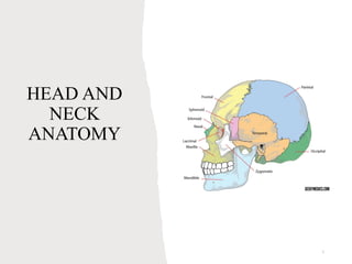
  • 4. 4 Gross Anatomy Osteology of the Skull: Cranium
  • 5. Skull 5 • (1) Frontal Bone • (2) Temporal Bones • (2) Parietal Bones • (1) Occipital Bone • (1) Sphenoid Bone • (1) Ethmoid Bone Cranium: Consists of 8 bones • (2) Maxilla • (2) Zygomatic bones • (2) Lacrimal bones • (2) Palatine bones • (2) Nasal bones • (2) Inferior nasal conchae • (1) Vomer • (1) Mandible Face: Consists of 14 bones (Studied in lecture 2)
  • 6. 6 Bones of the Cranium
  • 8. Frontal Bone 8 The large bone that makes up the forehead and supplies the upper edge and roof of the orbit (eye socket). The frontal bone articulates (comes together) with a number of other bones including the parietal, nasal, ethmoid, maxillary, and zygomatic bones. Landmarks: • Squama: flat portion that forms the forehead • Supraorbital margin: ridge under the eyebrow, forming the upper part of the orbit (eye socket) • Supraorbital foramen: small hole within supraorbital margin for blood vessels and nerves • Frontal sinuses: hollow spaces behind the squama, act as sound chambers to give the voice resonance.
  • 9. • Frontal Bone (Anterior View) (Blue Colored Bone) 9
  • 10. 10 Frontal Bone (Lateral View) (Blue Colored Bone)
  • 12. 12 Temporal Bones • A large irregular bone situated at the base and side of the skull. The temporal bone is connected with the mandible (the jaw bone) via the temporomandibular (TM) joint. • The temporal bone is formed of three parts (squamous, tympanic and petrous) that are distinct at birth but then fuse. The petrous portion of the temporal bone contains the structures of the inner ear. • Landmarks: – Squama: flat portion of the temporal bone forming the anterior and superior part of the temple – Zygomatic process: process forming part of the cheek – Petrous portion: internal, forming part of the floor of the cranium. Contains the ear canal and internal ear structures. – Mandibular fossa: socket between squama and petrous portion, articulates with the condyle of the mandible (TMJ) – External auditory meatus: opening to the ear canal – Mastoid process: bony prominence behind the external auditory meatus – Styloid process: looks like an elephant’s tusk located between the mastoid process and the jaw. Acts as a point of attachment for muscles and ligaments.
  • 13. 13 Temporal Bone (Lateral View) (Purple Colored Bone)
  • 14. 14 Parietal Bones • The main bone on the side of the skull. • The word "parietal" comes from the Latin "parietalis" meaning "belonging to the wall." • It articulates (joins) with the other parietal bone in the midline (top of the head), with the frontal bone in front of it, with the occipital bone behind it, and with the sphenoid and temporal bones lower down on the side of the skull.
  • 15. 15 Parietal Bone (Lateral View) (Red Colored Bone)
  • 17. 17 Occipital • From the Latin, meaning the part of the head opposite the front. • The bone that forms the rear and the rear bottom of the skull. • The occipital bone articulates (joins) with the parietal and temporal bones of the skull, the sphenoid bone in front of it, and the first cervical vertebra (the atlas) beneath it • Landmarks: – Foramen magnum: large hole, allowing passage of the spinal cord – External occipital protuberance (EOP): prominent projection on back of occipital – Nuchal lines: a superior and inferior line running laterally from the midline, serve as a point of muscle attachment
  • 18. 18 Occipital Bone (Posterior View) (Orange Colored Bone)
  • 19. 19 Occipital Bone (Lateral View) (Orange Bone)
  • 21. 21 Sphenoid Bone • A prominent, irregular, wedge-shaped bone at the base of the skull. The sphenoid bone has been called the "keystone" of the cranial floor since it is in contact with all of the other cranial bones. • The Greek physician Galan wrote that the sphenoid bone was "like a wedge thrust between the skull and the superior maxilla." • Landmarks – Greater wings: large lateral projections of bone that help to form the lateral border of the skull – Lesser wings: smaller lateral projections of bone above the greater wings – Pterygoid processes: two long downward projections from the greater wings that act as a point of muscle attachment. – Sella turcica: known as the Turkish Saddle which cradles the pituitary gland.
  • 22. 22 Sphenoid Bone (Lateral View) (Green Colored Bone)
  • 23. 23 Sphenoid Bone (Floor of Cranium) (Green Colored Bone)
  • 24. 24 Ethmoid Bone • An irregularly shaped, spongy bone that provides the floor of the front part of the skull and the roof of the nasal cavity. • The ethmoid consists of two masses of thin plates enclosing air cells and looks like a sieve. • Landmarks: – Lateral masses: form most of the wall between the nasal cavity and the orbits – Perpendicular plate: forms the superior portion of the nasal septum – Cribriform plate: forms the roof of the nasal cavity – Olfactory foramina: small holes within the cribriform plate for passage of the first cranial nerve (for smell) – Crista Galli: upward extension of bone above the cribriform plate, acts as an anchoring point for one of the coverings of the brain. – Nasal concha (turbinates): two scroll-shaped projections with a mucus membrane on either side of the nasal septum. Function to cause air turbulence and trap inhaled particles.
  • 25. 25
  • 28. 28 Maxilla • The largest bones of the face, except for the mandible and form, by their union, the whole of the upper jaw. • They hold the upper teeth, and connect on the left and right to the zygomatic bones (cheek bones). • Each assists in forming the boundaries of three cavities, namely, the roof of the mouth, the floor and lateral wall of the nose, and the floor of the orbit. • Landmarks: – Infra Orbital foramen: hole below the orbit, for blood vessels and nerves – Alveolar process: arch of the maxilla containing the upper teeth – Palatine process: horizontal projection of the maxilla forming the anterior Âľ of the hard palate.
  • 29. 29 Maxilla (Anterior View) (Yellow Colored Bones)
  • 30. 30 Zygomatic Bones Commonly referred to as the cheekbone. It is situated at the upper and lateral part of the face: it forms the prominence of the cheek and part of the lateral wall and floor of the orbit. It articulates with the zygomatic arch of the temporal bone.
  • 31. 31 Zygomatic Bones (Anterior View) (Pink Colored Bones)
  • 32. 32 Lacrimal Bones Smallest and most fragile bone of the face, is situated at the front part of the Medial of the orbit. Contains the lacrimal sac and the naso- lacrimal duct. Lacrimal bone
  • 33. 33 Lacrimal Bones (Anterior View) (Blue Colored Bones below frontal bone)
  • 34. 34 Palatine bones It contributes to the walls of three cavities: the floor and lateral wall of the nasal cavity, the roof of the mouth, and the floor of the orbit
  • 35. 35 Nasal Bones Varying in size and form in different individuals They are placed side by side at the middle and upper part of the face and form, by their junction, "the bridge" of the nose
  • 36. 36 Nasal Bones (Anterior View) (Bridge of the nose, below frontal bone)
  • 37. 37 Inferior Nasal Conchae Extends horizontally along the lateral wall of the nasal cavity and consists of a lamina of spongy bone, curled upon itself like a scroll. Inferior Nasal Conchae
  • 38. 38 Inferior Nasal Conchae (Anterior View) (Inside nasal cavity on lateral walls)
  • 39. 39 Vomer One of the unpaired facial bones of the skull. Located in the midsagittal line, and touches the sphenoid, the ethmoid, the left and right palatine bones, and the left and right maxillary bones.
  • 40. 40 Vomer (Anterior View) (Center wall in nasal cavity)
  • 41. 41 Mandible Largest and strongest bone of the face. Forms the lower jaw and holds the lower teeth in place. The mandible consists of a curved, horizontal portion, the body, and two perpendicular portions, the rami, which unite with the ends of the body nearly at right angles.
  • 42. 42 Mandible • Largest and strongest bone of the face. • Forms the lower jaw and holds the lower teeth in place. • Landmarks: – Body: curved horizontal portion of the mandible – Rami: two upward projections of bone that are perpendicular to the body of the mandible. – Angle of the mandible: angle formed where the body meets the ramus – Condylar process: a condyle on the posterior portion of the ramus that articulates with the mandibular fossa of the temporal bone. – Coronoid process: a sharp projection of bone on the anterior portion of the ramus that acts as a point of muscle attachment. – Alveolar process: arch of bone containing the lower teeth – Mental foramen: small hole on the side of the body for blood vessels and nerves.
  • 45. 45 Palpation of the Cranium Occipital: Prone; place hands on the back of the head between partner’s ears. Slide your fingers superiorly to the External Occipital Protuberance (EOP) two to three inches. Then slide fingers laterally to the mastoid process behind the ears. Superior Nuchal Lines: Prone or supine; locate the EOP and then slide your fingers laterally moving your finger pads up and down feeling for the edge of the superior nuchal line. Parietal Bone: Prone or supine; place both hands on the top of the cranium. Palpate the sagittal suture between the parietals. From the suture, palpate the parietal bones down towards the ears Temporal Bone: Supine; locate the mastoid process by placing your fingers behind the ear lobe. The zygomatic arch can be palpated by placing your fingers anterior to the external auditory meatus. Palpate anteriorly along the arch with your finger and thumb. The flat squamous portion can be palpated superior to the mastoids and external auditory meatus. The styloid process can be palpated between the mandible and the mastoid process (palpate very gently)
  • 46. 46 Frontal bone: Supine; palpate the region of the forehead from the eyebrows up toward the coronal sutures Mandible: Supine: place your fingers inferior to the bottom teeth and palpate the body of the mandible. Move inferiorly and palpate the base of the mandible from the chin to the angle of the mandible. Then curl your fingertips underneath the edge to palpate the submandibular fossa. To palpate the angle of the mandible slide posterior alone the base of the mandible. The angle is located between the body and the ramus. To palpate the mandibular condyle place your finger anterior to the ear canal and below the zygomatic arch. Ask your partner to open his/her mouth fully, the condyle will protrude laterally and become more palpable. Nasal bones: Supine; locate the bridge of the nose Zygomatic bone: Supine, return to the zygomatic arch of the temporal bone and continue to move anteriorly until you reach the zygomatic (cheek) bone. Maxilla: Supine; palpate inferior to the zygomatic bone down to the mouth. The maxilla forms the center of the face. The alveolar processes can also be palpated where the teeth insert into the maxilla.
  • 48. HYIOD BONE • the hyoid is suspended between the mandible and the larynx • it does not articulate with any other bone • is suspended from the styloid process of the temporal bone by two stylohyoid ligaments. 48
  • 49. HYIOD BONE CONTD. • It is shaped like a horseshoe • consists of a central body with two lateral projections • functions as a primary support for the tongue and floor of the mouth 49
  • 53. stapes • The stapes or stirrup is a bone in the middle ear of humans and other animals which is involved in the conduction of sound vibrations to the inner ear. • This bone is connected to the oval window by its annular ligament, which allows the footplate to transmit sound energy through the oval window into the inner ear. • The stapes is the smallest and lightest bone in the human body. 53
  • 55. 55
  • 57. TMJ Composed of 3 bony parts Glenoid fossa Articular eminence Condyle process 57
  • 59. TMJ CONTD 59 • It is the only movable joint in the skull • It have 2 type of movement • Gliding action and hinge action • Hinge action allows for opening and closing to the jaw • Gliding allows the side to side movement, protrusion and retrusion
  • 60. MALE VS FEMALE SKULL 60
  • 61. 61
  • 63. 63
  • 64. Pterion The pterion is the region where the frontal, parietal, temporal, and sphenoid bones join. • The pterion is known as the weakest part of the skull. The anterior division of the middle meningeal artery runs underneath the pterion. • A traumatic blow to the pterion may rupture the middle meningeal artery causing an epidural hematoma. • The pterion may also be fractured indirectly by blows to the top or back of the head that place sufficient force on the skull to fracture the pterion. 64
  • 66. LAMBDA • The lambda is the meeting point of the sagittal suture and the lambdoid suture. This is also the point of the occipital angle. • In the fetus, the lambda is membranous, and is called the posterior fontanelle. 66
  • 68. BREGMA • The bregma is the anatomical point on the skull at which the coronal suture is intersected perpendicularly by the sagittal suture. • The bregma is known as the anterior fontanelle during infancy. The anterior fontanelle is membranous and closes in the first 18-36 months of life. 68
  • 71. • Muscles of facial expression • Muscles of mastication • Muscles of the floor of the mouth • Muscles of the soft palate • Muscles of the tongue (extrinsic) • Muscles of the tongue (intrinsic) • Muscles of the neck • Muscles of the pharynx 71
  • 74. buccinator • Origin: alveolar processes of the maxilla and mandible • Insertion: orbicularis oris • Function: compresses the cheeks against the teeth and is used in acts such as blowing. It assists muscle of chewing in newborns it is used to suckle. 74
  • 75. 75
  • 77. Nasalis • Origin: maxilla • Insertion: nasal bone • Function: flares the nostrils, compresses the bridge of the nose & depresses the tip of the nose 77
  • 78. 78
  • 80. Orbicularis oculi 80 Origin: nasal part of the frontal bone, maxilla and lacrimal bone Insertion: skin of the orbital region Function: voluntary closing of eyelids (winking and forced squeezing) and pumping of tears
  • 81. 81
  • 83. frontalis • Origin: Galea aponeurotica (epicranial aponeurosis) • Insertion: orbicularis oculi • Function: elevating eyebrows 83
  • 85. mentalis • Origin: Anterior mandible • Insertion: Chin • Function: Elevates and wrinkles the chin , protrudes the lower lip 85
  • 87. RISORIUS • Origin: Parotid fascia • Insertion: Modiolus • Function: draws back the angles of the mouth (sometimes referred to as the smiling muscles) 87
  • 89. Zygomaticus major • Origin: anterior of the zygomatic • Insertion: modiolus of the mouth • Function: draws angles of the mouth upwards laterally 89
  • 90. Zygomaticus minor • Origin: zygomatic bone • Insertion: skin of the upper lip • Function: elevates three upper lip 90
  • 92. Orbicularis oris • Origin: maxilla and mandible • Insertion: skin around the lips • Function: puckers the lips ( sometimes referred to as the kissing muscle) 92
  • 93. 93
  • 95. Depressor anguli oris • Origin: tuberosity of the mandible • Insertion: modiolus of the mouth • Function: depresses the angles of the mouth 95
  • 97. Depressor labii inferioris • Origin: oblique line of the mandible, between the symphysis and the mental foramen • Insertion: orbicularis oris fibers • Function: depresses the lower lip 97
  • 99. Levator labii superioris • Origin: medial infraorbital margin • Insertion: skin of the upper lip • Function: elevates the upper lip 99
  • 101. Levator labii superioris aleaque nasi • Origin: nasal bone • Insertion: nostril and upper lip • Function: dilates the nostrils, elevates the upper lip & wing of the nose 101
  • 103. Depressor supercilli • Origin: medial orbital rim • Insertion: medial aspects of the bony orbits • Function: depressing the eyebrows 103
  • 105. Corrugator supercilli • Origin: supraorbital ridge • Insertion: skin of the forehead near the eyebrows • Function: wrinkles the foreheads and draws the eyebrows medially 105
  • 108. temporalis • Origin: temporal lines of the parietal bone and the superior temporal surface of the sphenoid bone • Insertion: coronoid process of the mandible and the retromolar fossa • Function: elevation and retraction of the mandible 108
  • 110. masseter • Origin: zygomatic arch and maxillary process of the zygomatic • Insertion: coronoid process, angle and lateral surfaces of ramus • Function: elevation (closing) and protrusion of the mandible 110
  • 112. Medial pterygoid • Origin: deep head-medial side of the lateral pterygoid plate superficial head- process of the palatine bone and maxillary tuberosity • Insertion: angle of the mandible • Function: elevates the mandible and closes the jaw and side to side movements 112
  • 114. Lateral pterygoid • Origin: superior head- sphenoid bone inferior head- lateral pterygoid plate • Insertion: superior head- anterior side of the condyle inferior head- pterygoid fossa • Function: depresses and protrudes the jaw side to side movement of the mandible 114
  • 117. digastric • Origin: anterior body- mandible posterior body- mastoid of the temporal bone • insertion: hyoid bone • Function: opens the jaw when the masseter and temporalis are relaxed 117
  • 119. mylohyoid • Origin: mylohyoid line of the mandible • Insertion: body of the hyoid bone • Function: raises the floor of the oral cavity elevates the tongue depresses the mandible 119
  • 121. geniohyoid • Origin: genial tubercle • Insertion: hyoid bone • Function: draws the tongue and the hyoid bone forward 121
  • 123. stylohyoid • Origin: styloid process of temporal bone • Insertion: hyoid process • Function: Elevates the hyoid bone during swallowing 123
  • 126. palatoglossus • Origin: arises from the soft palate • Forms the anterior arch on each side of the throat • Insertion: tongue • Function: arching the tongue against the soft palate depressing the soft palate towards the tongue 126
  • 128. palatopharyngeal • Origin: soft palate, posterior border of the thyroid cartilage • Insertion: upper borders of the thyroid cartilage • Function: helps to shut the nasopharynx • Forms the posterior arch of the throat 128
  • 130. Isthumus fauces • The fauces is a part of the oropharynx Between these two arches on the lateral walls of the oropharynx is the tonsillar fossa which is the location of the palatine tonsil. The arches are also known together as the palatine arches. 130
  • 132. tonsilitis • Tonsillitis is inflammation of the tonsils in the upper part of the throat. It can be acute or chronic. • Acute tonsillitis typically has a rapid onset. Symptoms may include sore throat, fever, enlargement of the tonsils, trouble swallowing, and enlarged lymph nodes around the neck. • Can be caused by viral or bacterial infections 132
  • 133. TONSILLAR HYPERTROPHY • Tonsillar hypertrophy is the enlargement of the tonsils, but without the history of inflammation. Obstructive tonsillar hypertrophy is currently the most common reason for tonsillectomy. These patients symptoms of loud snoring, irregular breathing, nocturnal choking and coughing, frequent awakenings& sleep apnea. 133
  • 135. Extrinsic muscles of the tongue 135
  • 137. styloglossus • Origin: styloid process of the temporal bone • Insertion: the sides of the tongue • Function: retraction and elevation the tongue 137
  • 139. genioglossus • Origin: superior part of the symphysis menti ( genial tubercle) • Insertion: under surface of the tongue • Function: protrudes and depresses the tongue 139
  • 141. hyoglossus • Origin: hyoid bone • Insertion: side of the tongue • Function: depresses and retracts the tongue 141
  • 142. Intrinsic muscles of the tongue 142
  • 144. Transverse muscle • Origin: median fibrous septum • Insertion: sides of the tongue • Function: makes the tongue narrow and elongated 144
  • 145. Vertical muscle • Origin: Submucosal fibrous layer of the dorsum of the tongue • Insertion: inferior surface borders of the tongue • Function: flattens and broadens the tongue 145
  • 147. Superior longitudinal muscle • Origin: median fibrous septum • Insertion: edges of the tongue • Function: retracts the tongue making the tongue short and thick 147
  • 148. Inferior longitudinal muscle • Origin: root of the tongue • Insertion: apex of the tongue • Function: retracts the tongue making it short and thick 148
  • 149. Muscles of the neck 149
  • 151. sternocleidomastoid • Origin: sternum and clavicle • Insertion: mastoid process • Function: divide the neck into anterior and posterior triangles important in extraoral examination 151
  • 153. trapezius • Origin: occipital bone • Insertion: clavicle and scapula • Function: shrugging of the shoulders, rotation, retraction and elevation of the scapula 153
  • 155. platysma • Origin: clavicle • Insertion: base of the mandible, orbicularis oris, angle of the mouth, skin of the cheeks and lips • Function: draws the angles of the mouth inferiorly 155
  • 157. 157
  • 158. Cranial nerves 158 NERVE TYPE FUNCTION I. OLFACTORY SENSORY SENSE OF SMELL II. OPTIC SENSORY SENSE OF SIGHT III. OCULOMOTOR MOTOR EYE MUSCLES IV. TROCHEAR MOTOR EYE MUSCLES V. TRIGEMINAL MOTOR& SENSORY MUSCLES OF MASTICATION, SENSATION IN THE MOUTH.TEETH, TONGUE & FACE VI. ABDUCENS MOTOR EYE MUSCLES VII. FACIAL MOTOR & SENSORY FACIAL EXPRESSION MUSCLES, SALIVARY GLANDS AND SENSE OF TASTE VIII. VESTIBULOCONCLEAR SENSORY SENSE OF BALANCE IX. GLOSSOPHARYNGEAL MOTOR & SENSORY PAROTID GLAND SENSATION AROUND THE EARS X. VAGUS MOTOR & SENSORY SOFT PALATE, PHARYNIX & LARYNIX MUSCLES SENSE OF TASTE, SENSATION AROUND EARS XI. ACESSORY MOTOR MUSCLES OF THE NECK &
  • 159. 159
  • 161. Trigeminal branches 16 1 • V1 Ophthalmic nerve • V2 Maxillary nerve • V3 Mandibular nerve
  • 163. V1 (ophthalmic nerve) • The ophthalmic nerve is purely a sensory nerve • Supplying sensory innervation to certain parts of the eye, the lacrimal gland & some paranasal sinuses • Also supplies the upper eyelid, dorsum of nose and anterior part of the scalp 163
  • 164. 164
  • 165. V2 (maxillary) • Has 5 branches that innervates the oral cavity 1. Anterior Superior Alveolar nerve 2. Middle Superior Alveolar Nerve 3. Posterior Superior Alveolar Nerve 4. Greater palatine Nerve 5. Nasopalatine Nerve 165
  • 166. 166
  • 167. 167
  • 168. 168 MAXILLARY NASOPALATINE MUCOPERIOSTEUM OG ANTERIOR TEETH GREATER PALATINE MUCOPERIOSTEUM OF ANTERIOR TEETH ANTERIOR SUPERIOR ALVEOLAR NERVE INCISORS AND CANINES MAXILLARY SINUSES MIDDLE SUPERIOR ALVEOLAR NERVE 1ST & 2ND PREMOLARS MESIOBUCCAL ROOT OF 1ST MOLAR POSTERIOR SUPERIOR ALVEOLAR NERVE 2 ROOTS OF 1ST MOLAR, 2ND AND 3RD MOLARS
  • 169. V3 (mandibular) • Has 3 branches that innervates the oral cavity 1. Inferior alveolar nerve( branches out into) • mylohyoid nerve • incisive nerve • mental nerve 2. Lingual nerve 3. Buccal nerve 169
  • 170. 170
  • 172. Trigeminal neuralgia • This chronic pain condition affects the trigeminal nerve, which carries sensation from your face to your brain. Symptoms may include: • Severe shooting pain that may feel like an electric shock • Pain or attacks activated by touching the face, biting, talking or brushing • Pain areas include the ear, eyes, forehead, jaw, or mouth and face • Over sensitivity, sensitivity to pain, or uncomfortable tingling and burning • Can be only one attack of pain, some may experience sharp pain every hour or every few seconds 172
  • 174. CAROTID ARTERY • Rises from the aorta • Branches into 1. internal carotid- supplies the brain and eyes 2. external carotid- oral cavity. Sinuses , nose and tongue 174
  • 175. 175
  • 176. External carotid branches • Facial artery- supplies muscles of the face, nasal septum, tonsils and posterior part of the tongue • Lingual artery- supplies the tongue, soft palate and tonsils • Maxillary artery – oral cavity and the teeth 176
  • 177. 177
  • 178. Maxillary branches • It divides into 3 branches 1. Pterygoid – supplies muscles of mastication and the upper jaw 2. Pterygopalatine- gingiva, maxillary sinuses. Palate & nasal septum 3. Mandibular- supplies the lower jaw, chin and lower lip 178
  • 179. 179
  • 180. Pterygoid artery branches • Greater palatine artery- supplies the hard palate & nasal septum • infraorbital artery- • Anterior superior alveolar artery • Anterior and middle superior alveolar artery • Posterior superior alveolar artery 180
  • 181. Mandibular artery branches • Divides into 5 branches 1. Inferior alveolar artery 2. Lingual artery 3. Mylohyoid artery 4. Incisive artery 5. Mental artery 181
  • 182. 182
  • 183. Veins • Maxillary vein corresponds with the maxillary artery • The retromandibular vein is formed by the union of the maxillary vein and the temporal vein. • The retromandibular has 2 branches 1. The anterior branch which joins the facial vein 183
  • 184. Veins contid 2. Posterior vein joins the auricular vein to make the external jugular vein • The facial vein joins the anterior division of the retromandibular vein to form the common facial vein • The external jugular vein empties into the subclavian vein 184
  • 185. Veins contid • The common facial vein and the deep facial vein enter the internal jugular which empties into the superior vena cava • The superior vena cava returns blood from the upper body to the right atrium of the heart 185
  • 186. 186
  • 187. 187
  • 188. 188
  • 190. DEEP AND SUPERFICIAL NODES • The superficial cervical lymph nodes are lymph nodes that lie near the surface of the neck. • The deep cervical lymph nodes are a group of cervical lymph nodes found near the internal jugular vein in the neck. 190
  • 192. • Cervical lymph node swelling can be a reliable indicator of infection or other inflammation in the area • Generally, swollen cervical lymph nodes (lymphadenopathy) are nonthreatening. Many things can cause cervical lymph node swelling, including: • bronchitis • common cold • ear infection • scalp infection • strep throat • Tonsillitis • Oral infections 192
  • 194. • also known as adenopathy • Unilateral swelling is often (but not always) a symptom of an infection or disease on that side of the body. • Bilateral swelling tends to point to systemic illness— that is, an illness affecting the entire body. • Common causes of axillary lymphadenopathy include; • Localized infection and short-term inflammation(e.g., a tattoo on arm) • HIV • Breast cancer • Lymphoma • Vaccinations 194
  • 196. • More often than not, swollen inguinal lymph nodes are caused by infections or injury affecting the lower body. This can include the: • groin • genitals • urinary tract • leg • Foot • In rare cases, swollen lymph nodes in the groin could be due to cancer. Some of these types of cancer include: • melanoma • testicular cancer • ovarian cancer 196
  • 198. Importance of saliva • Lubricates and cleanses the oral cavity • Aids in digestion through enzymatic process • Helps to soften and mix food in mechanical digestion • Maintains the integrity of tooth surfaces through chemical protection • Prevents infections and promotes healing in the oral cavity through antibacterial protection 198
  • 199. Types of saliva • Serous saliva (protein fluid) • Predominantly produced by the parotid gland • Mucous saliva (carbohydrate fluid) • Produced by the sublingual and submandibular gland (these two are seromucous) 199
  • 200. Saliva • Serous saliva is watery, thin and contains enzymes, antibodies and inorganic ions involved in digestion and immune defense • Mucous saliva is this and viscous involved in lubrication 200
  • 201. Minor salivary glands • Located through out the mouth • Produces 5% of saliva in the mouth • The von Ebner gland Is located on the posterior surfaces of the tongue alongside the circumvallate papillae
  • 203. Parotid gland • Found of both sides of the mouth in front of the ear • connects to the stensen’s duct which empties saliva into the mouth • Produces 25% of saliva • Produces serous saliva, rich in enzymes • A small projection called the parotid papillae is the opening to the parotid duct can be felt on the buccal vestibule 203
  • 205. Sublingual • Located under the tongue, it is the smallest of the major glands and has the most dispersal • Produces 10% of saliva • Associated with the Bartholin’s duct • Has small ducts known as Rivinus ducts 205
  • 206. RIVINUS DUCTS • Small ducts associated with the sublingual gland 20 6
  • 208. Submandibular gland • Located below the mandible • Produces 65% of saliva • Releases saliva through the Wharton’s duct 208
  • 209. Salivary glands diseases • Sialolithiasis : occurs when stones made of calcium form in the salivary glands. These stones can block the glands, and that can partially or completely stop the flow of saliva. • Sialadenitis (or sialoadenitis) is an infection involving a salivary gland. It often results from stones blocking the gland. Staph or strep bacteria can cause this infection. Older adults and infants are most likely to develop this condition. 209
  • 212. Sjogren syndrome • the immune system primarily attacks the glands that produce tears (the lacrimal glands) and saliva (the salivary glands), impairing the glands' ability to secrete these fluids. 212
  • 213. 213
  • 214. Cancerous and noncancerous tumors • Salivary gland tumors, also known as mucous gland adenomas or neoplasms, are tumors that form in the tissues of salivary glands. 214
  • 215. Parotid gland tumor • Could be benign or malignant 215
  • 217. Paranasal sinuses • Paranasal sinuses are a group of four paired air-filled spaces that surround the nasal cavity. • The paranasal sinuses have several functions of which reducing the weight of the head is the most important. Other functions include air humidification and aiding in voice resonance . 217
  • 219. MAXILLARY SINUSES • They are the largest of the paranasal sinuses • They are the first of the paranasal sinuses to form • Account for both transverse and vertical growth of the face • At birth, its 5-8 cubic cm in volume and continues to increase in size up to the age of 18years 219
  • 221. FRONTAL SINUSES • Frontal sinuses are absent at birth but are developed and well functioning by ag 6-8 years • Maximum growth is achieved after puberty • 5% of people have absent frontal sinuses • Assists in immune defense and air filtration performed by the nose 221
  • 223. ETHMOID SINUSES • Present at birth and is complete by 5 years • Acute ethmoiditis in childhood and ethmoidal carcinoma may spread superiorly causing meningitis and cerebrospinal fluid leakage 223
  • 225. Sphenoid sinuses • They are not present at birth, they slowly develop with the growth of the skull. Just after puberty the sinuses finish development. 225
  • 226. Paranasal sinuses disease • characterized by decreased aeration, mucosal thickening, soft tissue masses (e.g., mucus retention cyst, polyp, mucocele, tumor), air-fluid levels, and demineralization or bone destruction. • Inflammation in the posterior ethmoid and sphenoid sinus causes optic nerve dysfunction 226