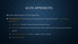
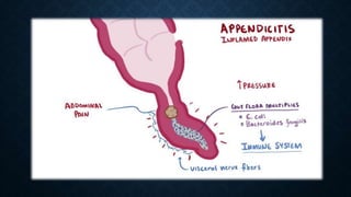
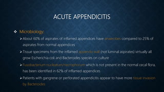
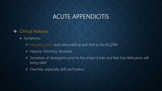
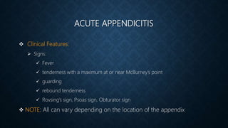
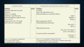
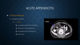
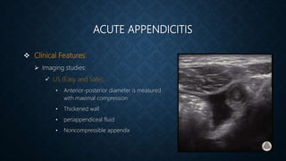
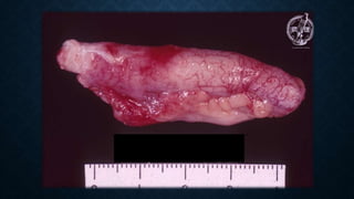
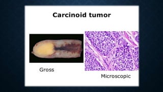
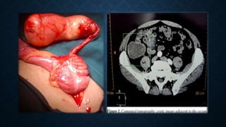
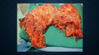
APPENDIX
OBJECTIVES
• History
• Anatomy, and Variants
• Appendiceal Diseases
HISTORY
1492: Leonardo da Vinci depicted the appendix in his anatomic drawings
1544: Jean Fernel for first describing appendiceal disease in a paper published
After death of a girl treated with quince for diarrhea
1736: The first known appendectomy was performed by Claudius Amyand in London
During hernia repair
RLQ pain Thought to be due to typhlitis and perityphlitis
It was recognized that most instances of appendicitis could resolve without surgical
treatment
EMBRYOLOGY
Starts to develop at the 6th week
First noticed at the 8th week
Elongate at the 5th month
The subsequent unequal growth of the lateral wall of the cecum causes the appendix to
find its adult position on the posterior medial wall, just below the ileocecal valve
located by following the longitudinally oriented taeniae coli to their confluence on the
cecum
EMBRYOLOGY
The tip of the appendix can be located anywhere in the right lower quadrant of the
abdomen, pelvis, or retroperitoneum
Gut malrutation: RUQ appendix
Situs inversus: LLQ appendix
ANATOMY
Appendix length: <1 to >30 cm with average of 6-9cm
Outer diameter: varies between 3 and 8 mm
luminal diameter: varies between 1 and 3 mm
Blood supply: ileocolic artery and vein
Innervation: sympathetic by the superior mesenteric plexus (T10-L1) and
parasympathetic via the vagus nerves
ANATOMY
McBurney’s point
Grid Iron incision
Rocky-Davis incision
ANATOMY
Normal variants in appendix
location
Same as colon but with much
more prominent lymphoid
aggregates
Less well developed muscularis
HISTOLOGY
PHYSIOLOGY
Immunologic organ, secretes IgA
A recent meta-analysis demonstrated a significant risk of Crohn’s disease early following
appendicitis
This risk diminishes later, which suggests that a diagnostic rather than a physiologic
relationship exists between appendectomy and Crohn’s disease
function as a reservoir to recolonize the colon with healthy bacteria
ACUTE APPENDICITIS
Acute inflammation of the appendix
 Pathogenesis: The etiology and pathogenesis of appendicitis are not completely
understood
I. Obstruction: fecalith, hypertrophied lymphoid tissue
II. Distention: continuing normal secretion by the appendiceal mucosa and bacterial
growth
III. Vascular compromise: venous, capillaries then arterial
IV. Bacterial invasion
ACUTE APPENDICITIS
 Microbiology:
About 60% of aspirates of inflamed appendices have anaerobes compared to 25% of
aspirates from normal appendices
Tissue specimens from the inflamed appendix wall (not luminal aspirates) virtually all
grow Escherichia coli and Bacteroides species on culture
Fusobacterium nucleatum/necrophorum which is not present in the normal cecal flora,
has been identified in 62% of inflamed appendices
Patients with gangrene or perforated appendicitis appear to have more tissue invasion
by Bacteroides
ACUTE APPENDICITIS
 Natural History:
 Not all patients with appendicitis will progress to perforation
 resolution may be a common event
 two randomized trials comparing early laparoscopy with conservative management of
patients with acute abdominal pain. These studies found three to five times more
patients with appendicitis in the group of patients who were randomized to
laparoscopy
 it has been proposed that nonperforated and perforated appendicitis may, in fact, be
different diseases
ACUTE APPENDICITIS
 Clinical Features:
 Symptoms:
 Migrating Pain: start periumbilical and shift to the RLQ/RIF
 Nausea, Vomiting, Anorexia
 Sensation of obstipation prior to the onset of pain and feel that defecation will
bring relief
 Diarrhea: especially with perforation
ACUTE APPENDICITIS
 Clinical Features:
 Signs:
 Fever
 tenderness with a maximum at or near McBurney’s point
 guarding
 rebound tenderness
 Rovsing’s sign, Psoas sign, Obturator sign
 NOTE: All can vary depending on the location of the appendix
ACUTE APPENDICITIS
 Clinical Features:
 Labs:
 CBC: Leukocytosis (low in lymphopenia or septic reaction ), Lift shift
 ESR
 CRP
 Urinalysis (several white or red blood cells can be present from irritation of the
ureter or bladder, Bacteriuria is generally not seen)
 NOTE: decreasing inflammatory response may indicate spontaneous resolution
ACUTE APPENDICITIS
 Clinical Features:
 The Alvarado score: Better to rule out appendicitis
 Appendicitis Inflammatory Response Score: Better to diagnose appendicitis
ACUTE APPENDICITIS
 Clinical Features:
 Imaging studies:
 CT (Best):
 periappendiceal fat stranding
 thickened mesoappendix,
 Periappendiceal phlegmon
 free fluid
ACUTE APPENDICITIS
 Clinical Features:
 Imaging studies:
 US (Easy and Safe):
• Anterior-posterior diameter is measured
with maximal compression
• Thickened wall
• periappendiceal fluid
• Noncompressible appendix
ACUTE APPENDICITIS
 Clinical Features:
 Imaging studies:
 CXR and AXR
 Technetium-99m–labeled leukocyte scan
ACUTE APPENDICITIS
 DDx:
 The differential diagnosis of acute appendicitis depends on:
 the anatomic location of the inflamed appendix
 the stage of the process (uncomplicated or complicated)
 the patient’s age
 patient’s gender
ACUTE APPENDICITIS
 DDx:
 GI: Mesenteric lymphadenitis, Intussusception, Gastroenteritis, Diverticulitis
 No organic pathologic condition
 GU: Ovarian torsion, PID, ruptured graafian follicle, Endometriosis, Ruptured
ectopic pregnancy, mittelschmerz, Ureteric stones, Pyelonephritis
ACUTE APPENDICITIS
 DDx:
 Pediatric Patient: Mesenteric lymphadenitis (M.C) (associated with RTI, Self-
limiting)
 Elderly Patient: Diverticulitis, Invasive tumors
 Immunosuppressed Patient: appendicitis presents typically, but with relative
leukocytosis, Neutropenic enterocolitis (typhlitis)
ACUTE APPENDICITIS
 Initial Management:
 Uncomplicated Appendicitis
 Non-operative treatment: ATB, 87-91% success but recurrence may occur with
higher rate of complications
 Urgent versus emergent appendectomy: No much difference if surgery was
delayed more that 12h assuming the appendicitis was early diagnosed without
presence of complications
ACUTE APPENDICITIS
 Initial Management:
 Complicated Appendicitis (perforation, abscess, phlegmon)
 Operative versus Nonoperative Management:
 If perforated with peritonitis or sepsis, surgery is a must
 If contained abscess or phlegmon with limited peritonitis , operatic and nonoperative
treatments are options
 Some studies shows that the nonoperative treatment is superior while other studies
shows the opposite
ACUTE APPENDICITIS
 Initial Management:
 Complicated Appendicitis (perforation, abscess, phlegmon)
 Interval Appendectomy Following Nonoperative Management:
 Some studies shows that it reduces the morbidity but not sufficient to clearly show
interval appendectomy rule following nonoperative treatment
ACUTE APPENDICITIS
 Operative Interventions for the Appendix:
 Open Appendectomy:
 By McBurney’s incision or Rocky-Davis incision even in pregnancy
 Expand the same incision if needed, midline incision may be required
 If appendix is normal, examine the whole abdomen
 Valentino’s appendicitis
 Always put a drain
 Remove the mucosa of the remnant of the appendix
ACUTE APPENDICITIS
 Operative Interventions for the Appendix:
 Laparoscopic Appendectomy:
 Appendiceal critical view
 Reduces in hospital stay, pain, scar formation and hernia
 Increase cost, intraoperative time
 Better if the diagnosis is in question
 Laparoscopic Single-Incision Appendectomy (no difference)
ACUTE APPENDICITIS
 Operative Interventions for the Appendix:
 Natural Orifice Transluminal Endoscopic Surgery (NOTES):
 Flexible endoscopes in the abdominal cavity
 Transgastric or Transvaginal
ACUTE APPENDICITIS
 Special Circumstances:
 Acute Appendicitis in the Young:
 diagnosis of acute appendicitis is more difficult in young children
 Higher rate of complications
 If nonperforated give ATB for 24-48hrs, if perforated give ATB up to 24hrs after the
WBC normalize and the patient become afebrile
ACUTE APPENDICITIS
 Special Circumstances:
 Acute Appendicitis in the Young:
 Diagnosis of acute appendicitis is more difficult in young children
 Higher rate of complications
 If nonperforated give ATB for 24-48hrs, if perforated give ATB up to 24hrs after the
WBC normalize and the patient become afebrile followed by appendectomy
ACUTE APPENDICITIS
 Special Circumstances:
 Acute Appendicitis in the Elderly:
 Atypical presentation
 Higher complication rate
ACUTE APPENDICITIS
 Special Circumstances:
 Acute Appendicitis during Pregnancy:
 Commonly in 1st and 2nd trimesters
 Harder to diagnose
 Laboratory evaluation is not helpful in establishing the diagnosis of acute
appendicitis during pregnancy (physiologic leukocytosis “>16,000cell/mm3”)
 No CT, do US or MRI, if still unclear do laparoscopy
 Risk for early delivery and fetal loss
ACUTE APPENDICITIS
 Postoperative care and complications:
 Uncomplicated appendectomy:
 Put on a diet
 No need for ATB
 Discharge within 1 day
ACUTE APPENDICITIS
 Postoperative care and complications:
 Complicated appendectomy:
 Diet depending on clinical exam (ileus or not)
 ATB for 4-7 days
 Surgical site infection: open the incision, culture, I&D
 Stump Appendicitis: recurrent appendicitis approximately 9 years after their initial
surgery due to incomplete resection, may require colectomy
 Incidental appendectomy:
ACUTE APPENDICITIS
 Postoperative care and complications:
 Complicated appendectomy:
 Incidental appendectomy:
 It was estimated that 36 incidental appendectomies had to be performed to prevent one
patient from developing appendicitis
 some special patient groups in whom it should be performed during laparotomy (children
about to undergo chemotherapy, the disabled who cannot describe symptoms or react
normally to abdominal pain, patients with Crohn’s disease in whom the cecum is free of
macroscopic disease, and individuals who are about to travel to remote places where there is
no access to medical or surgical care, malrotation)
NEOPLASMS OF THE APPENDIX
 Prevalence of Neoplasms:
 <1% of appendectomy specimens
 Appendiceal carcinoid and appendiceal adenomas are the
most common
 No clear age relationship
NEOPLASMS OF THE APPENDIX
 Carcinoid:
 Yellow, firm, bulbar
 Carcinoid syndrome in 2.9%
 The tumor can occasionally obstruct the appendiceal lumen much like a fecalith and
result in acute appendicitis
 Treatment:
 <1cm: appendectomy
 1-2cm at base, mesentery or LN involvement: Right hemicolectomy
NEOPLASMS OF THE APPENDIX
 Adenocarcinoma:
 Histologic subtypes: mucinous adenocarcinoma, colonic adenocarcinoma, and
adenocarcinoid
 The most common mode of presentation for appendiceal carcinoma is that of acute
appendicitis
 significant risk for both synchronous and metachronous neoplasms (50% GI)
 Treatment:
 Right hemicolectomy
NEOPLASMS OF THE APPENDIX
 Mucocele:
 obstructive dilatation by intraluminal accumulation of mucoid material, by one of four
processes:
 Retention cysts
 Mucosal hyperplasia
 Cystadenomas
 Cystadenocarcinomas
 When a mucocele is visualized at the time of laparoscopic examination, conversion to
open laparotomy is recommended
NEOPLASMS OF THE APPENDIX
 Mucocele:
 Treatment:
 resection of the appendix, wide resection of the mesoappendix to include all the
appendiceal lymph nodes, collection and cytologic examination of all
intraperitoneal mucus, and careful inspection of the base of the appendix
 Right hemicolectomy if positive margin
NEOPLASMS OF THE APPENDIX
 Pseudomyxoma Peritonei:
 Diffuse collections of gelatinous fluid are associated with mucinous implants on
peritoneal surfaces and omentum
 Caused by neoplastic mucus-secreting cells within the peritoneum, majority originating
in the appendix
 Present with abdominal pain, distention, or a mass
 Progresses slowly and in which recurrences may take years to develop or become
symptomatic
NEOPLASMS OF THE APPENDIX
 Pseudomyxoma Peritonei:
 Treatment:
 Surgical debulking
 Appendectomy
 Hysterectomy with bilateral salpingo-oophorectomy is performed in women
 Adjuvant intraperitoneal hyperthermic chemotherapy
 Radical cytoreductive surgery
 5 year-survival: improved from 30% up to 78%
NEOPLASMS OF THE APPENDIX
 Lymphoma:
 Primary lymphoma of the appendix accounts for 1% to 3% of gastrointestinal
lymphomas
 presents as acute appendicitis and is rarely suspected preoperatively
 Treatment:
 Confined to the appendix: Appendectomy
 Extends beyond the appendix onto the cecum or mesentery: Right hemicolectomy
 Adjuvant therapy: is not indicated for lymphoma confined to the appendix

Appendix

  • 1.
  • 2.
    OBJECTIVES • History • Anatomy,and Variants • Appendiceal Diseases
  • 3.
    HISTORY 1492: Leonardo daVinci depicted the appendix in his anatomic drawings 1544: Jean Fernel for first describing appendiceal disease in a paper published After death of a girl treated with quince for diarrhea 1736: The first known appendectomy was performed by Claudius Amyand in London During hernia repair RLQ pain Thought to be due to typhlitis and perityphlitis It was recognized that most instances of appendicitis could resolve without surgical treatment
  • 4.
    EMBRYOLOGY Starts to developat the 6th week First noticed at the 8th week Elongate at the 5th month The subsequent unequal growth of the lateral wall of the cecum causes the appendix to find its adult position on the posterior medial wall, just below the ileocecal valve located by following the longitudinally oriented taeniae coli to their confluence on the cecum
  • 5.
    EMBRYOLOGY The tip ofthe appendix can be located anywhere in the right lower quadrant of the abdomen, pelvis, or retroperitoneum Gut malrutation: RUQ appendix Situs inversus: LLQ appendix
  • 6.
    ANATOMY Appendix length: <1to >30 cm with average of 6-9cm Outer diameter: varies between 3 and 8 mm luminal diameter: varies between 1 and 3 mm Blood supply: ileocolic artery and vein Innervation: sympathetic by the superior mesenteric plexus (T10-L1) and parasympathetic via the vagus nerves
  • 7.
    ANATOMY McBurney’s point Grid Ironincision Rocky-Davis incision
  • 8.
  • 9.
    Same as colonbut with much more prominent lymphoid aggregates Less well developed muscularis HISTOLOGY
  • 10.
    PHYSIOLOGY Immunologic organ, secretesIgA A recent meta-analysis demonstrated a significant risk of Crohn’s disease early following appendicitis This risk diminishes later, which suggests that a diagnostic rather than a physiologic relationship exists between appendectomy and Crohn’s disease function as a reservoir to recolonize the colon with healthy bacteria
  • 11.
    ACUTE APPENDICITIS Acute inflammationof the appendix  Pathogenesis: The etiology and pathogenesis of appendicitis are not completely understood I. Obstruction: fecalith, hypertrophied lymphoid tissue II. Distention: continuing normal secretion by the appendiceal mucosa and bacterial growth III. Vascular compromise: venous, capillaries then arterial IV. Bacterial invasion
  • 13.
    ACUTE APPENDICITIS  Microbiology: About60% of aspirates of inflamed appendices have anaerobes compared to 25% of aspirates from normal appendices Tissue specimens from the inflamed appendix wall (not luminal aspirates) virtually all grow Escherichia coli and Bacteroides species on culture Fusobacterium nucleatum/necrophorum which is not present in the normal cecal flora, has been identified in 62% of inflamed appendices Patients with gangrene or perforated appendicitis appear to have more tissue invasion by Bacteroides
  • 14.
    ACUTE APPENDICITIS  NaturalHistory:  Not all patients with appendicitis will progress to perforation  resolution may be a common event  two randomized trials comparing early laparoscopy with conservative management of patients with acute abdominal pain. These studies found three to five times more patients with appendicitis in the group of patients who were randomized to laparoscopy  it has been proposed that nonperforated and perforated appendicitis may, in fact, be different diseases
  • 15.
    ACUTE APPENDICITIS  ClinicalFeatures:  Symptoms:  Migrating Pain: start periumbilical and shift to the RLQ/RIF  Nausea, Vomiting, Anorexia  Sensation of obstipation prior to the onset of pain and feel that defecation will bring relief  Diarrhea: especially with perforation
  • 16.
    ACUTE APPENDICITIS  ClinicalFeatures:  Signs:  Fever  tenderness with a maximum at or near McBurney’s point  guarding  rebound tenderness  Rovsing’s sign, Psoas sign, Obturator sign  NOTE: All can vary depending on the location of the appendix
  • 17.
    ACUTE APPENDICITIS  ClinicalFeatures:  Labs:  CBC: Leukocytosis (low in lymphopenia or septic reaction ), Lift shift  ESR  CRP  Urinalysis (several white or red blood cells can be present from irritation of the ureter or bladder, Bacteriuria is generally not seen)  NOTE: decreasing inflammatory response may indicate spontaneous resolution
  • 18.
    ACUTE APPENDICITIS  ClinicalFeatures:  The Alvarado score: Better to rule out appendicitis  Appendicitis Inflammatory Response Score: Better to diagnose appendicitis
  • 20.
    ACUTE APPENDICITIS  ClinicalFeatures:  Imaging studies:  CT (Best):  periappendiceal fat stranding  thickened mesoappendix,  Periappendiceal phlegmon  free fluid
  • 21.
    ACUTE APPENDICITIS  ClinicalFeatures:  Imaging studies:  US (Easy and Safe): • Anterior-posterior diameter is measured with maximal compression • Thickened wall • periappendiceal fluid • Noncompressible appendix
  • 22.
    ACUTE APPENDICITIS  ClinicalFeatures:  Imaging studies:  CXR and AXR  Technetium-99m–labeled leukocyte scan
  • 24.
    ACUTE APPENDICITIS  DDx: The differential diagnosis of acute appendicitis depends on:  the anatomic location of the inflamed appendix  the stage of the process (uncomplicated or complicated)  the patient’s age  patient’s gender
  • 25.
    ACUTE APPENDICITIS  DDx: GI: Mesenteric lymphadenitis, Intussusception, Gastroenteritis, Diverticulitis  No organic pathologic condition  GU: Ovarian torsion, PID, ruptured graafian follicle, Endometriosis, Ruptured ectopic pregnancy, mittelschmerz, Ureteric stones, Pyelonephritis
  • 26.
    ACUTE APPENDICITIS  DDx: Pediatric Patient: Mesenteric lymphadenitis (M.C) (associated with RTI, Self- limiting)  Elderly Patient: Diverticulitis, Invasive tumors  Immunosuppressed Patient: appendicitis presents typically, but with relative leukocytosis, Neutropenic enterocolitis (typhlitis)
  • 27.
    ACUTE APPENDICITIS  InitialManagement:  Uncomplicated Appendicitis  Non-operative treatment: ATB, 87-91% success but recurrence may occur with higher rate of complications  Urgent versus emergent appendectomy: No much difference if surgery was delayed more that 12h assuming the appendicitis was early diagnosed without presence of complications
  • 28.
    ACUTE APPENDICITIS  InitialManagement:  Complicated Appendicitis (perforation, abscess, phlegmon)  Operative versus Nonoperative Management:  If perforated with peritonitis or sepsis, surgery is a must  If contained abscess or phlegmon with limited peritonitis , operatic and nonoperative treatments are options  Some studies shows that the nonoperative treatment is superior while other studies shows the opposite
  • 29.
    ACUTE APPENDICITIS  InitialManagement:  Complicated Appendicitis (perforation, abscess, phlegmon)  Interval Appendectomy Following Nonoperative Management:  Some studies shows that it reduces the morbidity but not sufficient to clearly show interval appendectomy rule following nonoperative treatment
  • 30.
    ACUTE APPENDICITIS  OperativeInterventions for the Appendix:  Open Appendectomy:  By McBurney’s incision or Rocky-Davis incision even in pregnancy  Expand the same incision if needed, midline incision may be required  If appendix is normal, examine the whole abdomen  Valentino’s appendicitis  Always put a drain  Remove the mucosa of the remnant of the appendix
  • 31.
    ACUTE APPENDICITIS  OperativeInterventions for the Appendix:  Laparoscopic Appendectomy:  Appendiceal critical view  Reduces in hospital stay, pain, scar formation and hernia  Increase cost, intraoperative time  Better if the diagnosis is in question  Laparoscopic Single-Incision Appendectomy (no difference)
  • 32.
    ACUTE APPENDICITIS  OperativeInterventions for the Appendix:  Natural Orifice Transluminal Endoscopic Surgery (NOTES):  Flexible endoscopes in the abdominal cavity  Transgastric or Transvaginal
  • 33.
    ACUTE APPENDICITIS  SpecialCircumstances:  Acute Appendicitis in the Young:  diagnosis of acute appendicitis is more difficult in young children  Higher rate of complications  If nonperforated give ATB for 24-48hrs, if perforated give ATB up to 24hrs after the WBC normalize and the patient become afebrile
  • 34.
    ACUTE APPENDICITIS  SpecialCircumstances:  Acute Appendicitis in the Young:  Diagnosis of acute appendicitis is more difficult in young children  Higher rate of complications  If nonperforated give ATB for 24-48hrs, if perforated give ATB up to 24hrs after the WBC normalize and the patient become afebrile followed by appendectomy
  • 35.
    ACUTE APPENDICITIS  SpecialCircumstances:  Acute Appendicitis in the Elderly:  Atypical presentation  Higher complication rate
  • 36.
    ACUTE APPENDICITIS  SpecialCircumstances:  Acute Appendicitis during Pregnancy:  Commonly in 1st and 2nd trimesters  Harder to diagnose  Laboratory evaluation is not helpful in establishing the diagnosis of acute appendicitis during pregnancy (physiologic leukocytosis “>16,000cell/mm3”)  No CT, do US or MRI, if still unclear do laparoscopy  Risk for early delivery and fetal loss
  • 37.
    ACUTE APPENDICITIS  Postoperativecare and complications:  Uncomplicated appendectomy:  Put on a diet  No need for ATB  Discharge within 1 day
  • 38.
    ACUTE APPENDICITIS  Postoperativecare and complications:  Complicated appendectomy:  Diet depending on clinical exam (ileus or not)  ATB for 4-7 days  Surgical site infection: open the incision, culture, I&D  Stump Appendicitis: recurrent appendicitis approximately 9 years after their initial surgery due to incomplete resection, may require colectomy  Incidental appendectomy:
  • 39.
    ACUTE APPENDICITIS  Postoperativecare and complications:  Complicated appendectomy:  Incidental appendectomy:  It was estimated that 36 incidental appendectomies had to be performed to prevent one patient from developing appendicitis  some special patient groups in whom it should be performed during laparotomy (children about to undergo chemotherapy, the disabled who cannot describe symptoms or react normally to abdominal pain, patients with Crohn’s disease in whom the cecum is free of macroscopic disease, and individuals who are about to travel to remote places where there is no access to medical or surgical care, malrotation)
  • 40.
    NEOPLASMS OF THEAPPENDIX  Prevalence of Neoplasms:  <1% of appendectomy specimens  Appendiceal carcinoid and appendiceal adenomas are the most common  No clear age relationship
  • 41.
    NEOPLASMS OF THEAPPENDIX  Carcinoid:  Yellow, firm, bulbar  Carcinoid syndrome in 2.9%  The tumor can occasionally obstruct the appendiceal lumen much like a fecalith and result in acute appendicitis  Treatment:  <1cm: appendectomy  1-2cm at base, mesentery or LN involvement: Right hemicolectomy
  • 43.
    NEOPLASMS OF THEAPPENDIX  Adenocarcinoma:  Histologic subtypes: mucinous adenocarcinoma, colonic adenocarcinoma, and adenocarcinoid  The most common mode of presentation for appendiceal carcinoma is that of acute appendicitis  significant risk for both synchronous and metachronous neoplasms (50% GI)  Treatment:  Right hemicolectomy
  • 44.
    NEOPLASMS OF THEAPPENDIX  Mucocele:  obstructive dilatation by intraluminal accumulation of mucoid material, by one of four processes:  Retention cysts  Mucosal hyperplasia  Cystadenomas  Cystadenocarcinomas  When a mucocele is visualized at the time of laparoscopic examination, conversion to open laparotomy is recommended
  • 46.
    NEOPLASMS OF THEAPPENDIX  Mucocele:  Treatment:  resection of the appendix, wide resection of the mesoappendix to include all the appendiceal lymph nodes, collection and cytologic examination of all intraperitoneal mucus, and careful inspection of the base of the appendix  Right hemicolectomy if positive margin
  • 47.
    NEOPLASMS OF THEAPPENDIX  Pseudomyxoma Peritonei:  Diffuse collections of gelatinous fluid are associated with mucinous implants on peritoneal surfaces and omentum  Caused by neoplastic mucus-secreting cells within the peritoneum, majority originating in the appendix  Present with abdominal pain, distention, or a mass  Progresses slowly and in which recurrences may take years to develop or become symptomatic
  • 49.
    NEOPLASMS OF THEAPPENDIX  Pseudomyxoma Peritonei:  Treatment:  Surgical debulking  Appendectomy  Hysterectomy with bilateral salpingo-oophorectomy is performed in women  Adjuvant intraperitoneal hyperthermic chemotherapy  Radical cytoreductive surgery  5 year-survival: improved from 30% up to 78%
  • 50.
    NEOPLASMS OF THEAPPENDIX  Lymphoma:  Primary lymphoma of the appendix accounts for 1% to 3% of gastrointestinal lymphomas  presents as acute appendicitis and is rarely suspected preoperatively  Treatment:  Confined to the appendix: Appendectomy  Extends beyond the appendix onto the cecum or mesentery: Right hemicolectomy  Adjuvant therapy: is not indicated for lymphoma confined to the appendix