SlideShare a Scribd company logo
1 of 52
Urinary system – overview
Excretory Removal of metabolic toxins and wastes
Chemical Maintenance of 1) water volume; 2) electrolyte concentration in ECF; 3) acid-base balance
Metabolic Vitamin D activation; gluconeogenesis
Endocrine Renin (blood pressure) and erythropoietin (RBC production)
Kidneys Excretion
Ureters, urethra Urine transport
Urinary bladder Urine storage
Kidney anatomy
• Retroperitoneal, superior lumbar region; ~ T12 to L5
• Right is lower; adrenal gland atop; bean-shape; medial side – renal hilum
• Ureters, renal blood vessels, lymphatics, and nerves enter and exit at hilum
• Surrounded by (from superficial to deep): renal fascia, perirenal fat capsule, fibrous
capsule
• Layers: cortex and medulla
• Renal pyramids (medulla) produce urine; separated by renal columns (cortex)
• Urine from papilla goes to minor calyx → major calyx → renal pelvis → ureter
• Pyramid and surrounding columns = lobe
Renal anatomy
Blood and nerve supply
• Kidneys cleanse blood  rich blood supply
• Renal arteries – ~ ¼ (1200 ml) of cardiac output
• Similar paths for arterial and venous flow
• Nerve supply – sympathetic fibers from renal plexus
Renal anatomy
Nephron
• Structural and functional units that form urine; > 1 million per
kidney
• Filtration, reabsorption, and secretion (see bottom left)
• Renal corpuscle (RC): glomerulus and glomerular capsule
• Glomerulus (G): fenestrated capillaries  filtrate
• Like plasma but without proteins and cells; goes into
glomerular capsule
• Renal tubule: proximal convoluted tubule (PCT), nephron
loop, distal convoluted tubule (DCT); reabsorption and
secretion
• DCT enters collecting duct (not the part of a nephron)
Blood/ECF Filtrate
Filtration (RC)
Reabsorption (RT)
Secretion (RT)
Renal anatomy
Urine formation
• 180 L fluid processed daily (60x entire plasma
volume) → only 1.5 L urine
• 20-25% oxygen consumption
Three processes:
• Glomerular filtration
• Cell- and protein-free filtrate of blood plasma
minus proteins
• Tubular reabsorption
• Selectively returns 99% of substances from
filtrate to blood in renal tubules and
collecting ducts
• Tubular secretion
• Selectively moves substances from blood to
filtrate in renal tubules and collecting ducts
• Eventually urine is produced → <1% of
filtrate
• Contains metabolic wastes and unneeded
substances
Renal physiology
Nephron epithelia
Part Type Function and features
Glomerular capsule
(parietal)
Simple
squamous
Structural
Glomerular capsule
(visceral)
Branching
cuboidal
Filtration via filtration slits; located
around capillaries
PCT Cuboidal with
microvilli
↑ surface area; Reabsorption and
secretion; only in cortex
NL (thick
descending limb
Cuboidal with
microvilli
↑ surface area; reabsorption
NL (thin descending
limb)
Simple
squamous
Reabsorption
NL (thick ascending
limb)
Cuboidal to
columnar
Reabsorption
DCT Cuboidal; few
microvilli
Secretion > reabsorption; only cortex
Collecting duct Principal and
intercalated
cells – cuboidal
Principal – sparse, short microvilli; H2O
and Na+
balance
Intercalated (A and B) – many
microvilli; acid-base balance
Types of nephrons
• Cortical – 85% of nephrons; in cortex
• Glomerular capillaries: filtration
• Peritubular capillaries: reabsorption and secretion
• Juxtamedullary: long nephron loops; in the medulla
• Glomerulus: filtration; capillaries are the same as in cortical N.
• Vasa recta and nephron loop form countercurrent system; together with the collecting duct produce
concentrated urine
Nephron capillary beds
Capillary bed Blood flow Blood pressure Location Structure Function
Glomerular afferent a. →
glomerulus →
efferent a.
High (Ø of
afferent a. > Ø
of efferent a.)
Glomerulus Fenestrated,
surrounded by
podocytes
Filtration
Peritubular efferent a. →
capillaries →
venules
Low Around renal
tubule
Fenestrated,
thin-walled
Reabsorption,
secretion
Vasa recta efferent a. →
capillaries →
venules
Low Around
nephron loop
Fenestrated,
thin-walled
Formation of
concentrated
urine
Renal physiology
Filtration pressures
• Passive process; no energy required; from blood to capsule via filtration membrane; no reabsorption
• Capillary hydrostatic pressure pushes fluid into the filtrate; equals BP
• 55 mm Hg; due to the larger afferent a. diameter
• Capsular hydrostatic pressure pushes fluid back into the blood; 15 mm Hg
• Capillary osmotic pressure pulls fluid back into the blood; due to the plasma proteins; 30 mm Hg
• Sum of pressures → net filtration pressure (NFP) = 10 mm Hg (55 one way – 45 other way)
• Determines glomerular filtration rate
• Why there is no capsular osmotic pressure?
Glomerular filtration
Filtration mechanism
• Filtration membrane: fenestrated capillary endothelium; basement
membrane; foot processes of podocytes with filtration slits
• Molecules smaller than 3 nm pass (water, glucose, amino acids,
nitrogenous wastes) – everything but proteins and large biological
macromolecules
• Macromolecules "stuck" in filtration membrane engulfed by glomerular
mesangial cells
• Plasma proteins remain in blood  maintain osmotic pressure 
prevents loss of all water to filtrate
• Proteins in filtrate indicate membrane problem
Glomerular filtration
Glomerular filtration rate (GFR)
• Volume of filtrate formed per minute by both
kidneys (normal = 120–125 ml/min)
• GFR NFP, filtration membrane surface≡
(controlled by mesangial cells) and permeability
(very high)
• Constant GFR = normal filtration → ECF
homeostasis
• GFR affects BP: ↑BP → ↑GFR  ↑urine output
 ↓BP, and vice versa
• Goal of intrinsic controls – maintain GFR in
kidney, not BP
• Goal of extrinsic controls – maintain systemic BP
• Intrinsic controls: local within kidney; maintain
GFR; dominant at normal BP
• Extrinsic controls: nervous, endocrine; maintain
BP; can ↓ kidney function
• Preside over intrinsic if systemic BP < 80 or > 180
mm Hg
• Controlled via glomerular hydrostatic pressure
• If rises  NFP rises  GFR rises; 18% ↓ of
systemic BP → GFR = 0
NFP
Membrane permeability
Surface area
GFR
BP increases
GFR increases
Urine output increases
Blood volume decreases
BP decreases
Glomerular filtration
GFR ↑
Glomerular
blood
flow ↓
GFR ↓
INTRINSIC EXTRINSIC
Juxtaglomerular complex (JGC)
• One per nephron; portions of: 1) ascending limb of nephron loop; afferent and efferent a.
• Regulates GFR and BP
• Macula densa cells (MDC): in ascending limb; sense NaCl in the filtrate
• Granular/juxtaglomerular cells (JC): arteriolar SMC; dilate/constrict afferent a.
• Respond to ↓ stretch of afferent a. releasing renin (BP regulation)
• Extraglomerular mesangial cells (EMC): connected by gap junctions
• Signaling between MDC and JC
Glomerular filtration
Intrinsic controls of GFR
• Myogenic mechanism
• Smooth muscle contracts in response to stretch
• Both help maintain normal GFR despite normal
fluctuations in blood pressure
• Protects glomeruli from ↑ BP
• Tubuloglomerular feedback mechanism
• Directed by macula densa cells; respond to filtrate
NaCl concentration
• Opposite for ↓ GFR
BP
GFR ↑
Stretch↑
GFR ↓
Stretch ↓
Afferent a. constrict
Efferent a. dilate
Afferent a. dilate
Efferent a. constrict
Blood flow ↓ Blood flow ↑
GFR ↓ GFR ↑
↑ ↓
GFR ↑
Filtrate flow ↑
Reabsorption time ↓
Filtrate NaCl ↑
Afferent a. constrict
GFR ↓
Reabsorption time ↑
Filtrate NaCl ↓
Glomerular filtration
Extrinsic controls: BP regulation
BP ↓
Sympathetic response Adrenal medulla Juxtaglomerular cells Macula densa cells Kidneys
Angiotensin
Aldosterone
Na/water retention
BP ↑
Afferent a.
constriction
↓ urine
Reduced stretch
BV ↑
Norepinephrine
Vasoconstriction
Epinephrine
Filtrate NaCl ↓
Adenosine
Prostaglandin E2
Renin
1
1,2
2
• Under normal conditions blood vessels dilated, intrinsic controls prevail
• Three major mechanisms of BP regulation: 1) direct sympathetic stimulation; 2) renin-angiotensin-
aldosterone cascade; 3) kidney paracrine/endocrine signaling
Glomerular filtration
Transendothelial process
• Most but not all of chemicals in filtrate are reabsorbed (selective process)
• Organic nutrients (sugars, aminoacids) reabsorbed ≈completely; ions/water hormonally regulated
• Active and passive transport via transcellular or paracellular routes
• Transcellular: apical membrane → cytoplasm → basolateral membrane → ECF → peritubular capillaries
• Paracellular: between cells, controlled by tight junctions
Tubular reabsorption
Roles for 1° and 2° active
transport
• Requires ATP; 1° transport of Na+
at basal membrane → ↓
intracellular Na+
, (-) intracellular potential → Na+
diffusion via
brush border
• Na+
diffusion via brush border → coupled with 2° active
transport → reabsorption of glucose (sodium-glucose co-
transporter, SGLT), AA, H+
(sodium-hydrogen exchanger, NHE)
etc.
• Na+
reabsorption → H2O reabsorption by osmosis → ↓
chemical concentrations in filtrate → concentration gradient
→ passive reabsorption of Cl-
, urea, lipid-soluble substances
Tubular reabsorption
PCT
Chemical Apical membrane Basolateral membrane
Na+
Facilitated diffusion 1° AT (Na+
/K+
pump)
Water Osmosis (obligate water reabsorption via aquaporins)
K+
, Cl-
, Ca2+
, Mg2+
, HCO3
–
, PO4
3-
2° AT (Na+
symport), facilitated
diffusion (Mg2+
)
Facilitated diffusion, paracellular
diffusion (K+
, Ca2+
, Mg2+
)
AA, glucose, fructose, galactose,
lactate, succinate, citrate
2° AT (Na+
symport) Facilitated diffusion
• All nutrients, e.g., glucose
and amino acids
• 65% of Na+
and H2O; other
ions
• ~ all uric acid; ½ urea (later
secreted back into filtrate)
Nephron loop
Thin descending limb
Water Osmosis (obligate reabsorption via aquaporins)
Thick ascending limb
Chemical Apical membrane Basolateral membrane
Na+
Facilitated diffusion 1° AT (Na+
/K+
pump), paracellular diffusion
K+
, Cl-
, Ca2+
, Mg2+
2° AT (Na+
cotransporter, except Mg2+
) Paracellular diffusion ( K+
, Ca2+
, Mg2+
)
• Descending thin limb: H2O
(main function), also Na+
and
urea
• Ascending thick limb: ions
(Na+, Ca2+
, Mg2+
, HCO3
-
etc)
• Ascending thick limb is
impermeable to water
• Diluted tubular fluid at the
end of the ascending limb
• Allows kidneys to regulate
urine concentration
• Loop diuretics inhibit (1-Na+
,
2-Cl-
, 1-K+
) cotransporter
Tubular reabsorption
DCT
Chemical Apical membrane Basolateral membrane
Na+
2° AT (Na+
/Cl-
cotransporter)
1° AT (Na+
/K+
pump),
paracellular diffusion
Cl-
2° AT (Na+
/Cl-
cotransporter)
Facilitated diffusion
HCO3
-
Forms intracellularly HCO3
-
/Cl-
cotransporter
• Early DCT: ions (Na+
, K+
, Cl-
etc.); no water or urea
• Thiazide diuretics inhibit
Na+
/Cl-
symport
• Late DCT: Na+
reabsorption,
K+
excretion (aldosterone)
• K+
-sparing diuretics:
• Aldosterone antagonists XXX
A. receptor
• Na+
channel blockers
indirectly XXX Na+
/K+
pump
• DCT is important for acid-
base balance
Tubular reabsorption
Collecting duct. Summary
Ion Apical membrane Basolateral
membrane
Na+
2° AT (Na+
/Cl-
cotransporter)
1° AT (Na+
/K+
pump),
paracellular diffusion
Cl-
2° AT (Na+
/Cl-
cotransporter)
Facilitated diffusion
HCO3
-
Forms intracellularly HCO3
-
/Cl-
cotransporter
• Permeable to H2O via aquaporins (number is
regulated by ADH)
• Permeable to urea (diffuses into medullary
interstitium); helps to regulate urine
concentration
• H+
secretion → acid-base balance regulation
Summary of tubular reabsorption (<1 – net
reabsorption, >1 – net secretion; H2O – 1)
Tubular reabsorption
Transport maximum
• Transport maximum (Tm) for ~ many reabsorbed
substances (see below)
• Reflects number of carriers/pumps in the
membranes of tubular cells
• When carriers saturated, excess excreted in urine
• E.g., hyperglycemia  filtered load is higher than
reabsorption → exceed Tm  glucose in urine
• Some – gradient-time transport (Vd = D*(C1-C2); Vd –
diffusion rate; D = diffusion coefficient)
• Example: Na+
in the PCT
Tubular reabsorption
Summary and regulation
(3)
(1)
(2)
(4)
(5)
1 2 3 4 5
Adrenal gland Liver Posterior pituitary gland Heart Parathyroid gland
Some compounds
Regulation
Tubular reabsorption
Functions and features
• From ECF to tubular fluid; almost all in PCT
• K+
, H+,
NH4
+
, creatinine, organic acids and bases, drugs, urea,
uric acid
• Some are from tubular cells, e.g. HCO3
-
• Disposes of substances (e.g., drugs) bound to plasma
proteins
• Disposing of undesirable passively reabsorbed substances
(e.g., urea and uric acid)
• Disposing of excess K+
(aldosterone)
• Acid-base regulation by secreting H+
or HCO3
–
in urine
Location Compounds secreted
PCT Bile salts, oxalate, urate, catecholamines,
drugs (e.g., penicillin), salicylates, H+, NH
4
+
Nephron loop Urea (thin ascending limb), H+
(thick
ascending limb)
DCT K+
(aldosterone-regulated), H+
Collecting duct H+
Tubular secretion
Urine concentration and volume
• Osmolality: number of solute particles in 1 kg of H2O; in milliosmols (mOsm); ability to cause osmosis
• Osmolality of plasma – ~300 mOsm via regulating urine concentration and volume
• Countercurrent mechanism: fluid flows in opposite directions in two adjacent segments of same tube
with hair pin turn
• Countercurrent multiplier – filtrate flow in the nephron loops of juxtamedullary nephrons
• Countercurrent exchanger - blood flow in vasa recta
• Establish and maintain osmotic gradient (300 mOsm to 1200 mOsm) from renal cortex through medulla →
allows to vary urine concentration
Osmolality. Countercurrent mechanism
Countercurrent multiplier: nephron loop
• Descending limb: freely permeable to H2O; medullary IF is hyperosmotic → H2O from filtrate to medullary
IF → filtrate osmolality ↑ to ~1200 mOsm
• Ascending limb: impermeable to H2O, selectively permeable to solutes → Na+
, Cl–
reabsorbed → filtrate
osmolality ↓ to 100 mOsm
• Constant 200 mOsm difference between two limbs of nephron loop and between ascending limb and IF
• Difference "multiplied" along length of loop to ~ 900 mOsm
Urine concentration and volume
Countercurrent exchanger: vasa recta (VR)
• Ascending vasa recta remove reabsorbed H2O from 1) descending VR; 2) nephron loop (descending limb);
3) collecting duct; → volume of blood at end of vasa recta higher than at beginning
• NaCl: diffuses from concentrated IF into descending VR → goes into ascending VR → diffuses from
ascending VR into diluted IF → preservation of medullary gradient by recycling NaCl
• Gradient allows H2O reabsorption from collecting duct if aquaporins are present (regulated by ADH)
Urine concentration and volume
ADH regulates urine concentration Urine concentration and volume
Urea helps to maintain medullary osmotic gradient
• Descending limb: water reabsorption → urea in filtrate ↑
• Ascending thin limb: passive diffusion of urea from the IF, water reabsorption → urea in filtrate ↑
• Ascending thick limb: no water or urea transport
• Cortical collecting duct: water reabsorption reabsorbs water → urea in filtrate ↑
• Medullary collecting duct: high filtrate urea  diffusion into medullary IF  high osmolality in medulla
Urine concentration and volume
Diuretics increase urinary output
Acetazolamide (1) PCT XXX carbonic anhydrase; ↓ blood pH, excretion of HCO3
-
, Na+
; H2O follows
Osmotic d. (2) PCT, DL of NL E.g., mannitol; not reabsorbed, ↑ filtrate osmolality; prevent H2O reabsorption
Loop d. (3) AL of NL Inhibit 1-Na+
, 2-Cl-
, 1-K+
cotransporter → ↑ Na+
excretion → H2O follows
Thiazid d. (4) DCT Inhibit Na+
/Cl-
symport → ↑ Na+
excretion → H2O follows
K+
-sparing d. (5) DCT, CD Aldosterone inhibitors → ↑ Na+
excretion → H2O follows, K+
stays (sparing)
Na+
channel inhibitors DCT, CD Indirectly inhibit Na+
/K+
basolateral pump → ↑ Na+
excretion → H2O follows
ADH inhibitors CD Decrease water reabsorption → ↑ H2Oexcretion
Urine concentration and volume
Evaluation of kidney function. Renal clearance
• Kidney function is to filter out from/reabsorb chemicals in the
blood
• To determine, one has to know both plasma and urine
concentrations
• Renal clearance of a substance is the volume of plasma that
kidneys clear of this substance in a given time
• Used to determine GFR; helps to detect glomerular damage
• C = UV/P
• Measured using Inulin (plant polysaccharide)
• Freely filtered; neither reabsorbed nor secreted by kidneys; its renal clearance = GFR = 125 ml/min
C renal clearance rate (ml/min)
U concentration (mg/ml) of substance in urine
V flow rate of urine formation (ml/min)
P concentration of same substance in plasma
If C… Substance is…
= 0 completely reabsorbed or not filtered
= 125 ml/min no net reabsorption/secretion; freely filtered
< 125 ml/min reabsorbed
> 125 ml/min secreted
Clinical evaluation
Chronic kidney disease
• Several stages with varying degrees of ↓ GFR; chronic renal disease: < 60 ml/min for 3 months
• May be caused by diabetes mellitus, hypertension, damage to kidneys after infection
• Renal failure: GFR < 15 ml/min; leads to uremia – ↑ urea in blood, ionic and hormonal imbalances;
metabolic abnormalities
• Treated with hemodialysis or transplant
Clinical evaluation
Control of water balance Electrolyte balance
• In = out = ~ 2500 ml/day
• Obligatory water losses: urine (60%),
skin and lungs, perspiration, and feces
(2.5 L/day)
• Water intake: beverages, food,
metabolic water (2.5 L/day)
• Regulated vi ADH (hypothalamus); ↑
osmolality → ↑ thirst
• Dehydration (diarrhea, burns, fever
etc.) also increases ADH
Dehydration
•Hemorrhage, severe burns, vomiting, diarrhea,
sweating, water deprivation, diuretics, endocrine
diseases
•Signs and symptoms: "cottony" oral mucosa, thirst,
dry flushed skin, oliguria
•Weight loss, fever, mental confusion, hypovolemic
shock, and loss of electrolytes
Hypotonic hydration
•Renal insufficiency or overhydration
•ECF osmolality ↓ → hyponatremia → net osmosis
into tissue cells → swelling of cells → nausea,
vomiting, muscular cramping, cerebral edema →
death
•Treated with hypertonic saline
Edema
•↑ fluid out of blood: ↑ HPC (incompetent venous
valves, localized blood vessel blockage, congestive
heart failure, ↑ blood volume) and permeability
(inflammatory response)
•↓ fluid into blood: ↓OPC (hypoproteinemia),
malnutrition, liver disease, glomerulonephritis
Control of water balance Electrolyte balance
• Blocked (or surgically removed) lymphatic vessels: proteins accumulate in IF, ↑ OPIF
• Increases diffusion distance for nutrients and oxygen; ↓ BP, impaired circulation
Control of sodium ECF content Electrolyte balance
• Main component of osmotic pressure; regulates ECF
volume; water distribution; acid-base control
• Concentration is stable (water moves between ECF and ICF
due to osmosis)
• Body content changes (changes in BP and BV)
• No receptors; regulation through the blood volume
• Na+
is reabsorbed in PCT, NL, and DCT; not secreted
• Water follows Na+
; ↑Na+
in filtrate → diluted urine
• Renin/angiotensin/aldosterone system if ↑K+
or ↓Na+
→
Na+
reabsorption, K+
secretion; BV ↑; slow
• ANP in response to ↑ BP → natriuresis, water
secretion, ↓BV and BP; vasodilation
• Inhibits renin, ADH, and aldosterone release
• Estrogen: ↑ NaCl reabsorption → H2O retention
(menstrual cycle, pregnancy) → edema
• Progesterone: ↓ Na+
reabsorption (blocks
aldosterone); promotes Na+
and H2O loss
• Glucocorticoids: ↑ Na+
reabsorption → edema
Anions
• Cl–
is major anion in ECF; 99% are reabsorbed
under normal pH conditions
• Acidosis ↓ Cl-
reabsorption
• Other anions have transport maximums and
excesses are excreted in urine
Control of sodium ECF content. Anions Electrolyte balance
Control of potassium ECF concentration. Anions Electrolyte balance
• K+
concentration in plasma/ECF affects resting membrane potential
• ↑K+
– ↓ excitability; ↓K+
– hyperpolarization, nonresponsiveness
• Control is complicated – 98% K+
is in cells
• Cellular regulation: shift between ECF and ICF
• Renal regulation: reabsorption in PCT, secretion in NL and collecting duct
• ↑ K+
in ECF (diet intake) → ↑ secretion (without aldosterone)
• ↑ K+
in ECF → ↑ aldosterone → ↑ secretion (Na+
retained)
• ↑ Na+
intake → ↑ tubular flow rate → ↑ secretion
• If K+ intake is low, secretion ↓ - passive mechanism
Control of calcium ECF concentration Electrolyte balance
• Mostly in the bones; regulates blood clotting, membrane permeability,
secretion, neuromuscular excitability
• ↑ Ca2+
→ ↑ excitability and muscle tetany
• ↓ Ca2+
→ ↓ neurons and muscle cells, heart arrhythmias
• Controlled by PTH; ↑ Ca2+
renal reabsorption (98%)
• ↓ phosphate ion reabsorption (reabsorbed mainly in PCT), ↑ excretion
(passively)
• Via Ca2+
channels (apical membrane), Ca2+
pumps and antiporters (basal
membrane), transcellular route
• Insulin ↑ and glucagon ↓ Ca2+
reabsorption
Acid-base balance. Buffer systems
• Sources of H+
: protein breakdown (phosphoric acid), lactic acid, fatty acids, ketone bodies
• CO2 + H2O → H2CO3 → HCO3
-
+ H+
• H+ regulated by: buffer systems (rapid), respiratory centers (rapid), renal mechanisms (slow, most potent)
• Strong bases quickly bind H+
; strong acids quickly bind OH-
Bicarbonate buffer
• Mixture of H2CO3 (weak acid) and salts of HCO3
–
(e.g., NaHCO3, a medium base)
• Buffers ICF and ECF; only important ECF buffer
Arterial blood Venous blood, IF ICF Alkalosis Acidosis
pH 7.4 pH 7.35 pH 7.0 pH > 7.45 pH < 7.35
Phosphate buffer system
• NaH2PO4 , a weak acid; Na2HPO4, a weak base
• Buffers urine and ICF (PO4
3-
concentrations are high)
Protein buffer system
• Buffer both plasma and ICF
• Protein molecules are amphoteric (both weak acid and
week base)
• pH ↑ → COOH groups release H+
, become COO-
• pH ↓ → -NH2 groups bind H+
, become NH3+
Acid-base balance
Renal and respiratory compensatory systems
• Respiratory and renal systems are major (slow) regulators of pH; higher capacity than chemical buffers
• Chemical buffers cannot eliminate excess acids or bases from body
• Lungs: eliminate carbonic acid by eliminating CO2
• Kidneys: eliminate nonvolatile acids (phosphoric, uric, lactic acids; ketones); prevent metabolic acidosis
• Kidneys: regulate blood levels of bases; renew chemical buffers
• CO2 unloading → reaction shifts to left (and H+
incorporated into H2O)
• CO2 loading → reaction shifts to right (and H+
buffered by proteins)
CO2 ↑
H+
↑ medullary receptors
chemoreceptors
respiratory rate and depth ↑H+
↓
H+
↓ respiratory center ↓ H+
↑
• Hypercapnia → medullary chemoreceptors → ↑
respiratory rate and depth
• ↑ plasma H+
→ peripheral chemoreceptors → ↑
respiratory rate and depth
• CO2 is removed from blood; H+
↓
• Alkalosis (hypocapnea) → ↓ respiratory rate and
depth decrease; H+
↑
• Hypoventilation → respiratory acidosis
• Hyperventilation → respiratory alkalosis
Respiratory regulation
Acid-base balance
• Renal mechanisms are most important;
retain/secrete H+
• Cannot reabsorb HCO3
–
, have to go around
• H+
secretion in PCT and collecting duct
• H+
comes from H2CO3 produced inside cells
• As H+
secreted, Na+
reabsorbed
Acid-base regulation in kidneys (1) Acid-base balance
• Rate of H+
secretion changes with ECF CO2 levels; ↑ CO2 in peritubular capillary blood  ↑ H+
+ secretion
• To maintain alkaline reserve kidneys must replenish bicarbonate; cannot reabsorb
• PCT cells: CO2 + H2O → H2CO3 → H+
(secreted) + HCO3
–
(into capillary blood) – steps 1-3
• Filtrate: H+
+ HCO3
–
→ H2CO3 → CO2 (into cells, starts over) + H2O – steps 4-6
Acid-base regulation in kidneys (2) Acid-base balance
• New HCO3
–
has to be produced and added to alkaline reserve (balancing dietary H+
)
• Via renal excretion of acid (via secretion and excretion of H+
or NH4
+
)
• Excreted H+
is buffered by phosphate in the urine
Replenishing alkaline reserve Acid-base balance
Ammonium excretion
• NH4
+
– important mechanism for excreting acid
• Glutamine in PCT cells → 2 NH4
+
, 2 "new" HCO3
–
• HCO3
–
→ blood; NH4
+
→ in urine
• Replenishes alkaline reserve of blood
• In alkalosis, type B intercalated cells
secrete HCO3
–
and reclaim H+
• Mechanism is opposite of HCO3
–
ion
reabsorption type A intercalated cells
• Even in alkalosis, more HCO3
–
is
conserved than excreted
Acid-base balance
Type Acidosis Alkalosis
Respiratory Most important indicator is blood PCO2
PCO2 > 45 mm Hg
Decreased in ventilation or gas exchange
CO2 accumulates in blood
PCO2
<35 mm Hg
Common result of hyperventilation
Often due to stress or pain
CO2 eliminated faster than produced
Metabolic Abnormally ↓ HCO3
-
Ingestion of too much alcohol (→ acetic acid)
Excessive loss of HCO3
–
(e.g., persistent diarrhea)
Accumulation of lactic acid (exercise or shock)
Less common than metabolic acidosis
↑ blood pH and HCO3
–
Vomiting of acid contents of stomach
Intake of excess base (e.g., antacids)
Symptoms Diabetic ketosis, starvation, kidney failure
Blood pH below 6.8 → depression of CNS → coma
→ death
Blood pH above 7.8 → excitation of nervous system
→ muscle tetany, extreme nervousness, convulsions,
death often from respiratory arrest
• Failure of respiratory system to regulate pH – respiratory acidosis/alkalosis
• Other abnormalities other than caused by abnormal PCO2 – metabolic acidosis/alkalosis
• Respiratory system tries to compensate metabolic acid-base imbalances; renal system – respiratory ones
• Respiratory system cannot compensate for respiratory acidosis or alkalosis
• Renal system cannot compensate for acid-base imbalances caused by renal problems
Acid-base balance
Imbalance Respiratory Metabolic
Acidosis Alkalosis Acidosis Alkalosis
Responses Excretion of H+
Retention of H+
↑ H+
→ ↑ rate and
depth of breathing
↓ H+
→ ↓ rate and depth of
breathing
Results H+
excretion → ↑ HCO3
-
↑ PCO2 (acidosis)
H+
retention → ↓ HCO3
-
↓ PCO2 (alkalosis)
pH < 7.35 (acidosis)
HCO3
-
↓, PCO2 ↓
pH > 7.45 (alkalosis)
HCO3
-
↑, PCO2 ↑ (> 45 mm Hg)
Respiratory and renal compensation mechanisms
Physical and chemical characteristics
Property Normal Abnormal
Color • Pale to deep yellow (urochrome from
hemoglobin breakdown)
• More concentrated urine  deeper color
Pink, brown, smoky (food ingestion, bile
pigments, blood, drugs)
Transparency Clear Cloudy (may indicate UTI)
Odor • Slightly aromatic when fresh
• Ammonia odor upon standing (bacteria
metabolize solutes)
May be altered by some drugs and
vegetables
pH Slightly acidic (~pH 6, within 4.5 to 8.0) • Acidic diet (protein, whole wheat)  ↓
pH
• Alkaline diet (vegetarian), prolonged
vomiting, or urinary tract infections 
↑pH
Specific gravity 1.001 – 1.035 Out of the range
Composition Water – 95%
Solutes – 5%
Nitrogenous wastes
Urea (from amino acid breakdown) Uric acid
(from nucleic acid metabolism)
Creatinine (from creatine phosphate)
Other normal solutes: Na+
, K+,
PO4
3–
, and
SO4
2–
, Ca2+
, Mg2+
and HCO3
–
Abnormally ↑ or ↓ concentrations of any
constituent – pathology (e.g., glycosuria in
diabetes)
Abnormal components - pathology (e.g.,
blood proteins, WBCs, bile pigments)
Urine transport, storage, and elimination
Ureters
• Convey urine from kidneys to bladder; begin at L2 from renal pelvis; retroperitoneal
• Enter base of bladder through posterior wall; bladder pressure ↑ → distal ends of ureters close → no urine
backflow
Urine transport, storage, and elimination
Layer Structure Function
Mucosa Transitional epithelium Secretion, protection
Muscularis Smooth muscle Contraction in response to stretch; propels urine
Adventitia Fibrous connective tissue Support
Urinary bladder
• Muscular sac; temporary stores urine; retroperitoneal, on pelvic floor
• Posterior to pubic symphysis, superior to prostate (males), anterior to vagina and uterus (females)
• Ureters in, urethra out; trigone – between openings of ureters and urethra; most frequently infected
• Collapses when empty; expands and rises during filling; pressure remains stable
• ~ Full bladder 12 cm long; holds ~ 500 ml; can hold ~ twice that if necessary, but can burst
Urine transport, storage, and elimination
Layer Structure Function
Mucosa Transitional
epithelium
Secretion,
protection
Muscularis Smooth detrusor
muscle (3 layers)
Contraction;
propels urine
Adventitia Fibrous connective
tissue
Support
Urethra
Epithelium Where
Transitional Near bladder
Stratified
squamous
External
urethral
orifice
Sphincters Muscles Where Control
Internal
urethral
Smooth Bladder-urethra
junction
Involuntary
(ANS)
External
urethral
Skeletal At pelvic floor Voluntary
• Drains urinary bladder
• Female: 3–4 cm; bound to anterior vaginal wall; orifice
anterior to vaginal opening; posterior to clitoris
• Male: carries semen and urine; prostatic (2.5 cm),
membranous (2 cm), and spongy urethra (15 cm)
Urine transport, storage, and elimination
• Reflexive urination (urination in infants): pontine control (storage and micturition) centers develop only
between 2 and 3 yo (right part of the picture is absent in kids)
Micturition (urination) Urine transport, storage, and elimination
• Incontinence: usually weak pelvic muscles
• Stress incontinence: increased intra-abdominal
pressure forces urine through external sphincter
• Overflow incontinence: urine dribbles when
bladder overfills
• Urinary retention: bladder unable to expel urine;
common after general anesthesia; hypertrophy of
prostate; treatment is catheterization
Pathologies of micturition
• Frequent micturition in infants: small bladders
and less-concentrated urine
• Incontinence in infants: normal; control of
voluntary urethral sphincter develops with
nervous system
• Urinary tract infections: E. coli (≈80%), Klebsiella, Proteus mirabilis, Enterobacter, Streptococcus
saprophyticus, Staphylococcus aureus, Enterococcus faecalis
• Untreated infections may cause long-term renal damage → hypertension (?)
• Sexually transmitted diseases: gonorrhea, chlamydiasis etc. may lead to urinary tract inflammation and
kidney problems
• Elderly people: most have abnormal kidneys histologically
• Kidneys shrink; nephrons decrease in size and number; tubule cells less efficient
• GFR ½ that of young adult by age 80; atherosclerosis of renal arteries (?)
• Bladder shrinks; loss of bladder tone  nocturia and incontinence
Urine transport, storage, and elimination
Common congenital abnormalities
• Horseshoe kidney (left): two kidneys fuse across midline  single U-shaped kidney; usually asymptomatic
• Hypospadias (right): urethral orifice on ventral surface of penis; corrected surgically at ~ 12 months
• Polycystic kidney disease (left): many fluid-filled cysts interfere with function
• Autosomal dominant form – less severe, more common; autosomal recessive – more severe
• Cause unknown but involves defect in signaling proteins
Urinary diseases
Kidney infections. Renal calculi
• Urinary tract infections can reach the kidneys; cause inflammation of the pelvis, calyces, or entire organ
• Pyelitis – pelvis and calyces are affected (left top); pyelonephritis – whole organ is affected (left bottom)
• Treatment – antibiotics, sometimes surgical removal of affected kidney
• Renal calculi – kidney stones in renal pelvis; formed when calcium, magnesium, or uric acid salts crystalize
• Large stones block ureter  pressure & pain
• Causes: chronic bacterial infection, urine retention, ↑Ca2+ in blood, ↑pH of urine
• Treatment - shock wave lithotripsy – noninvasive; shock waves shatter calculi
Urinary diseases

More Related Content

What's hot

PHYSIOLOGY OF CONNECTING TUBULE AND COLLECTING DUCT
PHYSIOLOGY OF CONNECTING TUBULE AND COLLECTING DUCTPHYSIOLOGY OF CONNECTING TUBULE AND COLLECTING DUCT
PHYSIOLOGY OF CONNECTING TUBULE AND COLLECTING DUCTAhad Lodhi
 
counter-current-mechanism
counter-current-mechanism counter-current-mechanism
counter-current-mechanism Raghu Veer
 
RENAL PHYSIOLOGY REVISION NOTES
RENAL PHYSIOLOGY REVISION NOTESRENAL PHYSIOLOGY REVISION NOTES
RENAL PHYSIOLOGY REVISION NOTESTONY SCARIA
 
Urine formation and micturition
Urine formation and micturitionUrine formation and micturition
Urine formation and micturitionSoneeshah
 
Reabsorption In Renal Tubule (The Guyton and Hall physiology)
Reabsorption In Renal Tubule (The Guyton and Hall physiology)Reabsorption In Renal Tubule (The Guyton and Hall physiology)
Reabsorption In Renal Tubule (The Guyton and Hall physiology)Maryam Fida
 
Renal regulation of metabolic acidosis and alkalosis
Renal regulation of metabolic acidosis and alkalosis Renal regulation of metabolic acidosis and alkalosis
Renal regulation of metabolic acidosis and alkalosis Sana Arbab
 
Hepatic physiology & liver function tests
Hepatic physiology & liver function testsHepatic physiology & liver function tests
Hepatic physiology & liver function testsImran Sheikh
 
Body Fluid And Electrolyte Balance
Body Fluid And Electrolyte BalanceBody Fluid And Electrolyte Balance
Body Fluid And Electrolyte Balancemvraveendrambbs
 
urine concentration and dilution
urine concentration and dilutionurine concentration and dilution
urine concentration and dilutionDr.Nusrat Tariq
 
Renal phsyiology edited
Renal phsyiology editedRenal phsyiology edited
Renal phsyiology editedBrajesh Lahri
 
role of kindey in acid base balance
role of kindey in acid base balancerole of kindey in acid base balance
role of kindey in acid base balanceBharathiPriyan3
 
Urinary system.pdf
Urinary system.pdfUrinary system.pdf
Urinary system.pdfRAMDAS BHAT
 

What's hot (20)

PHYSIOLOGY OF CONNECTING TUBULE AND COLLECTING DUCT
PHYSIOLOGY OF CONNECTING TUBULE AND COLLECTING DUCTPHYSIOLOGY OF CONNECTING TUBULE AND COLLECTING DUCT
PHYSIOLOGY OF CONNECTING TUBULE AND COLLECTING DUCT
 
counter-current-mechanism
counter-current-mechanism counter-current-mechanism
counter-current-mechanism
 
Unit 4 Notes
Unit 4 NotesUnit 4 Notes
Unit 4 Notes
 
RENAL PHYSIOLOGY REVISION NOTES
RENAL PHYSIOLOGY REVISION NOTESRENAL PHYSIOLOGY REVISION NOTES
RENAL PHYSIOLOGY REVISION NOTES
 
Urine formation and micturition
Urine formation and micturitionUrine formation and micturition
Urine formation and micturition
 
Reabsorption In Renal Tubule (The Guyton and Hall physiology)
Reabsorption In Renal Tubule (The Guyton and Hall physiology)Reabsorption In Renal Tubule (The Guyton and Hall physiology)
Reabsorption In Renal Tubule (The Guyton and Hall physiology)
 
Renal regulation of metabolic acidosis and alkalosis
Renal regulation of metabolic acidosis and alkalosis Renal regulation of metabolic acidosis and alkalosis
Renal regulation of metabolic acidosis and alkalosis
 
general physiology - kidney
general physiology - kidneygeneral physiology - kidney
general physiology - kidney
 
Hepatic physiology & liver function tests
Hepatic physiology & liver function testsHepatic physiology & liver function tests
Hepatic physiology & liver function tests
 
Renal physiology
Renal physiology Renal physiology
Renal physiology
 
Urinary system
Urinary systemUrinary system
Urinary system
 
Body Fluid And Electrolyte Balance
Body Fluid And Electrolyte BalanceBody Fluid And Electrolyte Balance
Body Fluid And Electrolyte Balance
 
EXCRETORY SYSTEM
EXCRETORY SYSTEMEXCRETORY SYSTEM
EXCRETORY SYSTEM
 
Uninary System
Uninary SystemUninary System
Uninary System
 
urine concentration and dilution
urine concentration and dilutionurine concentration and dilution
urine concentration and dilution
 
Urinary system
Urinary systemUrinary system
Urinary system
 
Kidney127
Kidney127Kidney127
Kidney127
 
Renal phsyiology edited
Renal phsyiology editedRenal phsyiology edited
Renal phsyiology edited
 
role of kindey in acid base balance
role of kindey in acid base balancerole of kindey in acid base balance
role of kindey in acid base balance
 
Urinary system.pdf
Urinary system.pdfUrinary system.pdf
Urinary system.pdf
 

Similar to Renal system

Similar to Renal system (20)

702 kidney
702 kidney702 kidney
702 kidney
 
Physiology of kidney zamala.pdf
Physiology of kidney zamala.pdfPhysiology of kidney zamala.pdf
Physiology of kidney zamala.pdf
 
A Chapter 8- Renal Physiology-1.ppt
A Chapter 8- Renal Physiology-1.pptA Chapter 8- Renal Physiology-1.ppt
A Chapter 8- Renal Physiology-1.ppt
 
Renal Physiology.ppt
Renal Physiology.pptRenal Physiology.ppt
Renal Physiology.ppt
 
Urinary system
Urinary systemUrinary system
Urinary system
 
Urinary system
Urinary systemUrinary system
Urinary system
 
URINARY SYSTEM.ppt
URINARY SYSTEM.pptURINARY SYSTEM.ppt
URINARY SYSTEM.ppt
 
Renal anatomy and physiology in relation.pptx
Renal anatomy and physiology in relation.pptxRenal anatomy and physiology in relation.pptx
Renal anatomy and physiology in relation.pptx
 
A RENAL ppt.pptx
A RENAL ppt.pptxA RENAL ppt.pptx
A RENAL ppt.pptx
 
Renal Lectures
Renal LecturesRenal Lectures
Renal Lectures
 
Urinary
UrinaryUrinary
Urinary
 
Kidney
KidneyKidney
Kidney
 
Renal Physiology.pdf
Renal Physiology.pdfRenal Physiology.pdf
Renal Physiology.pdf
 
Renal physiology and its anesthetic implications
Renal physiology and its anesthetic implicationsRenal physiology and its anesthetic implications
Renal physiology and its anesthetic implications
 
EXCRETORY-PRODUCTS-THEIR-ELIMINATION.pptx
EXCRETORY-PRODUCTS-THEIR-ELIMINATION.pptxEXCRETORY-PRODUCTS-THEIR-ELIMINATION.pptx
EXCRETORY-PRODUCTS-THEIR-ELIMINATION.pptx
 
Functional anatomy and physiology of kideny
Functional anatomy and physiology of kidenyFunctional anatomy and physiology of kideny
Functional anatomy and physiology of kideny
 
Renal physiology-1
Renal physiology-1Renal physiology-1
Renal physiology-1
 
renal-physiology-1-1704071learihskhd.pdf
renal-physiology-1-1704071learihskhd.pdfrenal-physiology-1-1704071learihskhd.pdf
renal-physiology-1-1704071learihskhd.pdf
 
Renal system
Renal systemRenal system
Renal system
 
Nh lm322 renal_2006
Nh lm322 renal_2006Nh lm322 renal_2006
Nh lm322 renal_2006
 

Recently uploaded

Difference Between Search & Browse Methods in Odoo 17
Difference Between Search & Browse Methods in Odoo 17Difference Between Search & Browse Methods in Odoo 17
Difference Between Search & Browse Methods in Odoo 17Celine George
 
GRADE 4 - SUMMATIVE TEST QUARTER 4 ALL SUBJECTS
GRADE 4 - SUMMATIVE TEST QUARTER 4 ALL SUBJECTSGRADE 4 - SUMMATIVE TEST QUARTER 4 ALL SUBJECTS
GRADE 4 - SUMMATIVE TEST QUARTER 4 ALL SUBJECTSJoshuaGantuangco2
 
Influencing policy (training slides from Fast Track Impact)
Influencing policy (training slides from Fast Track Impact)Influencing policy (training slides from Fast Track Impact)
Influencing policy (training slides from Fast Track Impact)Mark Reed
 
call girls in Kamla Market (DELHI) 🔝 >༒9953330565🔝 genuine Escort Service 🔝✔️✔️
call girls in Kamla Market (DELHI) 🔝 >༒9953330565🔝 genuine Escort Service 🔝✔️✔️call girls in Kamla Market (DELHI) 🔝 >༒9953330565🔝 genuine Escort Service 🔝✔️✔️
call girls in Kamla Market (DELHI) 🔝 >༒9953330565🔝 genuine Escort Service 🔝✔️✔️9953056974 Low Rate Call Girls In Saket, Delhi NCR
 
Judging the Relevance and worth of ideas part 2.pptx
Judging the Relevance  and worth of ideas part 2.pptxJudging the Relevance  and worth of ideas part 2.pptx
Judging the Relevance and worth of ideas part 2.pptxSherlyMaeNeri
 
Incoming and Outgoing Shipments in 3 STEPS Using Odoo 17
Incoming and Outgoing Shipments in 3 STEPS Using Odoo 17Incoming and Outgoing Shipments in 3 STEPS Using Odoo 17
Incoming and Outgoing Shipments in 3 STEPS Using Odoo 17Celine George
 
Procuring digital preservation CAN be quick and painless with our new dynamic...
Procuring digital preservation CAN be quick and painless with our new dynamic...Procuring digital preservation CAN be quick and painless with our new dynamic...
Procuring digital preservation CAN be quick and painless with our new dynamic...Jisc
 
INTRODUCTION TO CATHOLIC CHRISTOLOGY.pptx
INTRODUCTION TO CATHOLIC CHRISTOLOGY.pptxINTRODUCTION TO CATHOLIC CHRISTOLOGY.pptx
INTRODUCTION TO CATHOLIC CHRISTOLOGY.pptxHumphrey A Beña
 
What is Model Inheritance in Odoo 17 ERP
What is Model Inheritance in Odoo 17 ERPWhat is Model Inheritance in Odoo 17 ERP
What is Model Inheritance in Odoo 17 ERPCeline George
 
4.18.24 Movement Legacies, Reflection, and Review.pptx
4.18.24 Movement Legacies, Reflection, and Review.pptx4.18.24 Movement Legacies, Reflection, and Review.pptx
4.18.24 Movement Legacies, Reflection, and Review.pptxmary850239
 
Q4 English4 Week3 PPT Melcnmg-based.pptx
Q4 English4 Week3 PPT Melcnmg-based.pptxQ4 English4 Week3 PPT Melcnmg-based.pptx
Q4 English4 Week3 PPT Melcnmg-based.pptxnelietumpap1
 
THEORIES OF ORGANIZATION-PUBLIC ADMINISTRATION
THEORIES OF ORGANIZATION-PUBLIC ADMINISTRATIONTHEORIES OF ORGANIZATION-PUBLIC ADMINISTRATION
THEORIES OF ORGANIZATION-PUBLIC ADMINISTRATIONHumphrey A Beña
 
HỌC TỐT TIẾNG ANH 11 THEO CHƯƠNG TRÌNH GLOBAL SUCCESS ĐÁP ÁN CHI TIẾT - CẢ NĂ...
HỌC TỐT TIẾNG ANH 11 THEO CHƯƠNG TRÌNH GLOBAL SUCCESS ĐÁP ÁN CHI TIẾT - CẢ NĂ...HỌC TỐT TIẾNG ANH 11 THEO CHƯƠNG TRÌNH GLOBAL SUCCESS ĐÁP ÁN CHI TIẾT - CẢ NĂ...
HỌC TỐT TIẾNG ANH 11 THEO CHƯƠNG TRÌNH GLOBAL SUCCESS ĐÁP ÁN CHI TIẾT - CẢ NĂ...Nguyen Thanh Tu Collection
 
USPS® Forced Meter Migration - How to Know if Your Postage Meter Will Soon be...
USPS® Forced Meter Migration - How to Know if Your Postage Meter Will Soon be...USPS® Forced Meter Migration - How to Know if Your Postage Meter Will Soon be...
USPS® Forced Meter Migration - How to Know if Your Postage Meter Will Soon be...Postal Advocate Inc.
 
ECONOMIC CONTEXT - PAPER 1 Q3: NEWSPAPERS.pptx
ECONOMIC CONTEXT - PAPER 1 Q3: NEWSPAPERS.pptxECONOMIC CONTEXT - PAPER 1 Q3: NEWSPAPERS.pptx
ECONOMIC CONTEXT - PAPER 1 Q3: NEWSPAPERS.pptxiammrhaywood
 
Science 7 Quarter 4 Module 2: Natural Resources.pptx
Science 7 Quarter 4 Module 2: Natural Resources.pptxScience 7 Quarter 4 Module 2: Natural Resources.pptx
Science 7 Quarter 4 Module 2: Natural Resources.pptxMaryGraceBautista27
 
MULTIDISCIPLINRY NATURE OF THE ENVIRONMENTAL STUDIES.pptx
MULTIDISCIPLINRY NATURE OF THE ENVIRONMENTAL STUDIES.pptxMULTIDISCIPLINRY NATURE OF THE ENVIRONMENTAL STUDIES.pptx
MULTIDISCIPLINRY NATURE OF THE ENVIRONMENTAL STUDIES.pptxAnupkumar Sharma
 
Full Stack Web Development Course for Beginners
Full Stack Web Development Course  for BeginnersFull Stack Web Development Course  for Beginners
Full Stack Web Development Course for BeginnersSabitha Banu
 

Recently uploaded (20)

Difference Between Search & Browse Methods in Odoo 17
Difference Between Search & Browse Methods in Odoo 17Difference Between Search & Browse Methods in Odoo 17
Difference Between Search & Browse Methods in Odoo 17
 
GRADE 4 - SUMMATIVE TEST QUARTER 4 ALL SUBJECTS
GRADE 4 - SUMMATIVE TEST QUARTER 4 ALL SUBJECTSGRADE 4 - SUMMATIVE TEST QUARTER 4 ALL SUBJECTS
GRADE 4 - SUMMATIVE TEST QUARTER 4 ALL SUBJECTS
 
Influencing policy (training slides from Fast Track Impact)
Influencing policy (training slides from Fast Track Impact)Influencing policy (training slides from Fast Track Impact)
Influencing policy (training slides from Fast Track Impact)
 
call girls in Kamla Market (DELHI) 🔝 >༒9953330565🔝 genuine Escort Service 🔝✔️✔️
call girls in Kamla Market (DELHI) 🔝 >༒9953330565🔝 genuine Escort Service 🔝✔️✔️call girls in Kamla Market (DELHI) 🔝 >༒9953330565🔝 genuine Escort Service 🔝✔️✔️
call girls in Kamla Market (DELHI) 🔝 >༒9953330565🔝 genuine Escort Service 🔝✔️✔️
 
Judging the Relevance and worth of ideas part 2.pptx
Judging the Relevance  and worth of ideas part 2.pptxJudging the Relevance  and worth of ideas part 2.pptx
Judging the Relevance and worth of ideas part 2.pptx
 
Incoming and Outgoing Shipments in 3 STEPS Using Odoo 17
Incoming and Outgoing Shipments in 3 STEPS Using Odoo 17Incoming and Outgoing Shipments in 3 STEPS Using Odoo 17
Incoming and Outgoing Shipments in 3 STEPS Using Odoo 17
 
Procuring digital preservation CAN be quick and painless with our new dynamic...
Procuring digital preservation CAN be quick and painless with our new dynamic...Procuring digital preservation CAN be quick and painless with our new dynamic...
Procuring digital preservation CAN be quick and painless with our new dynamic...
 
INTRODUCTION TO CATHOLIC CHRISTOLOGY.pptx
INTRODUCTION TO CATHOLIC CHRISTOLOGY.pptxINTRODUCTION TO CATHOLIC CHRISTOLOGY.pptx
INTRODUCTION TO CATHOLIC CHRISTOLOGY.pptx
 
What is Model Inheritance in Odoo 17 ERP
What is Model Inheritance in Odoo 17 ERPWhat is Model Inheritance in Odoo 17 ERP
What is Model Inheritance in Odoo 17 ERP
 
4.18.24 Movement Legacies, Reflection, and Review.pptx
4.18.24 Movement Legacies, Reflection, and Review.pptx4.18.24 Movement Legacies, Reflection, and Review.pptx
4.18.24 Movement Legacies, Reflection, and Review.pptx
 
Model Call Girl in Tilak Nagar Delhi reach out to us at 🔝9953056974🔝
Model Call Girl in Tilak Nagar Delhi reach out to us at 🔝9953056974🔝Model Call Girl in Tilak Nagar Delhi reach out to us at 🔝9953056974🔝
Model Call Girl in Tilak Nagar Delhi reach out to us at 🔝9953056974🔝
 
Q4 English4 Week3 PPT Melcnmg-based.pptx
Q4 English4 Week3 PPT Melcnmg-based.pptxQ4 English4 Week3 PPT Melcnmg-based.pptx
Q4 English4 Week3 PPT Melcnmg-based.pptx
 
THEORIES OF ORGANIZATION-PUBLIC ADMINISTRATION
THEORIES OF ORGANIZATION-PUBLIC ADMINISTRATIONTHEORIES OF ORGANIZATION-PUBLIC ADMINISTRATION
THEORIES OF ORGANIZATION-PUBLIC ADMINISTRATION
 
HỌC TỐT TIẾNG ANH 11 THEO CHƯƠNG TRÌNH GLOBAL SUCCESS ĐÁP ÁN CHI TIẾT - CẢ NĂ...
HỌC TỐT TIẾNG ANH 11 THEO CHƯƠNG TRÌNH GLOBAL SUCCESS ĐÁP ÁN CHI TIẾT - CẢ NĂ...HỌC TỐT TIẾNG ANH 11 THEO CHƯƠNG TRÌNH GLOBAL SUCCESS ĐÁP ÁN CHI TIẾT - CẢ NĂ...
HỌC TỐT TIẾNG ANH 11 THEO CHƯƠNG TRÌNH GLOBAL SUCCESS ĐÁP ÁN CHI TIẾT - CẢ NĂ...
 
USPS® Forced Meter Migration - How to Know if Your Postage Meter Will Soon be...
USPS® Forced Meter Migration - How to Know if Your Postage Meter Will Soon be...USPS® Forced Meter Migration - How to Know if Your Postage Meter Will Soon be...
USPS® Forced Meter Migration - How to Know if Your Postage Meter Will Soon be...
 
ECONOMIC CONTEXT - PAPER 1 Q3: NEWSPAPERS.pptx
ECONOMIC CONTEXT - PAPER 1 Q3: NEWSPAPERS.pptxECONOMIC CONTEXT - PAPER 1 Q3: NEWSPAPERS.pptx
ECONOMIC CONTEXT - PAPER 1 Q3: NEWSPAPERS.pptx
 
Science 7 Quarter 4 Module 2: Natural Resources.pptx
Science 7 Quarter 4 Module 2: Natural Resources.pptxScience 7 Quarter 4 Module 2: Natural Resources.pptx
Science 7 Quarter 4 Module 2: Natural Resources.pptx
 
MULTIDISCIPLINRY NATURE OF THE ENVIRONMENTAL STUDIES.pptx
MULTIDISCIPLINRY NATURE OF THE ENVIRONMENTAL STUDIES.pptxMULTIDISCIPLINRY NATURE OF THE ENVIRONMENTAL STUDIES.pptx
MULTIDISCIPLINRY NATURE OF THE ENVIRONMENTAL STUDIES.pptx
 
Full Stack Web Development Course for Beginners
Full Stack Web Development Course  for BeginnersFull Stack Web Development Course  for Beginners
Full Stack Web Development Course for Beginners
 
TataKelola dan KamSiber Kecerdasan Buatan v022.pdf
TataKelola dan KamSiber Kecerdasan Buatan v022.pdfTataKelola dan KamSiber Kecerdasan Buatan v022.pdf
TataKelola dan KamSiber Kecerdasan Buatan v022.pdf
 

Renal system

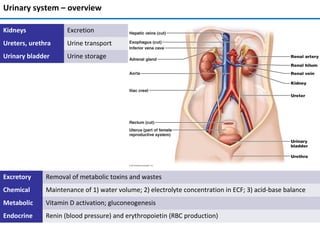
  • 1. Urinary system – overview Excretory Removal of metabolic toxins and wastes Chemical Maintenance of 1) water volume; 2) electrolyte concentration in ECF; 3) acid-base balance Metabolic Vitamin D activation; gluconeogenesis Endocrine Renin (blood pressure) and erythropoietin (RBC production) Kidneys Excretion Ureters, urethra Urine transport Urinary bladder Urine storage
  • 2. Kidney anatomy • Retroperitoneal, superior lumbar region; ~ T12 to L5 • Right is lower; adrenal gland atop; bean-shape; medial side – renal hilum • Ureters, renal blood vessels, lymphatics, and nerves enter and exit at hilum • Surrounded by (from superficial to deep): renal fascia, perirenal fat capsule, fibrous capsule • Layers: cortex and medulla • Renal pyramids (medulla) produce urine; separated by renal columns (cortex) • Urine from papilla goes to minor calyx → major calyx → renal pelvis → ureter • Pyramid and surrounding columns = lobe Renal anatomy
  • 3. Blood and nerve supply • Kidneys cleanse blood  rich blood supply • Renal arteries – ~ ¼ (1200 ml) of cardiac output • Similar paths for arterial and venous flow • Nerve supply – sympathetic fibers from renal plexus Renal anatomy
  • 4. Nephron • Structural and functional units that form urine; > 1 million per kidney • Filtration, reabsorption, and secretion (see bottom left) • Renal corpuscle (RC): glomerulus and glomerular capsule • Glomerulus (G): fenestrated capillaries  filtrate • Like plasma but without proteins and cells; goes into glomerular capsule • Renal tubule: proximal convoluted tubule (PCT), nephron loop, distal convoluted tubule (DCT); reabsorption and secretion • DCT enters collecting duct (not the part of a nephron) Blood/ECF Filtrate Filtration (RC) Reabsorption (RT) Secretion (RT) Renal anatomy
  • 5. Urine formation • 180 L fluid processed daily (60x entire plasma volume) → only 1.5 L urine • 20-25% oxygen consumption Three processes: • Glomerular filtration • Cell- and protein-free filtrate of blood plasma minus proteins • Tubular reabsorption • Selectively returns 99% of substances from filtrate to blood in renal tubules and collecting ducts • Tubular secretion • Selectively moves substances from blood to filtrate in renal tubules and collecting ducts • Eventually urine is produced → <1% of filtrate • Contains metabolic wastes and unneeded substances Renal physiology
  • 6. Nephron epithelia Part Type Function and features Glomerular capsule (parietal) Simple squamous Structural Glomerular capsule (visceral) Branching cuboidal Filtration via filtration slits; located around capillaries PCT Cuboidal with microvilli ↑ surface area; Reabsorption and secretion; only in cortex NL (thick descending limb Cuboidal with microvilli ↑ surface area; reabsorption NL (thin descending limb) Simple squamous Reabsorption NL (thick ascending limb) Cuboidal to columnar Reabsorption DCT Cuboidal; few microvilli Secretion > reabsorption; only cortex Collecting duct Principal and intercalated cells – cuboidal Principal – sparse, short microvilli; H2O and Na+ balance Intercalated (A and B) – many microvilli; acid-base balance
  • 7. Types of nephrons • Cortical – 85% of nephrons; in cortex • Glomerular capillaries: filtration • Peritubular capillaries: reabsorption and secretion • Juxtamedullary: long nephron loops; in the medulla • Glomerulus: filtration; capillaries are the same as in cortical N. • Vasa recta and nephron loop form countercurrent system; together with the collecting duct produce concentrated urine
  • 8. Nephron capillary beds Capillary bed Blood flow Blood pressure Location Structure Function Glomerular afferent a. → glomerulus → efferent a. High (Ø of afferent a. > Ø of efferent a.) Glomerulus Fenestrated, surrounded by podocytes Filtration Peritubular efferent a. → capillaries → venules Low Around renal tubule Fenestrated, thin-walled Reabsorption, secretion Vasa recta efferent a. → capillaries → venules Low Around nephron loop Fenestrated, thin-walled Formation of concentrated urine Renal physiology
  • 9. Filtration pressures • Passive process; no energy required; from blood to capsule via filtration membrane; no reabsorption • Capillary hydrostatic pressure pushes fluid into the filtrate; equals BP • 55 mm Hg; due to the larger afferent a. diameter • Capsular hydrostatic pressure pushes fluid back into the blood; 15 mm Hg • Capillary osmotic pressure pulls fluid back into the blood; due to the plasma proteins; 30 mm Hg • Sum of pressures → net filtration pressure (NFP) = 10 mm Hg (55 one way – 45 other way) • Determines glomerular filtration rate • Why there is no capsular osmotic pressure? Glomerular filtration
  • 10. Filtration mechanism • Filtration membrane: fenestrated capillary endothelium; basement membrane; foot processes of podocytes with filtration slits • Molecules smaller than 3 nm pass (water, glucose, amino acids, nitrogenous wastes) – everything but proteins and large biological macromolecules • Macromolecules "stuck" in filtration membrane engulfed by glomerular mesangial cells • Plasma proteins remain in blood  maintain osmotic pressure  prevents loss of all water to filtrate • Proteins in filtrate indicate membrane problem Glomerular filtration
  • 11. Glomerular filtration rate (GFR) • Volume of filtrate formed per minute by both kidneys (normal = 120–125 ml/min) • GFR NFP, filtration membrane surface≡ (controlled by mesangial cells) and permeability (very high) • Constant GFR = normal filtration → ECF homeostasis • GFR affects BP: ↑BP → ↑GFR  ↑urine output  ↓BP, and vice versa • Goal of intrinsic controls – maintain GFR in kidney, not BP • Goal of extrinsic controls – maintain systemic BP • Intrinsic controls: local within kidney; maintain GFR; dominant at normal BP • Extrinsic controls: nervous, endocrine; maintain BP; can ↓ kidney function • Preside over intrinsic if systemic BP < 80 or > 180 mm Hg • Controlled via glomerular hydrostatic pressure • If rises  NFP rises  GFR rises; 18% ↓ of systemic BP → GFR = 0 NFP Membrane permeability Surface area GFR BP increases GFR increases Urine output increases Blood volume decreases BP decreases Glomerular filtration GFR ↑ Glomerular blood flow ↓ GFR ↓ INTRINSIC EXTRINSIC
  • 12. Juxtaglomerular complex (JGC) • One per nephron; portions of: 1) ascending limb of nephron loop; afferent and efferent a. • Regulates GFR and BP • Macula densa cells (MDC): in ascending limb; sense NaCl in the filtrate • Granular/juxtaglomerular cells (JC): arteriolar SMC; dilate/constrict afferent a. • Respond to ↓ stretch of afferent a. releasing renin (BP regulation) • Extraglomerular mesangial cells (EMC): connected by gap junctions • Signaling between MDC and JC Glomerular filtration
  • 13. Intrinsic controls of GFR • Myogenic mechanism • Smooth muscle contracts in response to stretch • Both help maintain normal GFR despite normal fluctuations in blood pressure • Protects glomeruli from ↑ BP • Tubuloglomerular feedback mechanism • Directed by macula densa cells; respond to filtrate NaCl concentration • Opposite for ↓ GFR BP GFR ↑ Stretch↑ GFR ↓ Stretch ↓ Afferent a. constrict Efferent a. dilate Afferent a. dilate Efferent a. constrict Blood flow ↓ Blood flow ↑ GFR ↓ GFR ↑ ↑ ↓ GFR ↑ Filtrate flow ↑ Reabsorption time ↓ Filtrate NaCl ↑ Afferent a. constrict GFR ↓ Reabsorption time ↑ Filtrate NaCl ↓ Glomerular filtration
  • 14. Extrinsic controls: BP regulation BP ↓ Sympathetic response Adrenal medulla Juxtaglomerular cells Macula densa cells Kidneys Angiotensin Aldosterone Na/water retention BP ↑ Afferent a. constriction ↓ urine Reduced stretch BV ↑ Norepinephrine Vasoconstriction Epinephrine Filtrate NaCl ↓ Adenosine Prostaglandin E2 Renin 1 1,2 2 • Under normal conditions blood vessels dilated, intrinsic controls prevail • Three major mechanisms of BP regulation: 1) direct sympathetic stimulation; 2) renin-angiotensin- aldosterone cascade; 3) kidney paracrine/endocrine signaling Glomerular filtration
  • 15. Transendothelial process • Most but not all of chemicals in filtrate are reabsorbed (selective process) • Organic nutrients (sugars, aminoacids) reabsorbed ≈completely; ions/water hormonally regulated • Active and passive transport via transcellular or paracellular routes • Transcellular: apical membrane → cytoplasm → basolateral membrane → ECF → peritubular capillaries • Paracellular: between cells, controlled by tight junctions Tubular reabsorption
  • 16. Roles for 1° and 2° active transport • Requires ATP; 1° transport of Na+ at basal membrane → ↓ intracellular Na+ , (-) intracellular potential → Na+ diffusion via brush border • Na+ diffusion via brush border → coupled with 2° active transport → reabsorption of glucose (sodium-glucose co- transporter, SGLT), AA, H+ (sodium-hydrogen exchanger, NHE) etc. • Na+ reabsorption → H2O reabsorption by osmosis → ↓ chemical concentrations in filtrate → concentration gradient → passive reabsorption of Cl- , urea, lipid-soluble substances Tubular reabsorption
  • 17. PCT Chemical Apical membrane Basolateral membrane Na+ Facilitated diffusion 1° AT (Na+ /K+ pump) Water Osmosis (obligate water reabsorption via aquaporins) K+ , Cl- , Ca2+ , Mg2+ , HCO3 – , PO4 3- 2° AT (Na+ symport), facilitated diffusion (Mg2+ ) Facilitated diffusion, paracellular diffusion (K+ , Ca2+ , Mg2+ ) AA, glucose, fructose, galactose, lactate, succinate, citrate 2° AT (Na+ symport) Facilitated diffusion • All nutrients, e.g., glucose and amino acids • 65% of Na+ and H2O; other ions • ~ all uric acid; ½ urea (later secreted back into filtrate)
  • 18. Nephron loop Thin descending limb Water Osmosis (obligate reabsorption via aquaporins) Thick ascending limb Chemical Apical membrane Basolateral membrane Na+ Facilitated diffusion 1° AT (Na+ /K+ pump), paracellular diffusion K+ , Cl- , Ca2+ , Mg2+ 2° AT (Na+ cotransporter, except Mg2+ ) Paracellular diffusion ( K+ , Ca2+ , Mg2+ ) • Descending thin limb: H2O (main function), also Na+ and urea • Ascending thick limb: ions (Na+, Ca2+ , Mg2+ , HCO3 - etc) • Ascending thick limb is impermeable to water • Diluted tubular fluid at the end of the ascending limb • Allows kidneys to regulate urine concentration • Loop diuretics inhibit (1-Na+ , 2-Cl- , 1-K+ ) cotransporter Tubular reabsorption
  • 19. DCT Chemical Apical membrane Basolateral membrane Na+ 2° AT (Na+ /Cl- cotransporter) 1° AT (Na+ /K+ pump), paracellular diffusion Cl- 2° AT (Na+ /Cl- cotransporter) Facilitated diffusion HCO3 - Forms intracellularly HCO3 - /Cl- cotransporter • Early DCT: ions (Na+ , K+ , Cl- etc.); no water or urea • Thiazide diuretics inhibit Na+ /Cl- symport • Late DCT: Na+ reabsorption, K+ excretion (aldosterone) • K+ -sparing diuretics: • Aldosterone antagonists XXX A. receptor • Na+ channel blockers indirectly XXX Na+ /K+ pump • DCT is important for acid- base balance Tubular reabsorption
  • 20. Collecting duct. Summary Ion Apical membrane Basolateral membrane Na+ 2° AT (Na+ /Cl- cotransporter) 1° AT (Na+ /K+ pump), paracellular diffusion Cl- 2° AT (Na+ /Cl- cotransporter) Facilitated diffusion HCO3 - Forms intracellularly HCO3 - /Cl- cotransporter • Permeable to H2O via aquaporins (number is regulated by ADH) • Permeable to urea (diffuses into medullary interstitium); helps to regulate urine concentration • H+ secretion → acid-base balance regulation Summary of tubular reabsorption (<1 – net reabsorption, >1 – net secretion; H2O – 1) Tubular reabsorption
  • 21. Transport maximum • Transport maximum (Tm) for ~ many reabsorbed substances (see below) • Reflects number of carriers/pumps in the membranes of tubular cells • When carriers saturated, excess excreted in urine • E.g., hyperglycemia  filtered load is higher than reabsorption → exceed Tm  glucose in urine • Some – gradient-time transport (Vd = D*(C1-C2); Vd – diffusion rate; D = diffusion coefficient) • Example: Na+ in the PCT Tubular reabsorption
  • 22. Summary and regulation (3) (1) (2) (4) (5) 1 2 3 4 5 Adrenal gland Liver Posterior pituitary gland Heart Parathyroid gland Some compounds Regulation Tubular reabsorption
  • 23. Functions and features • From ECF to tubular fluid; almost all in PCT • K+ , H+, NH4 + , creatinine, organic acids and bases, drugs, urea, uric acid • Some are from tubular cells, e.g. HCO3 - • Disposes of substances (e.g., drugs) bound to plasma proteins • Disposing of undesirable passively reabsorbed substances (e.g., urea and uric acid) • Disposing of excess K+ (aldosterone) • Acid-base regulation by secreting H+ or HCO3 – in urine Location Compounds secreted PCT Bile salts, oxalate, urate, catecholamines, drugs (e.g., penicillin), salicylates, H+, NH 4 + Nephron loop Urea (thin ascending limb), H+ (thick ascending limb) DCT K+ (aldosterone-regulated), H+ Collecting duct H+ Tubular secretion
  • 24. Urine concentration and volume • Osmolality: number of solute particles in 1 kg of H2O; in milliosmols (mOsm); ability to cause osmosis • Osmolality of plasma – ~300 mOsm via regulating urine concentration and volume • Countercurrent mechanism: fluid flows in opposite directions in two adjacent segments of same tube with hair pin turn • Countercurrent multiplier – filtrate flow in the nephron loops of juxtamedullary nephrons • Countercurrent exchanger - blood flow in vasa recta • Establish and maintain osmotic gradient (300 mOsm to 1200 mOsm) from renal cortex through medulla → allows to vary urine concentration Osmolality. Countercurrent mechanism
  • 25. Countercurrent multiplier: nephron loop • Descending limb: freely permeable to H2O; medullary IF is hyperosmotic → H2O from filtrate to medullary IF → filtrate osmolality ↑ to ~1200 mOsm • Ascending limb: impermeable to H2O, selectively permeable to solutes → Na+ , Cl– reabsorbed → filtrate osmolality ↓ to 100 mOsm • Constant 200 mOsm difference between two limbs of nephron loop and between ascending limb and IF • Difference "multiplied" along length of loop to ~ 900 mOsm Urine concentration and volume
  • 26. Countercurrent exchanger: vasa recta (VR) • Ascending vasa recta remove reabsorbed H2O from 1) descending VR; 2) nephron loop (descending limb); 3) collecting duct; → volume of blood at end of vasa recta higher than at beginning • NaCl: diffuses from concentrated IF into descending VR → goes into ascending VR → diffuses from ascending VR into diluted IF → preservation of medullary gradient by recycling NaCl • Gradient allows H2O reabsorption from collecting duct if aquaporins are present (regulated by ADH) Urine concentration and volume
  • 27. ADH regulates urine concentration Urine concentration and volume
  • 28. Urea helps to maintain medullary osmotic gradient • Descending limb: water reabsorption → urea in filtrate ↑ • Ascending thin limb: passive diffusion of urea from the IF, water reabsorption → urea in filtrate ↑ • Ascending thick limb: no water or urea transport • Cortical collecting duct: water reabsorption reabsorbs water → urea in filtrate ↑ • Medullary collecting duct: high filtrate urea  diffusion into medullary IF  high osmolality in medulla Urine concentration and volume
  • 29. Diuretics increase urinary output Acetazolamide (1) PCT XXX carbonic anhydrase; ↓ blood pH, excretion of HCO3 - , Na+ ; H2O follows Osmotic d. (2) PCT, DL of NL E.g., mannitol; not reabsorbed, ↑ filtrate osmolality; prevent H2O reabsorption Loop d. (3) AL of NL Inhibit 1-Na+ , 2-Cl- , 1-K+ cotransporter → ↑ Na+ excretion → H2O follows Thiazid d. (4) DCT Inhibit Na+ /Cl- symport → ↑ Na+ excretion → H2O follows K+ -sparing d. (5) DCT, CD Aldosterone inhibitors → ↑ Na+ excretion → H2O follows, K+ stays (sparing) Na+ channel inhibitors DCT, CD Indirectly inhibit Na+ /K+ basolateral pump → ↑ Na+ excretion → H2O follows ADH inhibitors CD Decrease water reabsorption → ↑ H2Oexcretion Urine concentration and volume
  • 30. Evaluation of kidney function. Renal clearance • Kidney function is to filter out from/reabsorb chemicals in the blood • To determine, one has to know both plasma and urine concentrations • Renal clearance of a substance is the volume of plasma that kidneys clear of this substance in a given time • Used to determine GFR; helps to detect glomerular damage • C = UV/P • Measured using Inulin (plant polysaccharide) • Freely filtered; neither reabsorbed nor secreted by kidneys; its renal clearance = GFR = 125 ml/min C renal clearance rate (ml/min) U concentration (mg/ml) of substance in urine V flow rate of urine formation (ml/min) P concentration of same substance in plasma If C… Substance is… = 0 completely reabsorbed or not filtered = 125 ml/min no net reabsorption/secretion; freely filtered < 125 ml/min reabsorbed > 125 ml/min secreted Clinical evaluation
  • 31. Chronic kidney disease • Several stages with varying degrees of ↓ GFR; chronic renal disease: < 60 ml/min for 3 months • May be caused by diabetes mellitus, hypertension, damage to kidneys after infection • Renal failure: GFR < 15 ml/min; leads to uremia – ↑ urea in blood, ionic and hormonal imbalances; metabolic abnormalities • Treated with hemodialysis or transplant Clinical evaluation
  • 32. Control of water balance Electrolyte balance • In = out = ~ 2500 ml/day • Obligatory water losses: urine (60%), skin and lungs, perspiration, and feces (2.5 L/day) • Water intake: beverages, food, metabolic water (2.5 L/day) • Regulated vi ADH (hypothalamus); ↑ osmolality → ↑ thirst • Dehydration (diarrhea, burns, fever etc.) also increases ADH
  • 33. Dehydration •Hemorrhage, severe burns, vomiting, diarrhea, sweating, water deprivation, diuretics, endocrine diseases •Signs and symptoms: "cottony" oral mucosa, thirst, dry flushed skin, oliguria •Weight loss, fever, mental confusion, hypovolemic shock, and loss of electrolytes Hypotonic hydration •Renal insufficiency or overhydration •ECF osmolality ↓ → hyponatremia → net osmosis into tissue cells → swelling of cells → nausea, vomiting, muscular cramping, cerebral edema → death •Treated with hypertonic saline Edema •↑ fluid out of blood: ↑ HPC (incompetent venous valves, localized blood vessel blockage, congestive heart failure, ↑ blood volume) and permeability (inflammatory response) •↓ fluid into blood: ↓OPC (hypoproteinemia), malnutrition, liver disease, glomerulonephritis Control of water balance Electrolyte balance • Blocked (or surgically removed) lymphatic vessels: proteins accumulate in IF, ↑ OPIF • Increases diffusion distance for nutrients and oxygen; ↓ BP, impaired circulation
  • 34. Control of sodium ECF content Electrolyte balance • Main component of osmotic pressure; regulates ECF volume; water distribution; acid-base control • Concentration is stable (water moves between ECF and ICF due to osmosis) • Body content changes (changes in BP and BV) • No receptors; regulation through the blood volume • Na+ is reabsorbed in PCT, NL, and DCT; not secreted • Water follows Na+ ; ↑Na+ in filtrate → diluted urine • Renin/angiotensin/aldosterone system if ↑K+ or ↓Na+ → Na+ reabsorption, K+ secretion; BV ↑; slow
  • 35. • ANP in response to ↑ BP → natriuresis, water secretion, ↓BV and BP; vasodilation • Inhibits renin, ADH, and aldosterone release • Estrogen: ↑ NaCl reabsorption → H2O retention (menstrual cycle, pregnancy) → edema • Progesterone: ↓ Na+ reabsorption (blocks aldosterone); promotes Na+ and H2O loss • Glucocorticoids: ↑ Na+ reabsorption → edema Anions • Cl– is major anion in ECF; 99% are reabsorbed under normal pH conditions • Acidosis ↓ Cl- reabsorption • Other anions have transport maximums and excesses are excreted in urine Control of sodium ECF content. Anions Electrolyte balance
  • 36. Control of potassium ECF concentration. Anions Electrolyte balance • K+ concentration in plasma/ECF affects resting membrane potential • ↑K+ – ↓ excitability; ↓K+ – hyperpolarization, nonresponsiveness • Control is complicated – 98% K+ is in cells • Cellular regulation: shift between ECF and ICF • Renal regulation: reabsorption in PCT, secretion in NL and collecting duct • ↑ K+ in ECF (diet intake) → ↑ secretion (without aldosterone) • ↑ K+ in ECF → ↑ aldosterone → ↑ secretion (Na+ retained) • ↑ Na+ intake → ↑ tubular flow rate → ↑ secretion • If K+ intake is low, secretion ↓ - passive mechanism
  • 37. Control of calcium ECF concentration Electrolyte balance • Mostly in the bones; regulates blood clotting, membrane permeability, secretion, neuromuscular excitability • ↑ Ca2+ → ↑ excitability and muscle tetany • ↓ Ca2+ → ↓ neurons and muscle cells, heart arrhythmias • Controlled by PTH; ↑ Ca2+ renal reabsorption (98%) • ↓ phosphate ion reabsorption (reabsorbed mainly in PCT), ↑ excretion (passively) • Via Ca2+ channels (apical membrane), Ca2+ pumps and antiporters (basal membrane), transcellular route • Insulin ↑ and glucagon ↓ Ca2+ reabsorption
  • 38. Acid-base balance. Buffer systems • Sources of H+ : protein breakdown (phosphoric acid), lactic acid, fatty acids, ketone bodies • CO2 + H2O → H2CO3 → HCO3 - + H+ • H+ regulated by: buffer systems (rapid), respiratory centers (rapid), renal mechanisms (slow, most potent) • Strong bases quickly bind H+ ; strong acids quickly bind OH- Bicarbonate buffer • Mixture of H2CO3 (weak acid) and salts of HCO3 – (e.g., NaHCO3, a medium base) • Buffers ICF and ECF; only important ECF buffer Arterial blood Venous blood, IF ICF Alkalosis Acidosis pH 7.4 pH 7.35 pH 7.0 pH > 7.45 pH < 7.35 Phosphate buffer system • NaH2PO4 , a weak acid; Na2HPO4, a weak base • Buffers urine and ICF (PO4 3- concentrations are high) Protein buffer system • Buffer both plasma and ICF • Protein molecules are amphoteric (both weak acid and week base) • pH ↑ → COOH groups release H+ , become COO- • pH ↓ → -NH2 groups bind H+ , become NH3+ Acid-base balance
  • 39. Renal and respiratory compensatory systems • Respiratory and renal systems are major (slow) regulators of pH; higher capacity than chemical buffers • Chemical buffers cannot eliminate excess acids or bases from body • Lungs: eliminate carbonic acid by eliminating CO2 • Kidneys: eliminate nonvolatile acids (phosphoric, uric, lactic acids; ketones); prevent metabolic acidosis • Kidneys: regulate blood levels of bases; renew chemical buffers • CO2 unloading → reaction shifts to left (and H+ incorporated into H2O) • CO2 loading → reaction shifts to right (and H+ buffered by proteins) CO2 ↑ H+ ↑ medullary receptors chemoreceptors respiratory rate and depth ↑H+ ↓ H+ ↓ respiratory center ↓ H+ ↑ • Hypercapnia → medullary chemoreceptors → ↑ respiratory rate and depth • ↑ plasma H+ → peripheral chemoreceptors → ↑ respiratory rate and depth • CO2 is removed from blood; H+ ↓ • Alkalosis (hypocapnea) → ↓ respiratory rate and depth decrease; H+ ↑ • Hypoventilation → respiratory acidosis • Hyperventilation → respiratory alkalosis Respiratory regulation Acid-base balance
  • 40. • Renal mechanisms are most important; retain/secrete H+ • Cannot reabsorb HCO3 – , have to go around • H+ secretion in PCT and collecting duct • H+ comes from H2CO3 produced inside cells • As H+ secreted, Na+ reabsorbed Acid-base regulation in kidneys (1) Acid-base balance
  • 41. • Rate of H+ secretion changes with ECF CO2 levels; ↑ CO2 in peritubular capillary blood  ↑ H+ + secretion • To maintain alkaline reserve kidneys must replenish bicarbonate; cannot reabsorb • PCT cells: CO2 + H2O → H2CO3 → H+ (secreted) + HCO3 – (into capillary blood) – steps 1-3 • Filtrate: H+ + HCO3 – → H2CO3 → CO2 (into cells, starts over) + H2O – steps 4-6 Acid-base regulation in kidneys (2) Acid-base balance
  • 42. • New HCO3 – has to be produced and added to alkaline reserve (balancing dietary H+ ) • Via renal excretion of acid (via secretion and excretion of H+ or NH4 + ) • Excreted H+ is buffered by phosphate in the urine Replenishing alkaline reserve Acid-base balance
  • 43. Ammonium excretion • NH4 + – important mechanism for excreting acid • Glutamine in PCT cells → 2 NH4 + , 2 "new" HCO3 – • HCO3 – → blood; NH4 + → in urine • Replenishes alkaline reserve of blood • In alkalosis, type B intercalated cells secrete HCO3 – and reclaim H+ • Mechanism is opposite of HCO3 – ion reabsorption type A intercalated cells • Even in alkalosis, more HCO3 – is conserved than excreted Acid-base balance
  • 44. Type Acidosis Alkalosis Respiratory Most important indicator is blood PCO2 PCO2 > 45 mm Hg Decreased in ventilation or gas exchange CO2 accumulates in blood PCO2 <35 mm Hg Common result of hyperventilation Often due to stress or pain CO2 eliminated faster than produced Metabolic Abnormally ↓ HCO3 - Ingestion of too much alcohol (→ acetic acid) Excessive loss of HCO3 – (e.g., persistent diarrhea) Accumulation of lactic acid (exercise or shock) Less common than metabolic acidosis ↑ blood pH and HCO3 – Vomiting of acid contents of stomach Intake of excess base (e.g., antacids) Symptoms Diabetic ketosis, starvation, kidney failure Blood pH below 6.8 → depression of CNS → coma → death Blood pH above 7.8 → excitation of nervous system → muscle tetany, extreme nervousness, convulsions, death often from respiratory arrest • Failure of respiratory system to regulate pH – respiratory acidosis/alkalosis • Other abnormalities other than caused by abnormal PCO2 – metabolic acidosis/alkalosis • Respiratory system tries to compensate metabolic acid-base imbalances; renal system – respiratory ones • Respiratory system cannot compensate for respiratory acidosis or alkalosis • Renal system cannot compensate for acid-base imbalances caused by renal problems Acid-base balance Imbalance Respiratory Metabolic Acidosis Alkalosis Acidosis Alkalosis Responses Excretion of H+ Retention of H+ ↑ H+ → ↑ rate and depth of breathing ↓ H+ → ↓ rate and depth of breathing Results H+ excretion → ↑ HCO3 - ↑ PCO2 (acidosis) H+ retention → ↓ HCO3 - ↓ PCO2 (alkalosis) pH < 7.35 (acidosis) HCO3 - ↓, PCO2 ↓ pH > 7.45 (alkalosis) HCO3 - ↑, PCO2 ↑ (> 45 mm Hg) Respiratory and renal compensation mechanisms
  • 45. Physical and chemical characteristics Property Normal Abnormal Color • Pale to deep yellow (urochrome from hemoglobin breakdown) • More concentrated urine  deeper color Pink, brown, smoky (food ingestion, bile pigments, blood, drugs) Transparency Clear Cloudy (may indicate UTI) Odor • Slightly aromatic when fresh • Ammonia odor upon standing (bacteria metabolize solutes) May be altered by some drugs and vegetables pH Slightly acidic (~pH 6, within 4.5 to 8.0) • Acidic diet (protein, whole wheat)  ↓ pH • Alkaline diet (vegetarian), prolonged vomiting, or urinary tract infections  ↑pH Specific gravity 1.001 – 1.035 Out of the range Composition Water – 95% Solutes – 5% Nitrogenous wastes Urea (from amino acid breakdown) Uric acid (from nucleic acid metabolism) Creatinine (from creatine phosphate) Other normal solutes: Na+ , K+, PO4 3– , and SO4 2– , Ca2+ , Mg2+ and HCO3 – Abnormally ↑ or ↓ concentrations of any constituent – pathology (e.g., glycosuria in diabetes) Abnormal components - pathology (e.g., blood proteins, WBCs, bile pigments) Urine transport, storage, and elimination
  • 46. Ureters • Convey urine from kidneys to bladder; begin at L2 from renal pelvis; retroperitoneal • Enter base of bladder through posterior wall; bladder pressure ↑ → distal ends of ureters close → no urine backflow Urine transport, storage, and elimination Layer Structure Function Mucosa Transitional epithelium Secretion, protection Muscularis Smooth muscle Contraction in response to stretch; propels urine Adventitia Fibrous connective tissue Support
  • 47. Urinary bladder • Muscular sac; temporary stores urine; retroperitoneal, on pelvic floor • Posterior to pubic symphysis, superior to prostate (males), anterior to vagina and uterus (females) • Ureters in, urethra out; trigone – between openings of ureters and urethra; most frequently infected • Collapses when empty; expands and rises during filling; pressure remains stable • ~ Full bladder 12 cm long; holds ~ 500 ml; can hold ~ twice that if necessary, but can burst Urine transport, storage, and elimination Layer Structure Function Mucosa Transitional epithelium Secretion, protection Muscularis Smooth detrusor muscle (3 layers) Contraction; propels urine Adventitia Fibrous connective tissue Support
  • 48. Urethra Epithelium Where Transitional Near bladder Stratified squamous External urethral orifice Sphincters Muscles Where Control Internal urethral Smooth Bladder-urethra junction Involuntary (ANS) External urethral Skeletal At pelvic floor Voluntary • Drains urinary bladder • Female: 3–4 cm; bound to anterior vaginal wall; orifice anterior to vaginal opening; posterior to clitoris • Male: carries semen and urine; prostatic (2.5 cm), membranous (2 cm), and spongy urethra (15 cm) Urine transport, storage, and elimination
  • 49. • Reflexive urination (urination in infants): pontine control (storage and micturition) centers develop only between 2 and 3 yo (right part of the picture is absent in kids) Micturition (urination) Urine transport, storage, and elimination
  • 50. • Incontinence: usually weak pelvic muscles • Stress incontinence: increased intra-abdominal pressure forces urine through external sphincter • Overflow incontinence: urine dribbles when bladder overfills • Urinary retention: bladder unable to expel urine; common after general anesthesia; hypertrophy of prostate; treatment is catheterization Pathologies of micturition • Frequent micturition in infants: small bladders and less-concentrated urine • Incontinence in infants: normal; control of voluntary urethral sphincter develops with nervous system • Urinary tract infections: E. coli (≈80%), Klebsiella, Proteus mirabilis, Enterobacter, Streptococcus saprophyticus, Staphylococcus aureus, Enterococcus faecalis • Untreated infections may cause long-term renal damage → hypertension (?) • Sexually transmitted diseases: gonorrhea, chlamydiasis etc. may lead to urinary tract inflammation and kidney problems • Elderly people: most have abnormal kidneys histologically • Kidneys shrink; nephrons decrease in size and number; tubule cells less efficient • GFR ½ that of young adult by age 80; atherosclerosis of renal arteries (?) • Bladder shrinks; loss of bladder tone  nocturia and incontinence Urine transport, storage, and elimination
  • 51. Common congenital abnormalities • Horseshoe kidney (left): two kidneys fuse across midline  single U-shaped kidney; usually asymptomatic • Hypospadias (right): urethral orifice on ventral surface of penis; corrected surgically at ~ 12 months • Polycystic kidney disease (left): many fluid-filled cysts interfere with function • Autosomal dominant form – less severe, more common; autosomal recessive – more severe • Cause unknown but involves defect in signaling proteins Urinary diseases
  • 52. Kidney infections. Renal calculi • Urinary tract infections can reach the kidneys; cause inflammation of the pelvis, calyces, or entire organ • Pyelitis – pelvis and calyces are affected (left top); pyelonephritis – whole organ is affected (left bottom) • Treatment – antibiotics, sometimes surgical removal of affected kidney • Renal calculi – kidney stones in renal pelvis; formed when calcium, magnesium, or uric acid salts crystalize • Large stones block ureter  pressure & pain • Causes: chronic bacterial infection, urine retention, ↑Ca2+ in blood, ↑pH of urine • Treatment - shock wave lithotripsy – noninvasive; shock waves shatter calculi Urinary diseases