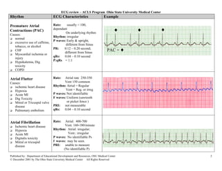
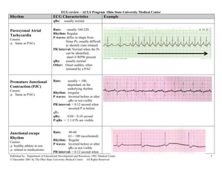
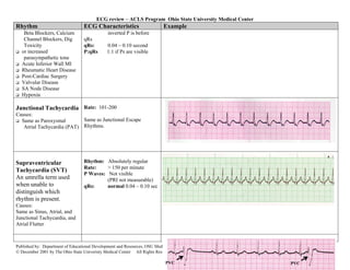
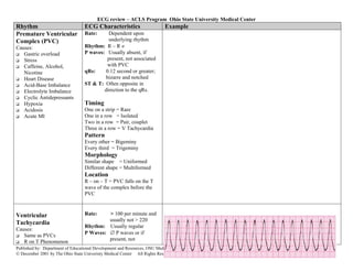
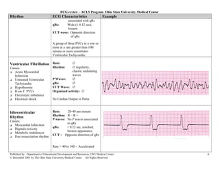
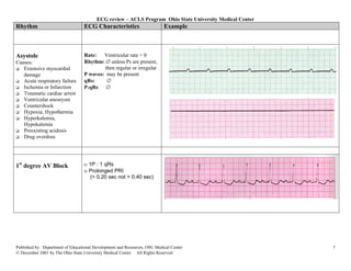
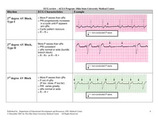
ECG review – ACLS Program Ohio State University Medical Center

Rhythm                            ECG Characteristics                      Example

Normal Sinus Rhythm               Rate:   60-100 per minute
                                  Rhythm: R- R =
(NSR)
                                  P waves: Upright, similar
                                  P-R:    0.12 -0 .20 second
                                              & consistent
                                  qRs:    0.04 – 0.10 second
                                  P:qRs: 1P:1qRs


Sinus Tachycardia                 Rate:    > 100
Causes:                           Rhythm: R- R =
   Exercise                       P waves: Upright, similar
   Hypovolemia                    P-R:     0.12 -0 .20 second
   Medications                              & consistent
   Fever                          qRs:    0.04 – 0.10 second
   Hypoxia                        P:qRs: 1P:1qRs
   Substances
   Anxiety, Fear
   Acute MI
   Fight or Flight
   Congestive Heart Failure


Sinus Bradycardia                 Rate: < 60
Causes:                           Rhythm: R- R =
   intrinsic sinus node           P waves: Upright; similar
   disease                        P-R:     0.12 -0 .20 second
   increased                                & consistent
   parasympathetic tone           qRs:     0.04 – 0.10 second
   drug effect.                   P:qRs: 1P:1qRs


Published by: Department of Educational Development and Resources, OSU Medical Center                     1
© December 2001 by The Ohio State Univeristy Medical Center All Rights Reserved
ECG review – ACLS Program Ohio State University Medical Center
Rhythm                            ECG Characteristics                      Example

Premature Atrial                  Rate:    usually < 100,
Contractions (PAC)                dependant
                                          On underlying rhythm
                                                                                   ♦     ♦                 ♦
Causes:
                                  Rhythm: irregular
   normal
                                  P waves: Early & upright,
   excessive use of caffeine,
                                          different from Sinus
   tobacco, or alcohol
   CHF
                                  PR:    0.12 – 0.20 second;
                                         different from Sinus
                                                                              PAC = ♦
   Myocardial ischemia or
                                  qRs:   0.04 – 0.10 second
   injury
                                  P:qRs = 1:1
   Hypokalemia, Dig
   toxicity
   COPD

Atrial Flutter                    Rate:   Atrial rate 250-350
Causes:                                  Vent 150 common
   ischemic heart disease         Rhythm: Atrial = Regular
   Hypoxia                                Vent = Reg. or irreg
   Acute MI                       P waves: Not identifiable
   Dig Toxicity                   F waves: Uniform (sawtooth
   Mitral or Tricuspid valve               or picket fence )
   disease                        PRI:   not measurable
   Pulmonary embolism             qRs:   0.04 – 0.10 second


Atrial Fibrillation               Rate:   Atrial: 400-700
    Ischemic heart disease                Vent. 160-180/minute
    Hypoxia                       Rhythm: Atrial: irregular;
    Acute MI                                Vent.: irregular
    Digitalis toxicity            P waves: No identifiable Ps
    Mitral or tricuspid           f waves: may be seen.
    disease                       PRI:    unable to measure
                                         (No identifiable P)

Published by: Department of Educational Development and Resources, OSU Medical Center                          2
© December 2001 by The Ohio State Univeristy Medical Center All Rights Reserved
ECG review – ACLS Program Ohio State University Medical Center
Rhythm                            ECG Characteristics                      Example
                                  qRs:     usually normal

Paroxysmal Atrial                 Rate:    usually 160-220
                                  Rhythm: Regular
Tachycardia
                                  P waves: differ in shape from
Causes:
                                          Sinus Ps; usually difficult
   Same as PACs
                                          to identify (rate related)
                                  PR Interval: Normal when the Ps
                                          can be identified;
                                           short if WPW present
                                  qRs:    usually normal
                                  Other: Onset sudden, often
                                          initiated by a PAC


Premature Junctional              Rate:    usually < 100,
                                           dependant on the
Contraction (PJC)
                                           underlying rhythm
Causes:
                                  Rhythm: irregular
   Same as PACs
                                  P waves: Inverted before or after
                                            qRs or not visible
                                  PR interval: < 0.12 second when
                                            inverted P is before
                                  qRs
                                  qRs:     0.04 – 0.10 second
                                  P:qRs = 1:1 if Ps are visible


Junctional escape                 Rate:     40-60
                                            61 – 100 (accelerated)
Rhythm
                                  Rhythm: Regular
Causes:
                                  P waves: Inverted before or after
  healthy athlete at rest
                                            qRs or not visible
  related to medications-
                                  PR interval: < 0.12 second when
Published by: Department of Educational Development and Resources, OSU Medical Center                      3
© December 2001 by The Ohio State Univeristy Medical Center All Rights Reserved
ECG review – ACLS Program Ohio State University Medical Center
Rhythm                            ECG Characteristics                      Example
    Beta Blockers, Calcium                     inverted P is before
    Channel Blockers, Dig         qRs
    Toxicity                      qRs:        0.04 – 0.10 second
   or increased                   P:qRs       1:1 if Ps are visible
    parasympathetic tone
   Acute Inferior Wall MI
   Rheumatic Heart Disease
   Post-Cardiac Surgery
   Valvular Disease
   SA Node Disease
   Hypoxia

Junctional Tachycardia Rate: 101-200
Causes:
  Same as Paroxysmal        Same as Junctional Escape
   Atrial Tachycardia (PAT) Rhythms.


Supraventricular                  Rhythm: Absolutely regular
                                  Rate:    > 150 per minute
Tachycardia (SVT)
                                  P Waves: Not visible
An umrella term used                       (PRI not measurable)
when unable to                    qRs:     normal 0.04 – 0.10 sec
distinguish which
rhythm is present.
Causes:
Same as Sinus, Atrial, and
Junctional Tachycardia, and
Atrial Flutter


Published by: Department of Educational Development and Resources, OSU Medical Center                           4
© December 2001 by The Ohio State Univeristy Medical Center All Rights Reserved

                                                                            PVC                           PVC
ECG review – ACLS Program Ohio State University Medical Center
Rhythm                            ECG Characteristics                      Example
Premature Ventricular             Rate:     Dependent upon
Complex (PVC)                               underlying rhythm
Causes:                           Rhythm: R – R ≠
   Gastric overload               P waves: Usually absent, if
   Stress                                  present, not associated
   Caffeine, Alcohol,                      with PVC
   Nicotine                       qRs:    0.12 second or greater;
   Heart Disease                          bizarre and notched
   Acid-Base Imbalance            ST & T: Often opposite in
   Electrolyte Imbalance                  direction to the qRs.
   Cyclic Antidepressants
   Hypoxia                        Timing
   Acidosis                       One on a strip = Rare
   Acute MI                       One in a row = Isolated
                                  Two in a row = Pair, couplet
                                  Three in a row = V Tachycardia
                                  Pattern
                                  Every other = Bigeminy
                                  Every third = Trigeminy
                                  Morphology
                                  Similar shape = Uniformed
                                  Different shape = Multiformed
                                  Location
                                  R – on – T = PVC falls on the T
                                  wave of the complex before the
                                  PVC


Ventricular                       Rate:    > 100 per minute and
                                           usually not > 220
Tachycardia
                                  Rhythm: Usually regular
Causes:
                                  P Waves: ∅ P waves or if
   Same as PVCs
                                           present, not
   R on T Phenomenon
Published by: Department of Educational Development and Resources, OSU Medical Center                      5
© December 2001 by The Ohio State Univeristy Medical Center All Rights Reserved
ECG review – ACLS Program Ohio State University Medical Center
Rhythm                            ECG Characteristics                      Example
                                            associated with qRs
                                  qRs:     Wide (≥ 0.12 sec),
                                            bizarre
                                  ST/T wave: Opposite direction
                                              of qRs

                                  A group of three PVCs in a row or
                                  more at a rate greater than 100/
                                  minute or more constitutes
                                  Ventricular Tachycardia.

Ventricular Fibrillation Rate:                 ∅
Causes:                           Rhythm:      ∅ regularity,
   Acute Myocardial                            chaotic undulating
   Infarction                                  waves
   Untreated Ventricular          P Waves:     ∅
   Tachycardia                    qRs:         ∅
   Hypothermia                    ST/T Wave: ∅
   R-on-T PVCs                    Organized activity: ∅
   Electrolyte imbalance
   Electrical shock               No Cardiac Output or Pulse


Idioventricular                   Rate:     20-40 per minute
                                  Rhythm: R – R =
Rhythm
                                  P waves: No P waves associated
Causes:
                                           to qRs
   Myocardial Infarction
                                  qRs:     > 0.12 sec, notched,
   Digitalis toxicity
                                           bizarre appearance
   Metabolic imbalances
                                  ST/T : Opposite direction of qRs
   Post resuscitation rhythm

                                  Rate > 40 to 100 = Accelerated

Published by: Department of Educational Development and Resources, OSU Medical Center                     6
© December 2001 by The Ohio State Univeristy Medical Center All Rights Reserved
ECG review – ACLS Program Ohio State University Medical Center
Rhythm                            ECG Characteristics                      Example


Asystole                          Rate:   Ventricular rate = 0
Causes:                           Rhythm: ∅ unless Ps are present,
   Extensive myocardial                    then regular or irregular
   damage                         P waves: may be present
   Acute respiratory failure      qRs:      ∅
   Ischemia or Infarction         P:qRs ∅
   Traumatic cardiac arrest
   Ventricular aneurysm
   Countershock
   Hypoxia, Hypothermia
   Hyperkalemia,
   Hypokalemia
   Preexisting acidosis
   Drug overdose


1st degree AV Block               υ 1P : 1 qRs
                                  υ Prolonged PRI
                                    (> 0.20 sec not > 0.40 sec)


Published by: Department of Educational Development and Resources, OSU Medical Center                     7
© December 2001 by The Ohio State Univeristy Medical Center All Rights Reserved
ECG review – ACLS Program Ohio State University Medical Center
Rhythm                            ECG Characteristics                      Example

2nd degree AV Block,              υ More P waves than qRs
Type I                            υ PRI progressively increases
                                    in a cycle until P appears
                                    w/o qRs.
                                  υ Cyclic pattern reoccurs
                                  υR–R≠                                                 = non-conducted P wave


2nd degree AV Block,              More P waves than qRs
                                  υ PRI consistent
Type II
                                  υ qRs normal or wide (bundle
                                  branch block)
                                  υ R - R≠ or R – R =


                                                                                        = non-conducted P wave


3rd degree AV Block               υ More P waves than qRs
                                  υ P not r/t qRs
                                    (P too close, P too far)
                                  υ PRI varies greatly
                                  υ qRs normal or wide
                                  υR–R=
                                                                                        = non-conducted P wave


Published by: Department of Educational Development and Resources, OSU Medical Center                            8
© December 2001 by The Ohio State Univeristy Medical Center All Rights Reserved
ECG review – ACLS Program Ohio State University Medical Center


Published by: Department of Educational Development and Resources, OSU Medical Center                     9
© December 2001 by The Ohio State Univeristy Medical Center All Rights Reserved

Ecg rhythms charex

  • 1.
    ECG review –ACLS Program Ohio State University Medical Center Rhythm ECG Characteristics Example Normal Sinus Rhythm Rate: 60-100 per minute Rhythm: R- R = (NSR) P waves: Upright, similar P-R: 0.12 -0 .20 second & consistent qRs: 0.04 – 0.10 second P:qRs: 1P:1qRs Sinus Tachycardia Rate: > 100 Causes: Rhythm: R- R = Exercise P waves: Upright, similar Hypovolemia P-R: 0.12 -0 .20 second Medications & consistent Fever qRs: 0.04 – 0.10 second Hypoxia P:qRs: 1P:1qRs Substances Anxiety, Fear Acute MI Fight or Flight Congestive Heart Failure Sinus Bradycardia Rate: < 60 Causes: Rhythm: R- R = intrinsic sinus node P waves: Upright; similar disease P-R: 0.12 -0 .20 second increased & consistent parasympathetic tone qRs: 0.04 – 0.10 second drug effect. P:qRs: 1P:1qRs Published by: Department of Educational Development and Resources, OSU Medical Center 1 © December 2001 by The Ohio State Univeristy Medical Center All Rights Reserved
  • 2.
    ECG review –ACLS Program Ohio State University Medical Center Rhythm ECG Characteristics Example Premature Atrial Rate: usually < 100, Contractions (PAC) dependant On underlying rhythm ♦ ♦ ♦ Causes: Rhythm: irregular normal P waves: Early & upright, excessive use of caffeine, different from Sinus tobacco, or alcohol CHF PR: 0.12 – 0.20 second; different from Sinus PAC = ♦ Myocardial ischemia or qRs: 0.04 – 0.10 second injury P:qRs = 1:1 Hypokalemia, Dig toxicity COPD Atrial Flutter Rate: Atrial rate 250-350 Causes: Vent 150 common ischemic heart disease Rhythm: Atrial = Regular Hypoxia Vent = Reg. or irreg Acute MI P waves: Not identifiable Dig Toxicity F waves: Uniform (sawtooth Mitral or Tricuspid valve or picket fence ) disease PRI: not measurable Pulmonary embolism qRs: 0.04 – 0.10 second Atrial Fibrillation Rate: Atrial: 400-700 Ischemic heart disease Vent. 160-180/minute Hypoxia Rhythm: Atrial: irregular; Acute MI Vent.: irregular Digitalis toxicity P waves: No identifiable Ps Mitral or tricuspid f waves: may be seen. disease PRI: unable to measure (No identifiable P) Published by: Department of Educational Development and Resources, OSU Medical Center 2 © December 2001 by The Ohio State Univeristy Medical Center All Rights Reserved
  • 3.
    ECG review –ACLS Program Ohio State University Medical Center Rhythm ECG Characteristics Example qRs: usually normal Paroxysmal Atrial Rate: usually 160-220 Rhythm: Regular Tachycardia P waves: differ in shape from Causes: Sinus Ps; usually difficult Same as PACs to identify (rate related) PR Interval: Normal when the Ps can be identified; short if WPW present qRs: usually normal Other: Onset sudden, often initiated by a PAC Premature Junctional Rate: usually < 100, dependant on the Contraction (PJC) underlying rhythm Causes: Rhythm: irregular Same as PACs P waves: Inverted before or after qRs or not visible PR interval: < 0.12 second when inverted P is before qRs qRs: 0.04 – 0.10 second P:qRs = 1:1 if Ps are visible Junctional escape Rate: 40-60 61 – 100 (accelerated) Rhythm Rhythm: Regular Causes: P waves: Inverted before or after healthy athlete at rest qRs or not visible related to medications- PR interval: < 0.12 second when Published by: Department of Educational Development and Resources, OSU Medical Center 3 © December 2001 by The Ohio State Univeristy Medical Center All Rights Reserved
  • 4.
    ECG review –ACLS Program Ohio State University Medical Center Rhythm ECG Characteristics Example Beta Blockers, Calcium inverted P is before Channel Blockers, Dig qRs Toxicity qRs: 0.04 – 0.10 second or increased P:qRs 1:1 if Ps are visible parasympathetic tone Acute Inferior Wall MI Rheumatic Heart Disease Post-Cardiac Surgery Valvular Disease SA Node Disease Hypoxia Junctional Tachycardia Rate: 101-200 Causes: Same as Paroxysmal Same as Junctional Escape Atrial Tachycardia (PAT) Rhythms. Supraventricular Rhythm: Absolutely regular Rate: > 150 per minute Tachycardia (SVT) P Waves: Not visible An umrella term used (PRI not measurable) when unable to qRs: normal 0.04 – 0.10 sec distinguish which rhythm is present. Causes: Same as Sinus, Atrial, and Junctional Tachycardia, and Atrial Flutter Published by: Department of Educational Development and Resources, OSU Medical Center 4 © December 2001 by The Ohio State Univeristy Medical Center All Rights Reserved PVC PVC
  • 5.
    ECG review –ACLS Program Ohio State University Medical Center Rhythm ECG Characteristics Example Premature Ventricular Rate: Dependent upon Complex (PVC) underlying rhythm Causes: Rhythm: R – R ≠ Gastric overload P waves: Usually absent, if Stress present, not associated Caffeine, Alcohol, with PVC Nicotine qRs: 0.12 second or greater; Heart Disease bizarre and notched Acid-Base Imbalance ST & T: Often opposite in Electrolyte Imbalance direction to the qRs. Cyclic Antidepressants Hypoxia Timing Acidosis One on a strip = Rare Acute MI One in a row = Isolated Two in a row = Pair, couplet Three in a row = V Tachycardia Pattern Every other = Bigeminy Every third = Trigeminy Morphology Similar shape = Uniformed Different shape = Multiformed Location R – on – T = PVC falls on the T wave of the complex before the PVC Ventricular Rate: > 100 per minute and usually not > 220 Tachycardia Rhythm: Usually regular Causes: P Waves: ∅ P waves or if Same as PVCs present, not R on T Phenomenon Published by: Department of Educational Development and Resources, OSU Medical Center 5 © December 2001 by The Ohio State Univeristy Medical Center All Rights Reserved
  • 6.
    ECG review –ACLS Program Ohio State University Medical Center Rhythm ECG Characteristics Example associated with qRs qRs: Wide (≥ 0.12 sec), bizarre ST/T wave: Opposite direction of qRs A group of three PVCs in a row or more at a rate greater than 100/ minute or more constitutes Ventricular Tachycardia. Ventricular Fibrillation Rate: ∅ Causes: Rhythm: ∅ regularity, Acute Myocardial chaotic undulating Infarction waves Untreated Ventricular P Waves: ∅ Tachycardia qRs: ∅ Hypothermia ST/T Wave: ∅ R-on-T PVCs Organized activity: ∅ Electrolyte imbalance Electrical shock No Cardiac Output or Pulse Idioventricular Rate: 20-40 per minute Rhythm: R – R = Rhythm P waves: No P waves associated Causes: to qRs Myocardial Infarction qRs: > 0.12 sec, notched, Digitalis toxicity bizarre appearance Metabolic imbalances ST/T : Opposite direction of qRs Post resuscitation rhythm Rate > 40 to 100 = Accelerated Published by: Department of Educational Development and Resources, OSU Medical Center 6 © December 2001 by The Ohio State Univeristy Medical Center All Rights Reserved
  • 7.
    ECG review –ACLS Program Ohio State University Medical Center Rhythm ECG Characteristics Example Asystole Rate: Ventricular rate = 0 Causes: Rhythm: ∅ unless Ps are present, Extensive myocardial then regular or irregular damage P waves: may be present Acute respiratory failure qRs: ∅ Ischemia or Infarction P:qRs ∅ Traumatic cardiac arrest Ventricular aneurysm Countershock Hypoxia, Hypothermia Hyperkalemia, Hypokalemia Preexisting acidosis Drug overdose 1st degree AV Block υ 1P : 1 qRs υ Prolonged PRI (> 0.20 sec not > 0.40 sec) Published by: Department of Educational Development and Resources, OSU Medical Center 7 © December 2001 by The Ohio State Univeristy Medical Center All Rights Reserved
  • 8.
    ECG review –ACLS Program Ohio State University Medical Center Rhythm ECG Characteristics Example 2nd degree AV Block, υ More P waves than qRs Type I υ PRI progressively increases in a cycle until P appears w/o qRs. υ Cyclic pattern reoccurs υR–R≠ = non-conducted P wave 2nd degree AV Block, More P waves than qRs υ PRI consistent Type II υ qRs normal or wide (bundle branch block) υ R - R≠ or R – R = = non-conducted P wave 3rd degree AV Block υ More P waves than qRs υ P not r/t qRs (P too close, P too far) υ PRI varies greatly υ qRs normal or wide υR–R= = non-conducted P wave Published by: Department of Educational Development and Resources, OSU Medical Center 8 © December 2001 by The Ohio State Univeristy Medical Center All Rights Reserved
  • 9.
    ECG review –ACLS Program Ohio State University Medical Center Published by: Department of Educational Development and Resources, OSU Medical Center 9 © December 2001 by The Ohio State Univeristy Medical Center All Rights Reserved