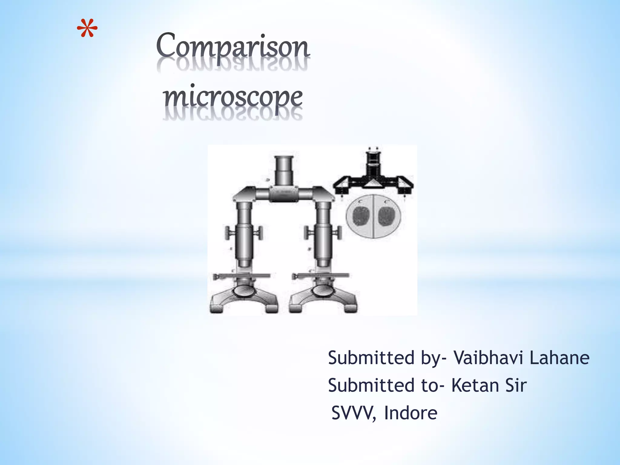
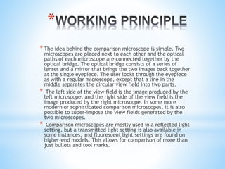
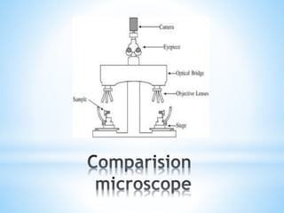
Submitted by- Vaibhavi Lahane
Submitted to- Ketan Sir
SVVV, Indore
*
*A comparison microscope is a device used to analyze side-by-
side specimens. It consists of two microscopes connected by
an optical bridge, which results in a split view window
enabling two separate objects to be viewed simultaneously.
This avoids the observer having to rely on memory when
comparing two objects under a conventional microscope.
*the primary use of this type of instrument is criminology as in
ballistics, other scientific fields including palaeontology and
archaeology utilize these special compound microscopes.
*In 1925, Major Calvin H. Goddard wrote an article for the
Army Ordnance titled “Forensic Ballistics.” In it, he
described the use of the comparison microscope in
firearms investigations. He is generally credited with the
conception of the term “forensic ballistics.”
*Philip O. Gravelle developed the comparison microscope
for the identification of fired bullets and cartridge cases
with the support and guidance of Goddard. It was a giant
leap in the science of firearms identification. The firearm
from which a bullet or cartridge case has been fired is
identified by a comparison of the unique striations left on
a projectile or cartridge.
*
* The idea behind the comparison microscope is simple. Two
microscopes are placed next to each other and the optical
paths of each microscope are connected together by the
optical bridge. The optical bridge consists of a series of
lenses and a mirror that brings the two images back together
at the single eyepiece. The user looks through the eyepiece
as with a regular microscope, except that a line in the
middle separates the circular view field into two parts.
* The left side of the view field is the image produced by the
left microscope, and the right side of the view field is the
image produced by the right microscope. In some more
modern or sophisticated comparison microscopes, it is also
possible to super-impose the view fields generated by the
two microscopes.
* Comparison microscopes are mostly used in a reflected light
setting, but a transmitted light setting is also available in
some instances, and fluorescent light settings are found on
higher-end models. This allows for comparison of more than
just bullets and tool marks.
*Forensics Examination of Spent Cartridges
*Micro-stamping Gun Firing Pins
*Forensics Examination of Bullets-
1.According to class characteristics
The three main class characteristics of all bullet
are
a)the lands and grooves
b)the calibre of the bullet, and
c)the rifling twist
2.According to individual characteristics
~ by individual striation mark on bullet
*
*Firing pin mark
Individual to each gun
Left on cartridge when firing pin
strikes the bottom of cartridge
Breech mark
Extractor or ejector mark
*Rifling marks
*
*
Land and groove marks
Rifling Twist
*Striation marks on bullet
*
* The device is very costly.
* It requires very accurate handling.
* The person using the device must be an
expert.
*
* Comparison microscope is a very important tool from forensic point
of view. It is very less time consuming device as it helps to
compare side by side instead of comparing the evidence separately.
Use of a comparison microscope is straightforward. The
incriminated impression, typically a bullet or casing found at a
crime scene or a tool mark's cast from a crime scene, is placed
under the left microscope and thus, appears in the left part of the
circular view field. A comparison impression, such as a bullet fired
from a revolver found on a suspect, is placed under the right
microscope and thus, appears in the right part of the view field.
When comparing striations, the forensic scientist moves the
comparison object until the striations match the ones present on
the incriminated object. If the striations do not present similarities,
then the two objects cannot be associated with a common origin. If
the striations match, then a common source between the two
objects is established. When comparing impression marks, the
forensic scientist can use the superimposition option and, again, by
moving the comparison object on the right, try to find common
characteristics between the two objects.

Comparison microscope

  • 1.
    Submitted by- VaibhaviLahane Submitted to- Ketan Sir SVVV, Indore *
  • 2.
    *A comparison microscopeis a device used to analyze side-by- side specimens. It consists of two microscopes connected by an optical bridge, which results in a split view window enabling two separate objects to be viewed simultaneously. This avoids the observer having to rely on memory when comparing two objects under a conventional microscope. *the primary use of this type of instrument is criminology as in ballistics, other scientific fields including palaeontology and archaeology utilize these special compound microscopes.
  • 3.
    *In 1925, MajorCalvin H. Goddard wrote an article for the Army Ordnance titled “Forensic Ballistics.” In it, he described the use of the comparison microscope in firearms investigations. He is generally credited with the conception of the term “forensic ballistics.” *Philip O. Gravelle developed the comparison microscope for the identification of fired bullets and cartridge cases with the support and guidance of Goddard. It was a giant leap in the science of firearms identification. The firearm from which a bullet or cartridge case has been fired is identified by a comparison of the unique striations left on a projectile or cartridge.
  • 4.
    * * The ideabehind the comparison microscope is simple. Two microscopes are placed next to each other and the optical paths of each microscope are connected together by the optical bridge. The optical bridge consists of a series of lenses and a mirror that brings the two images back together at the single eyepiece. The user looks through the eyepiece as with a regular microscope, except that a line in the middle separates the circular view field into two parts. * The left side of the view field is the image produced by the left microscope, and the right side of the view field is the image produced by the right microscope. In some more modern or sophisticated comparison microscopes, it is also possible to super-impose the view fields generated by the two microscopes. * Comparison microscopes are mostly used in a reflected light setting, but a transmitted light setting is also available in some instances, and fluorescent light settings are found on higher-end models. This allows for comparison of more than just bullets and tool marks.
  • 6.
    *Forensics Examination ofSpent Cartridges *Micro-stamping Gun Firing Pins *Forensics Examination of Bullets- 1.According to class characteristics The three main class characteristics of all bullet are a)the lands and grooves b)the calibre of the bullet, and c)the rifling twist 2.According to individual characteristics ~ by individual striation mark on bullet
  • 7.
    * *Firing pin mark Individualto each gun Left on cartridge when firing pin strikes the bottom of cartridge
  • 8.
  • 9.
  • 10.
  • 11.
    * Land and groovemarks Rifling Twist
  • 12.
  • 13.
    * * The deviceis very costly. * It requires very accurate handling. * The person using the device must be an expert.
  • 14.
    * * Comparison microscopeis a very important tool from forensic point of view. It is very less time consuming device as it helps to compare side by side instead of comparing the evidence separately. Use of a comparison microscope is straightforward. The incriminated impression, typically a bullet or casing found at a crime scene or a tool mark's cast from a crime scene, is placed under the left microscope and thus, appears in the left part of the circular view field. A comparison impression, such as a bullet fired from a revolver found on a suspect, is placed under the right microscope and thus, appears in the right part of the view field. When comparing striations, the forensic scientist moves the comparison object until the striations match the ones present on the incriminated object. If the striations do not present similarities, then the two objects cannot be associated with a common origin. If the striations match, then a common source between the two objects is established. When comparing impression marks, the forensic scientist can use the superimposition option and, again, by moving the comparison object on the right, try to find common characteristics between the two objects.