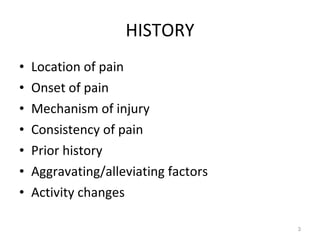
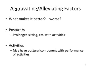
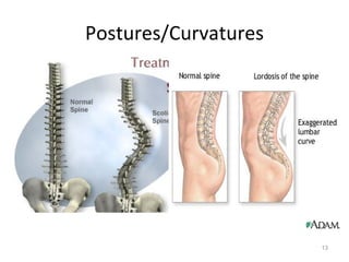
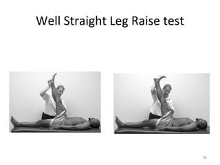
KIN 191A Advanced Assessment of  Lower Extremity Injuries THE THORACIC AND LUMBAR SPINE EVALUATION
INTRODUCTION HISTORY INSPECTION PALPATION ROM TESTS STRESS/STREE TESTS NEUROGIC TEST VASCULAR TEST
HISTORY Location of pain Onset of pain Mechanism of injury Consistency of pain Prior history Aggravating/alleviating factors Activity changes
Location of Pain Often difficult to isolate specific location of pain with low back injuries/conditions Radiating pain indicative of nerve root irritation Must have perspective on myotome/dermatome patterns for evaluation Regarding peripheral nerves, must have perspective on nerve root level origin
Onset of Pain Acute, traumatic onset of symptoms, while possible, is not a typical presentation of low back pain symptoms May be able to isolate one incident with onset of symptoms, but typically that incident is representative of accumulative stresses over time which reach their “breaking” point
Mechanism of Injury Direct trauma may indicate spinal fracture/s and/or internal organ injury Hyperextension activities (gymnastics, FB lineman, etc.) predispose to pars interarticularis injuries Multiple compression and shear forces depending upon activities engaged in Often difficult to isolate specific MOI
Consistency of Pain Constant pain If unable to affect pain with position change, associated with inflammation from injury (acute or chronic) – swelling from facet sprain or pars  interarticularis  fracture Intermittent pain If symptoms impacted (alleviated or aggravated) with spine position, associated with mechanical injury – compression/stretching of nerve root
Prior History Must question regarding previous history of low back injury/pain Structural or degenerative changes may predispose to symptoms Scar tissue may irritate tissues causing symptoms
Aggravating/Alleviating Factors What makes it better? …worse? Posture/s Prolonged sitting, etc. with activities Activities May have postural component with performance of activities
Activity Changes As with many lower extremity injuries/conditions, must be mindful of changes in Intensity of workouts Duration of workouts Training surface Footwear Sleeping arrangements
INSPECTION Posture/Curvatures Standing, sitting, with activities (e.g. – lifting) Lateral shift away from pain/nerve root impingement Frontal curvature – scoliosis  (see slide 14) Sagittal curvature – excessive/absent lordosis Muscular appearance Evaluate for spasm/atrophy – often readily visible
Gait Skin markings Café-au-lait spots (neurofibramatosis), Faun’s beard (spina bifida) Neurofibromatosis Increased cell growth of neutral tissues; normally a benign condition; pain possible secondary to pressure on the local nerves Spina bifida occulta Incomplete closure of the spinal vertebrae
Postures/Curvatures
Scoliosis Forward flexion test Posterior view of the spinal column while the patient flexes the spine; note the presence of  hump over the spine, suggesting scoliosis If functional, “hump” disappears If structural, “hump” present
PALPATION (Thoracic Spine) 1. Spinous processes 2. Supraspinous ligament 3. Costovertebral junction 4. Trapezius 5. Paravertebral muscles 6. Scapular muscles
PALPATION (Lumbar Spine) 1. Spinous processes 2. Step-off deformity 3. Paravertebral muscles
PALPATION (Sacrum & Pelvis) 1. Median sacral crest 2. Iliac crest 3. PSIS 4. Gluteals 5. Ischial tuberocity 6. Greater trochanter 7. Pubic symphysis
RANGE OF MOTION  TESTS
AROM Flexion Measure with distance from finger tips to floor Affected by hamstring/calf/paraspinal tightness Extension Lateral flexion/bending Measure finger tips to floor or at level on LE Rotation Best done in sitting to stabilize pelvis/LE Should be bilaterally equal
PROM Flexion Bring knees to chest in supine position Extension Prone position and “press-up” with pelvis on table Lateral flexion/bending Often referred to as side gliding – often eliminated Rotation Knees/hips flexed and rotate pelvis with shoulders on table
RROM Flexion Stabilize pelvis, resistance to sternum with “crunch” Extension Stabilize low back, resistance near scapula to “reverse crunch” Lateral flexion/bending – typically not assessed Rotation Stabilize opposite ASIS, resistance to opposite shoulder for “curl crunch”
SPECIAL TESTS Ligamentous testing Spring test for facet mobility/irritation Tests for nerve root impingement Valsalva maneuver Milgram test Kernig test Straight leg raise and well straight leg raise tests Quadrant test Slump test Test for malingering Hoover test
Spring Test
Increased Intrathecal Pressure Increased pressure may compress intervertebral disc forcing nucleus pulposus out causing nerve root irritation/impingement – radiating pain Valsalva Maneuver Seated, simulated bowel movement – hold breath Milgram Test Supine, SLR to a few inches and hold
Valsalva Test
Milgram Test
Kernig’s Test /Brudzinski’s Test Evaluative for nerve root impingement from disc bulge or herniation Supine, perform unilateral SLR with knee extended until pain occurs Flex knee at pain and symptoms should subside Brudzinski’s test – modification with cervical flexion to further stretch neural elements
Kernig ’ s test / Brudzinski ’ s test
SLR and Well SLR Tests Evaluative for nerve root impingement, typically discogenic in nature SLR test Supine, flex hip (SLR) on affected side to pain with knee extended, back off a little, DF ankle, if symptoms reoccur, + test Well SLR test SLR of opposite (uninvolved) leg, test + if pain felt on opposite side
Straight Leg Raise test
Well Straight Leg Raise test
Quadrant Test Often performed, not often named as such Standing, patient extends then laterally bends and rotates to affected side If radiating pain, indicative of nerve root irritation or impingement If local pain with no radiating symptoms, indicative of facet irritation/sprain
Quadrant Test
Slump Test Evaluative for nerve root, dural sheath or spinal cord irritation/inflammation Seated, cervical flexion and thoracic “slump” followed by knee extension and ankle dorsiflexion Pain and/or radiating symptoms are + test – due to neural element lengthening/stretch
Slump Test
Hoover Test Evaluative for malingering patient Supine, clinician holding both heels, patient instructed to perform active SLR on involved side Pressure should be noted in opposite heel by clinician – attempt to stabilize for movement Absence of pressure or inability to do SLR is + for malingering
Hoover Test

Kin191 A.Ch.10. Lumbar. Thoracic. Evaluation

  • 1.
    KIN 191A AdvancedAssessment of Lower Extremity Injuries THE THORACIC AND LUMBAR SPINE EVALUATION
  • 2.
    INTRODUCTION HISTORY INSPECTIONPALPATION ROM TESTS STRESS/STREE TESTS NEUROGIC TEST VASCULAR TEST
  • 3.
    HISTORY Location ofpain Onset of pain Mechanism of injury Consistency of pain Prior history Aggravating/alleviating factors Activity changes
  • 4.
    Location of PainOften difficult to isolate specific location of pain with low back injuries/conditions Radiating pain indicative of nerve root irritation Must have perspective on myotome/dermatome patterns for evaluation Regarding peripheral nerves, must have perspective on nerve root level origin
  • 5.
    Onset of PainAcute, traumatic onset of symptoms, while possible, is not a typical presentation of low back pain symptoms May be able to isolate one incident with onset of symptoms, but typically that incident is representative of accumulative stresses over time which reach their “breaking” point
  • 6.
    Mechanism of InjuryDirect trauma may indicate spinal fracture/s and/or internal organ injury Hyperextension activities (gymnastics, FB lineman, etc.) predispose to pars interarticularis injuries Multiple compression and shear forces depending upon activities engaged in Often difficult to isolate specific MOI
  • 7.
    Consistency of PainConstant pain If unable to affect pain with position change, associated with inflammation from injury (acute or chronic) – swelling from facet sprain or pars interarticularis fracture Intermittent pain If symptoms impacted (alleviated or aggravated) with spine position, associated with mechanical injury – compression/stretching of nerve root
  • 8.
    Prior History Mustquestion regarding previous history of low back injury/pain Structural or degenerative changes may predispose to symptoms Scar tissue may irritate tissues causing symptoms
  • 9.
    Aggravating/Alleviating Factors Whatmakes it better? …worse? Posture/s Prolonged sitting, etc. with activities Activities May have postural component with performance of activities
  • 10.
    Activity Changes Aswith many lower extremity injuries/conditions, must be mindful of changes in Intensity of workouts Duration of workouts Training surface Footwear Sleeping arrangements
  • 11.
    INSPECTION Posture/Curvatures Standing,sitting, with activities (e.g. – lifting) Lateral shift away from pain/nerve root impingement Frontal curvature – scoliosis (see slide 14) Sagittal curvature – excessive/absent lordosis Muscular appearance Evaluate for spasm/atrophy – often readily visible
  • 12.
    Gait Skin markingsCafé-au-lait spots (neurofibramatosis), Faun’s beard (spina bifida) Neurofibromatosis Increased cell growth of neutral tissues; normally a benign condition; pain possible secondary to pressure on the local nerves Spina bifida occulta Incomplete closure of the spinal vertebrae
  • 13.
  • 14.
    Scoliosis Forward flexiontest Posterior view of the spinal column while the patient flexes the spine; note the presence of hump over the spine, suggesting scoliosis If functional, “hump” disappears If structural, “hump” present
  • 15.
    PALPATION (Thoracic Spine)1. Spinous processes 2. Supraspinous ligament 3. Costovertebral junction 4. Trapezius 5. Paravertebral muscles 6. Scapular muscles
  • 16.
    PALPATION (Lumbar Spine)1. Spinous processes 2. Step-off deformity 3. Paravertebral muscles
  • 17.
    PALPATION (Sacrum &Pelvis) 1. Median sacral crest 2. Iliac crest 3. PSIS 4. Gluteals 5. Ischial tuberocity 6. Greater trochanter 7. Pubic symphysis
  • 18.
  • 19.
    AROM Flexion Measurewith distance from finger tips to floor Affected by hamstring/calf/paraspinal tightness Extension Lateral flexion/bending Measure finger tips to floor or at level on LE Rotation Best done in sitting to stabilize pelvis/LE Should be bilaterally equal
  • 23.
    PROM Flexion Bringknees to chest in supine position Extension Prone position and “press-up” with pelvis on table Lateral flexion/bending Often referred to as side gliding – often eliminated Rotation Knees/hips flexed and rotate pelvis with shoulders on table
  • 25.
    RROM Flexion Stabilizepelvis, resistance to sternum with “crunch” Extension Stabilize low back, resistance near scapula to “reverse crunch” Lateral flexion/bending – typically not assessed Rotation Stabilize opposite ASIS, resistance to opposite shoulder for “curl crunch”
  • 27.
    SPECIAL TESTS Ligamentoustesting Spring test for facet mobility/irritation Tests for nerve root impingement Valsalva maneuver Milgram test Kernig test Straight leg raise and well straight leg raise tests Quadrant test Slump test Test for malingering Hoover test
  • 28.
  • 29.
    Increased Intrathecal PressureIncreased pressure may compress intervertebral disc forcing nucleus pulposus out causing nerve root irritation/impingement – radiating pain Valsalva Maneuver Seated, simulated bowel movement – hold breath Milgram Test Supine, SLR to a few inches and hold
  • 30.
  • 31.
  • 32.
    Kernig’s Test /Brudzinski’sTest Evaluative for nerve root impingement from disc bulge or herniation Supine, perform unilateral SLR with knee extended until pain occurs Flex knee at pain and symptoms should subside Brudzinski’s test – modification with cervical flexion to further stretch neural elements
  • 33.
    Kernig ’ stest / Brudzinski ’ s test
  • 34.
    SLR and WellSLR Tests Evaluative for nerve root impingement, typically discogenic in nature SLR test Supine, flex hip (SLR) on affected side to pain with knee extended, back off a little, DF ankle, if symptoms reoccur, + test Well SLR test SLR of opposite (uninvolved) leg, test + if pain felt on opposite side
  • 35.
  • 36.
  • 37.
    Quadrant Test Oftenperformed, not often named as such Standing, patient extends then laterally bends and rotates to affected side If radiating pain, indicative of nerve root irritation or impingement If local pain with no radiating symptoms, indicative of facet irritation/sprain
  • 38.
  • 39.
    Slump Test Evaluativefor nerve root, dural sheath or spinal cord irritation/inflammation Seated, cervical flexion and thoracic “slump” followed by knee extension and ankle dorsiflexion Pain and/or radiating symptoms are + test – due to neural element lengthening/stretch
  • 40.
  • 41.
    Hoover Test Evaluativefor malingering patient Supine, clinician holding both heels, patient instructed to perform active SLR on involved side Pressure should be noted in opposite heel by clinician – attempt to stabilize for movement Absence of pressure or inability to do SLR is + for malingering
  • 42.