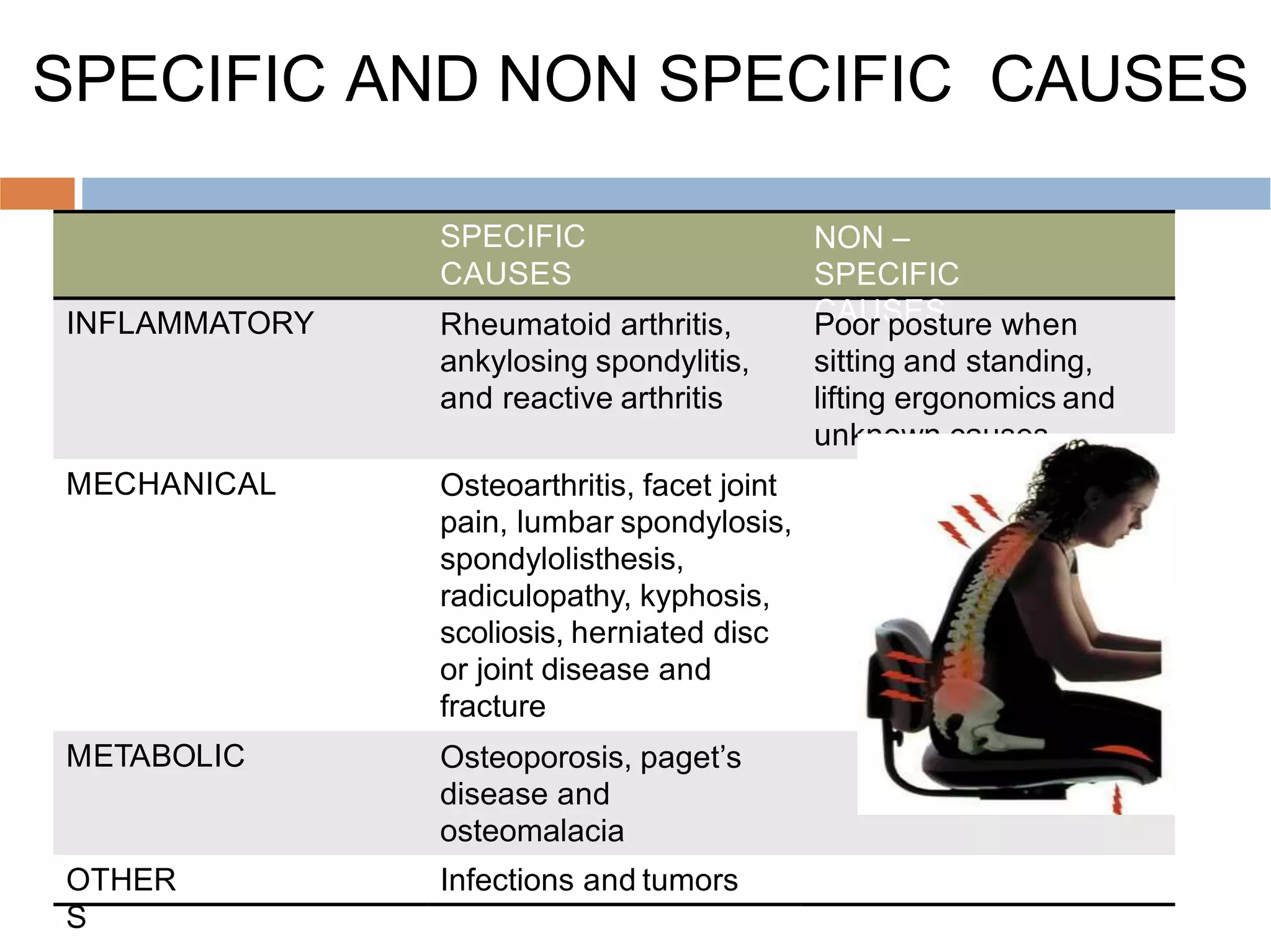
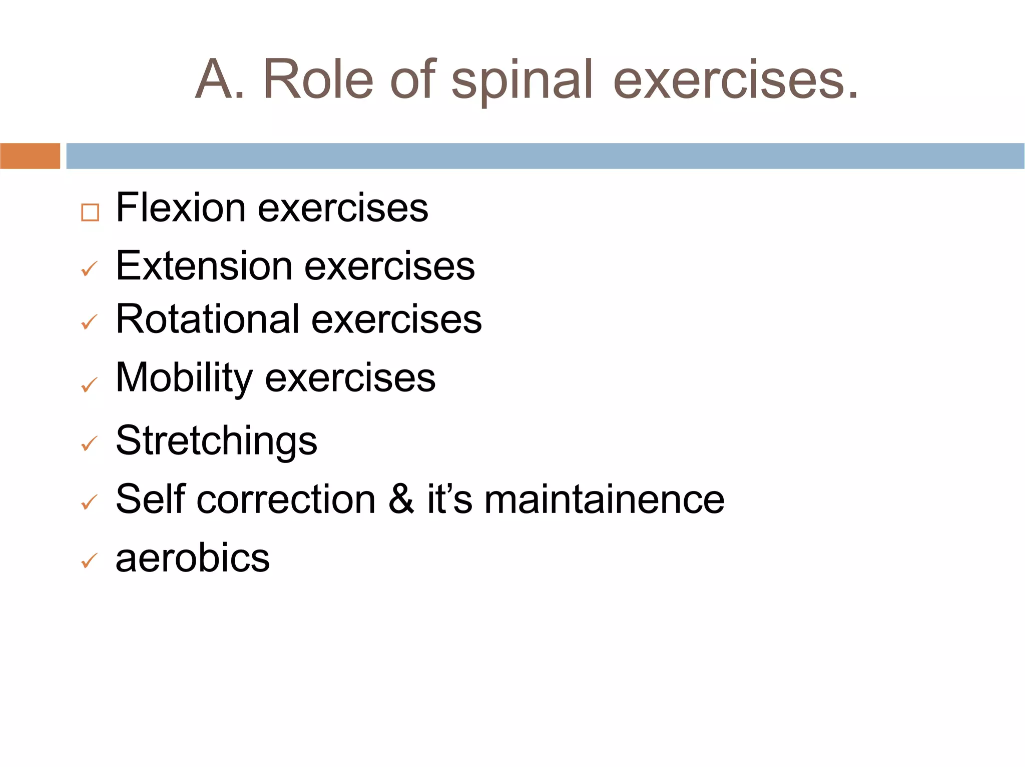
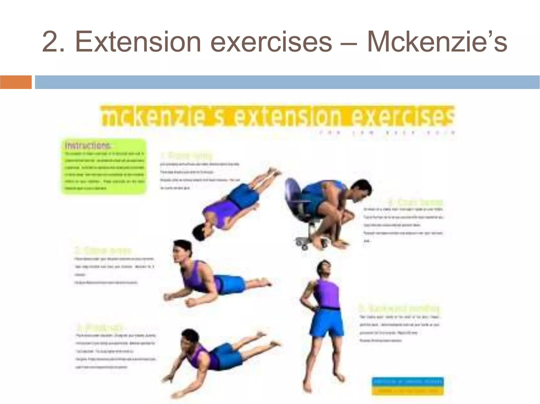
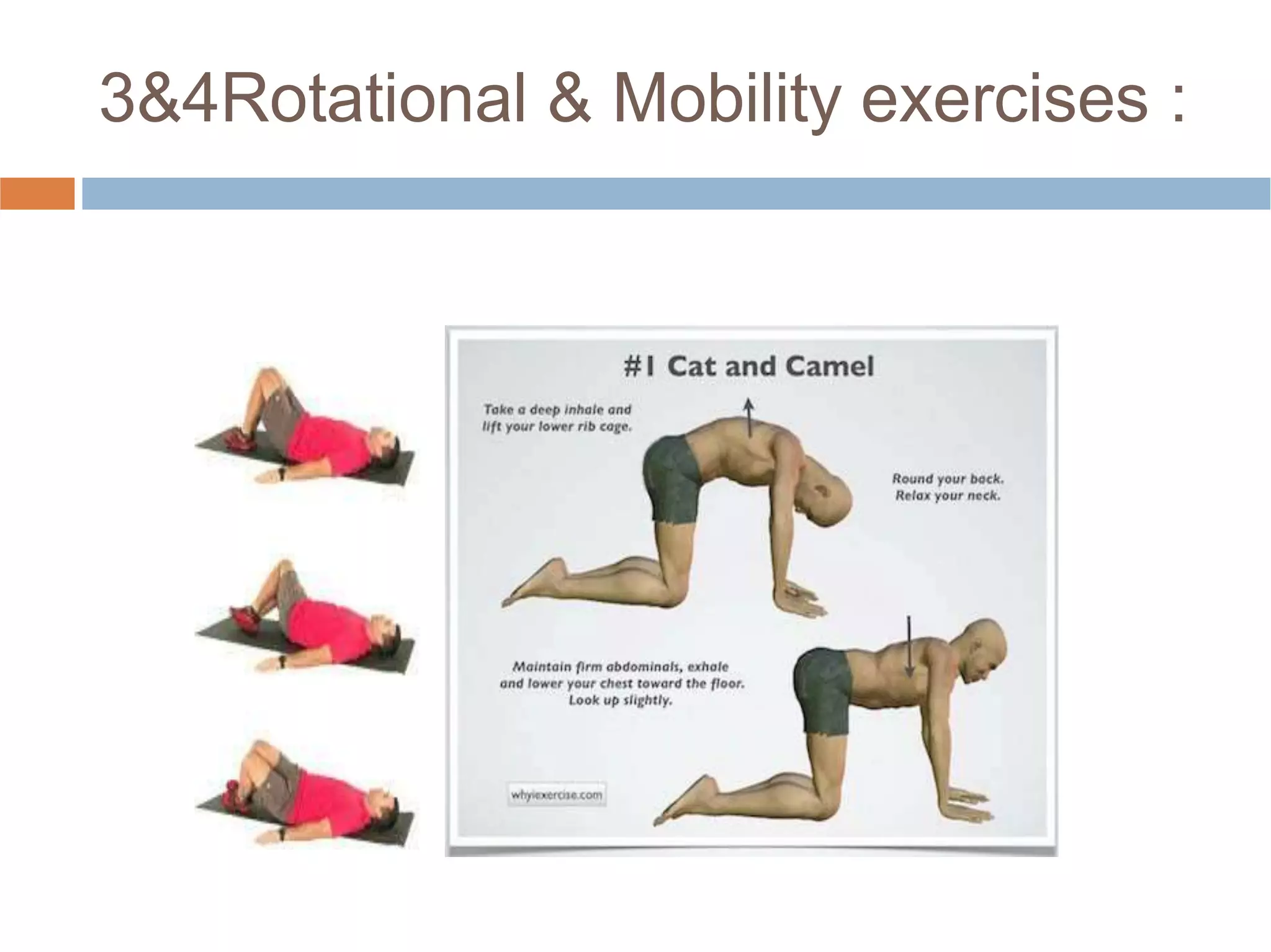
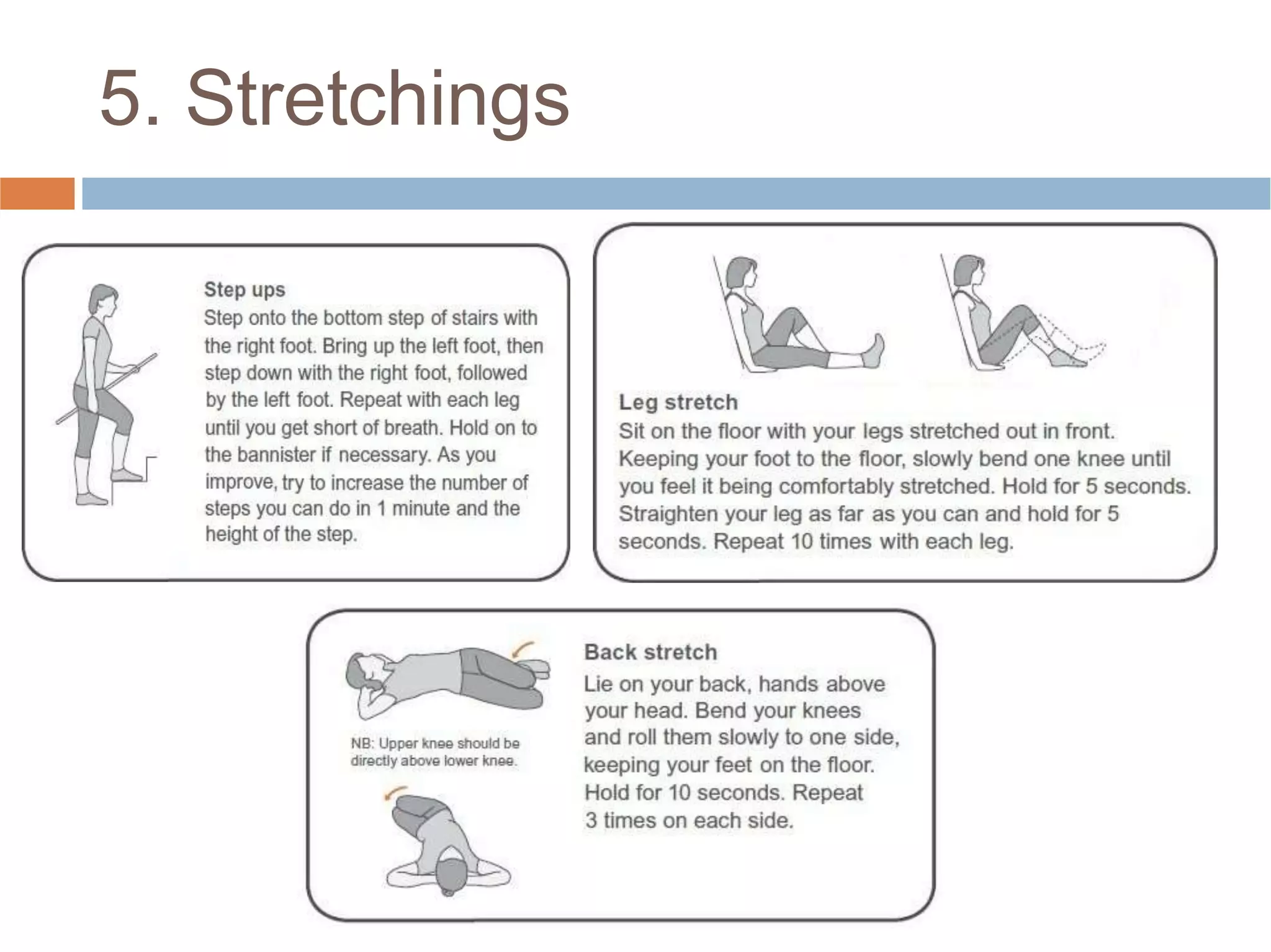
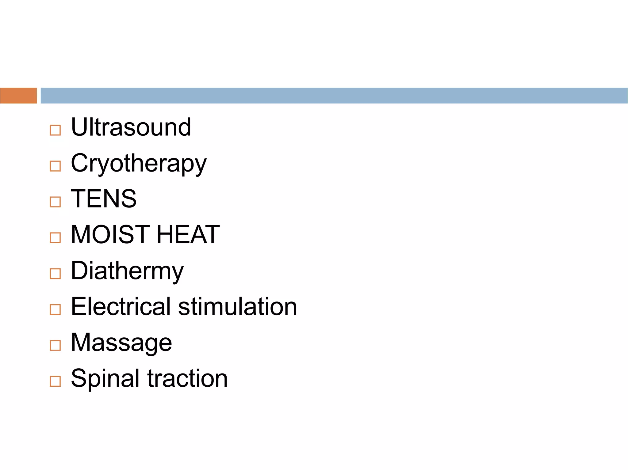
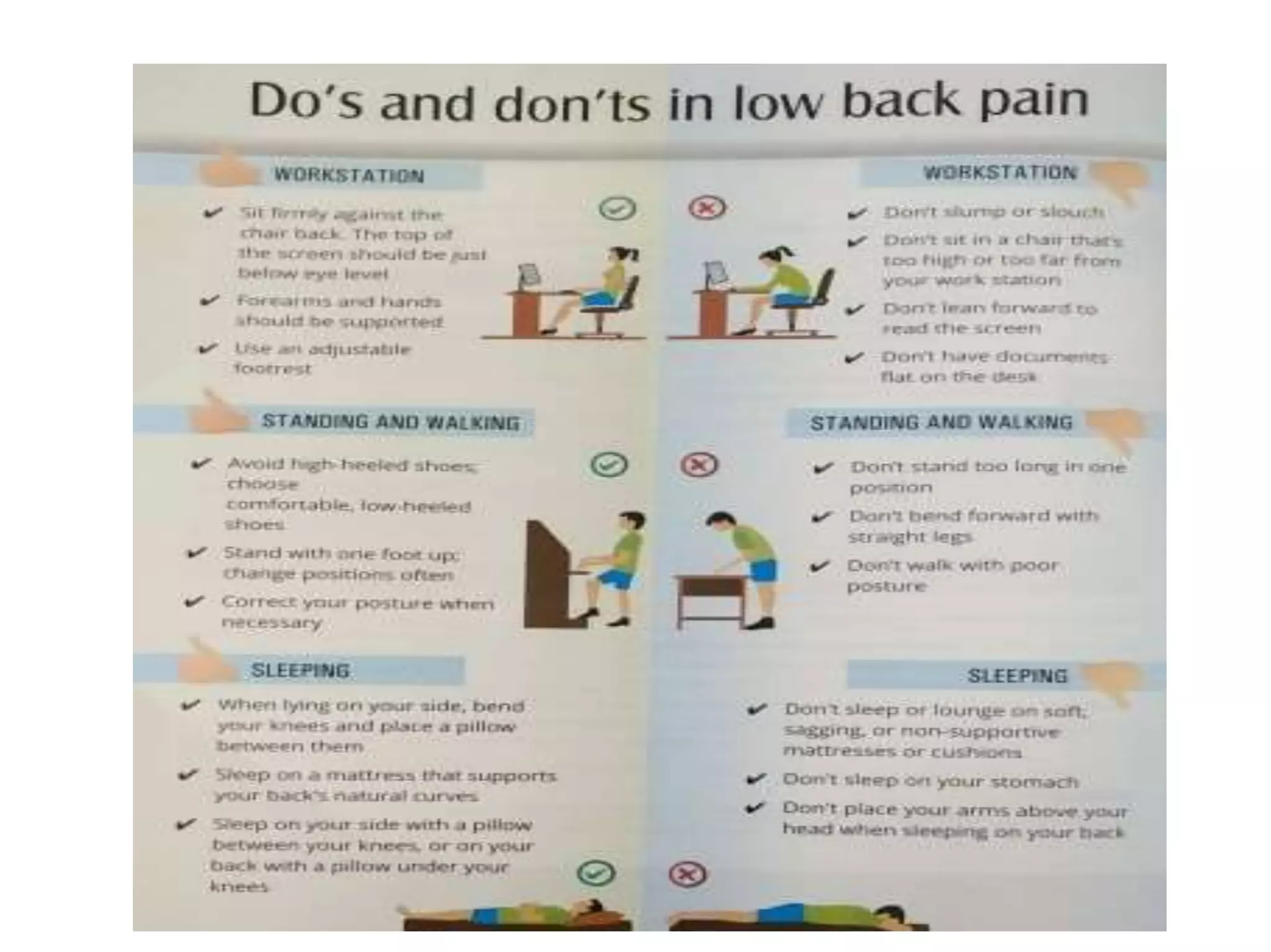
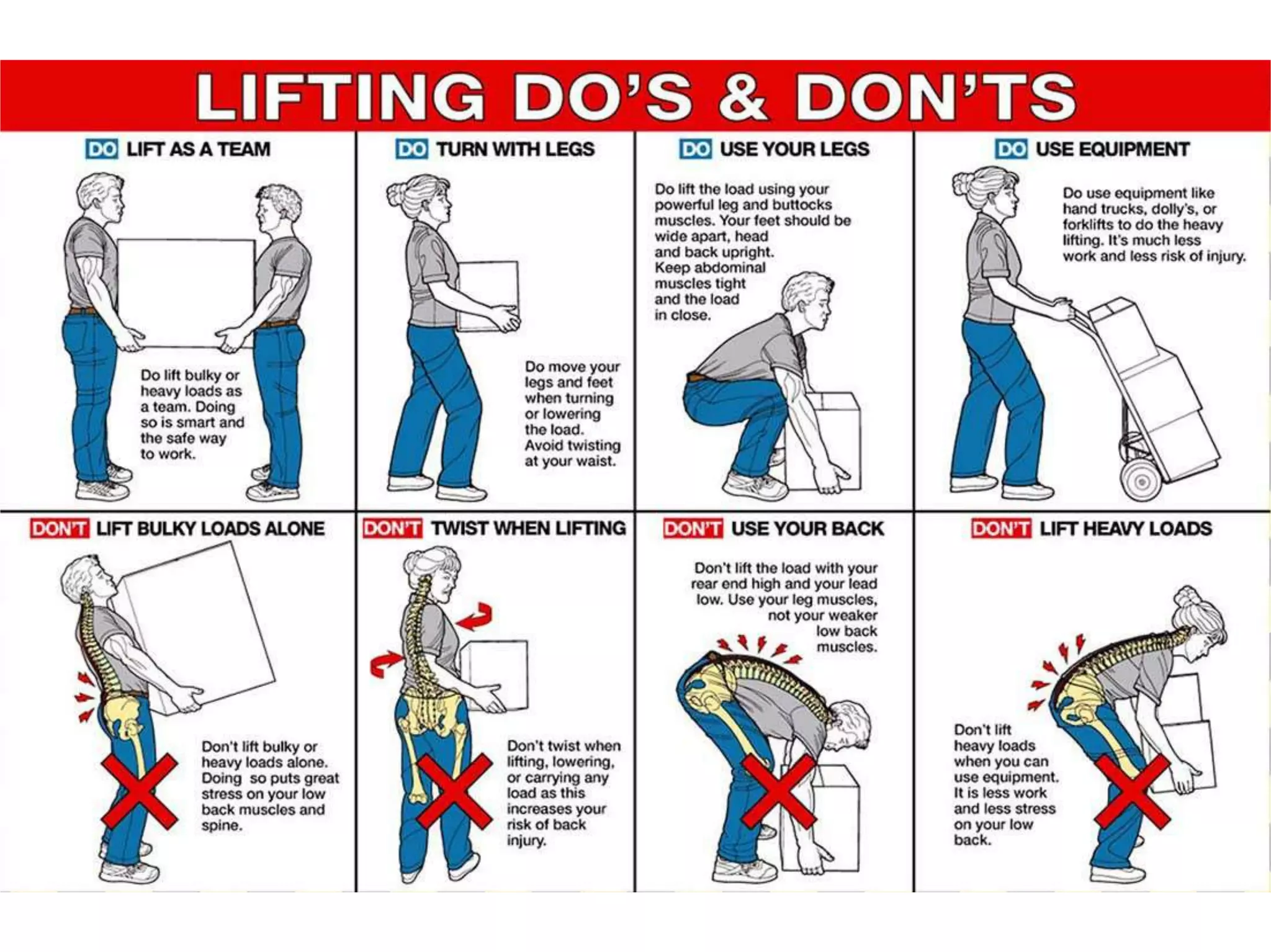
LOW BACK PAIN
& ERGONOMIC MEASURES
Dr M.Rajani Cartor BPT MPTVOrtho, DHA, DSPS,.
VAPMS COLLEGE OF PHYSIOTHERAPY, VISAKHAPAT
 INTRODUCTION
 ANATOMY
 PATHOPHYSIOLOGY
 CAUSES & RISK FACTORS
 SIGNS & SYMPTOMS
 PHYSICAL EXAMINATION
 PHYSIOTHERPEUTIC
MANAGEMENT
INTRODUCTION – LOW BACK PAIN
 Low back pain is a leading cause of disability. It occurs in
similar proportions in all cultures, interferes with quality of life
and work performance, and is the most common reason for
medical consultations. Most common cause of disability in
patients < 45 yearsold.

 Acute back pain is the most common presentation and is
usually self-limiting, lasting less than three monthsregardless
of treatment.
 Chronic back pain is a more difficult problem, which often has
strong psychological overlay: work dissatisfaction, boredom,
and a generous compensation system contribute toit.
Few cases of back pain are due to specific causes; most
cases are non-specific.
ANATOMY
 Spinal column is formed by 33 vertebrae and
divided into 5 regions :
 Cervical - 7
 Thoracic – 12
 Lumbar – 5
 Sacral – 5
 Coccygeal – 4
 It has also other components such as :
intervertebral discs ( shock absorbers),
paravertebral muscles (flexors, extensors and
obliques) & ligaments ( stabilizers).
LBA – Any pain in the low back region i.e., usually characterised by
dull, continous pain and tenderness in the lower lumbar, lumbosacral
or sacro-iliac regions, sometimes referred to leg, following the
distribution of the sciatic nerve.
 International Association for the study of pain (IASP)
Low Back Pain
 Lumbar spinal pain
 Sacral spinal pain
 Lumbosacral pain
Gluteal and Loin pain (not considered
LBP)
CLASSIFICATION OF LBA
 Mechanical ( OA, spinal stenosis, spondylolisthesis,
compression fracture)
 Nonmechanical
Tumor (metastasis, multiple myeloma, lymphoma)
Infection (osteomyelitis, diskitis)
Inflammatory arthritis (RA, AS)
 Visceral disease (nephrolithiasis, pancreatitis, prostatitis
 Complicated (“Red Flag” conditions)
 Specific Diagnosis
Lumbar Radiculopathy
Lumbar Spinal Stenosis
Others such as Ankylosing Spondylitis
 Uncomplicated (Non-Specific)
A diagnosis of exclusion
RED FLAGS
1.
2.
3.
4.
5.
6.
7.
Metastatic CA (History of cancer*, Unexplained weightloss,
Rest pain, Age >50)
Infection(Unexplained fever*, Recent bacterial infection,
Immunosuppression)
Fracture (Steroids*, Osteoporosis, Recent trauma, Age >70)
Cauda Equina Syndrome (Urinary retention or
incontinence, Saddle anesthesia, Decreased rectal tone,
Bilateral
lower extremity weakness/numbness)
Severe or progressive focal neurologic deficit
Failure to improve with therapy
Pain > 4 weeks
(* Most important in the condition)
RED FLAGS
YELLOW FLAGS
PATHO PHYSIOLOGY
 There are many structures in the lumbar spine that
can cause pain ; any irritation to the nerve roots that
exit the spine, joint problems, the discs, the bones
and the muscles.
 Many lumbar spine conditions are interrelated. For
example, joint instability can lead to disc degeneration,
which in turn can put pressure on the nerve roots.
 The most common cause of LBA is muscle strain or
other muscle problems. Strain due to heavy lifting,
bending, or other repetative use can be quite
painful, but muscle strain usually heal within few
days or weeks.
CAUSES AND RISK FACTORS
CAUSES OF ACUTE LBA CAUSES OF CHRONIC LBA
DURATION: <6weeks;
subacute lasts between
6-12 weeks.
More than 12 weeks
Sudden injury ( strain or
tears) to the musclesand
ligaments)
Arthritis
Compression fractures
(osteoporosis)
Extra wear and tear on the
spine from the work or sport
Cancer Past injuries
Herniated disc Fractures
Sciatica Past surgery
Spinal stenosis
Scoliosis or Kyphosis
Osteoarthritis
Herniated disc
Spinal stenosis
Scoliosis or kyphosis
SPECIFIC AND NON SPECIFIC CAUSES
SPECIFIC
CAUSES
NON –
SPECIFIC
CAUSES
INFLAMMATORY Rheumatoid arthritis,
ankylosing spondylitis,
and reactive arthritis
Poor posture when
sitting and standing,
lifting ergonomics and
unknown causes.
MECHANICAL Osteoarthritis, facet joint
pain, lumbar spondylosis,
spondylolisthesis,
radiculopathy, kyphosis,
scoliosis, herniated disc
or joint disease and
fracture
METABOLIC Osteoporosis, paget’s
disease and
osteomalacia
OTHER
S
Infections and tumors
OTHER NON – SPECIFIC CAUSES:
 Work that requires heavy lifting; bending and
twisting; or whole-body vibration, such as truck
driving
 Physical inactivity
 Obesity
 Arthritis or osteoporosis
 Pregnancy
 Age >30 years
 Bad posture
 Stress or depression
 Smoking
SIGNS AND SYMPTOMS
H/O an event that caused immediate low backpain:
Lifting and/or twisting while holding a heavy object
Operating a machine that vibrates eg;truck
Prolonged sitting
Involvement in a motor vehicle collision
Falls
Past H/O:
Arthritis, Infections, Surgery, Cancer or Degenerative diseases,
Vocational history.
Pain complaint:
Quality : sharp,dull,burning,intermittent or diffuse
Onset : sudden or insidious
Localisation or radiation
Exacerbating and relieving factors
Associated symptoms
Intensity
PHYSICAL EXAMINATION
 Changes in spinal alignment or sagital balance
 Restricted movements of the lumbar spine
 Evaluate disturbances of patellar and ankle
reflexes
 Asses strength and sensation of myotomes and
dermatomes to determine neural compression
 LBA can cause leg symptoms such as pain,
numbness or tingling and difficulty in standing
straight.
 Diagnostic tests : x-ray, CT scan, MRI
DIAGNOSTIC PHYSICAL TESTS :



SLR Or Laseque’s sciatic nerve test :- It’s an important
diagnostic protective reflex test which causes traction on
sciatic nerve, lumbosacral nerve roots and duramater.
It’s a passive test done in supine. Appearance of pain in
the distribution of sciatic nerve upto 45degrees of hip
flexion with knee extended indicates - +ve SLR
 If pain thus felt, is aggrevated by passive flexion of neck
and passive dorsiflexion of the foot, then only it os a
‘positive neural sign’
Real stretching of the inflammed dura is possible only with all
these three manoeuvrs.
 Note : while conducting SLR don’t confuse it with hamstrings
stretch,due to straight leg raises, especially in patients with
hamstrings tightness ( which is confirmed by dull pain over the
posterior aspect of the knee joint )
ALTERNATE SLR:
 Whenever there is doubt about the
genuineness of the test, ask the patient to sit
up with legs straight. If the sitting posture can
be assumed without flexing the knee, test is
negative.
Bowstring sign:
 In this test SLR is carried out untill the pain is
reproduced. At this point the knee is gradually
flexed till the pain disappears. The examiner
rests the limb on his/her shoulder and places
the thumb in the popliteal fossa over the
sciatic nerve. Sudden firm pressure on the
nerve produces pain in the back/pain radiating
down the legs indicating ‘+ve BOWSTRING
SIGN’ Or significant root tension
SLUMP TEST – For mobility at the
intervertebral foramen and the spinal cord




Passive neck flexion and straight leg raising (SLR) help in detecting any
reduce in mobility of pain sensitive structures within intervertebral
foramen or the vertebral canal.
If these prove negative, then the ultimate test for mobility of these
structures is done by slump test.
Test : the patient is made to sit in a slouch sitting with knees in relaxed
flexion at the edge of the table. The physiotherapist passively bends
the head and the trunk forward, as much as possbile, with total suport,
bringing the head down between the knees. The patient is then asked to
extend the knees alternately to the maximum, maintaining foot in
dorsiflexion. If pain is reproduced on attempting knee extension, the
limiting range is noted.
+ve test : indicates interfernce of the mobility at the intervertebral
foramen or at the vertebral canal.
Lumbar 3rd nerve root test
(Reverse Lasegue test )
 The patient is made to lie in prone, maintaining
the hip in neutral extension. The knee is
passively flexed. Pain in the distribution of
femoral nerve indicates irritation of 3rd lumbar
nerve root.
 The test is positive if the symptoms
aggrevated on passively extending the
hip.
Test for sacro-iliac irritation :
 Presence of tenderness on palpation at sacro-iliac joint
is tested further to confirm the lesion at this joint by two
tests.
 1. Gaenslen’s test : The patient is made to lie on the side
of the unaffected hip joint, and asked to flex the
unaffected hip to the knee chest position. The holds the
thigh firm against chest. The examiner passively extends
the other hip joint, keeping the knee straight. This
produces rotary strain on pelvis and tends to rotate half
of the ilium against sacrum, eliciting pain a SI joint in the
presence of SI joint pathology.
 2. Pelvic compression test : Pain is elicited in SI joint by
pelvic compression or by attempting to ‘open out’ the
pelvis. This is done by thumbs hooks around the ASIS.
PHYSIOTHERAPY MANAGEMENT
AIMS:
 To decrease pain
 To strengthen the weak muscles
 To improve endurance to the muscles
 To decrease mechanical stress to spinal
structures
 To stabilise hypomobile structures
 To improve posture
 To improve mobility and flexibility
 To improve fitness level to prevent the
recurrence.
A. Role of spinal exercises.

 Flexion exercises
 Extension exercises
 Rotational exercises
Mobility exercises
 Stretchings
 Self correction & it’s maintainence
 aerobics
1. Flexion exercises - WILLIAMS
2. Extension exercises – Mckenzie’s
3&4Rotational & Mobility exercises :
5. Stretchings
b. Physical agents
o
o
o
o
 Aims :
o To reduce pain
To control spasm
To reduce inflammation
To facilitate the use of specialised techniques
like mobilisation, traction and exercise
To reduce depression, tension or any other
psychological factor
 Ultrasound
 Cryotherapy
 TENS
 MOIST HEAT
 Diathermy
 Electrical stimulation
 Massage
 Spinal traction
Low back pain & ergonomics

Low back pain & ergonomics

  • 1.
    LOW BACK PAIN &ERGONOMIC MEASURES Dr M.Rajani Cartor BPT MPTVOrtho, DHA, DSPS,. VAPMS COLLEGE OF PHYSIOTHERAPY, VISAKHAPAT
  • 3.
     INTRODUCTION  ANATOMY PATHOPHYSIOLOGY  CAUSES & RISK FACTORS  SIGNS & SYMPTOMS  PHYSICAL EXAMINATION  PHYSIOTHERPEUTIC MANAGEMENT
  • 4.
    INTRODUCTION – LOWBACK PAIN  Low back pain is a leading cause of disability. It occurs in similar proportions in all cultures, interferes with quality of life and work performance, and is the most common reason for medical consultations. Most common cause of disability in patients < 45 yearsold.   Acute back pain is the most common presentation and is usually self-limiting, lasting less than three monthsregardless of treatment.  Chronic back pain is a more difficult problem, which often has strong psychological overlay: work dissatisfaction, boredom, and a generous compensation system contribute toit. Few cases of back pain are due to specific causes; most cases are non-specific.
  • 5.
    ANATOMY  Spinal columnis formed by 33 vertebrae and divided into 5 regions :  Cervical - 7  Thoracic – 12  Lumbar – 5  Sacral – 5  Coccygeal – 4  It has also other components such as : intervertebral discs ( shock absorbers), paravertebral muscles (flexors, extensors and obliques) & ligaments ( stabilizers).
  • 7.
    LBA – Anypain in the low back region i.e., usually characterised by dull, continous pain and tenderness in the lower lumbar, lumbosacral or sacro-iliac regions, sometimes referred to leg, following the distribution of the sciatic nerve.  International Association for the study of pain (IASP) Low Back Pain  Lumbar spinal pain  Sacral spinal pain  Lumbosacral pain Gluteal and Loin pain (not considered LBP)
  • 9.
    CLASSIFICATION OF LBA Mechanical ( OA, spinal stenosis, spondylolisthesis, compression fracture)  Nonmechanical Tumor (metastasis, multiple myeloma, lymphoma) Infection (osteomyelitis, diskitis) Inflammatory arthritis (RA, AS)  Visceral disease (nephrolithiasis, pancreatitis, prostatitis  Complicated (“Red Flag” conditions)  Specific Diagnosis Lumbar Radiculopathy Lumbar Spinal Stenosis Others such as Ankylosing Spondylitis  Uncomplicated (Non-Specific) A diagnosis of exclusion
  • 10.
    RED FLAGS 1. 2. 3. 4. 5. 6. 7. Metastatic CA(History of cancer*, Unexplained weightloss, Rest pain, Age >50) Infection(Unexplained fever*, Recent bacterial infection, Immunosuppression) Fracture (Steroids*, Osteoporosis, Recent trauma, Age >70) Cauda Equina Syndrome (Urinary retention or incontinence, Saddle anesthesia, Decreased rectal tone, Bilateral lower extremity weakness/numbness) Severe or progressive focal neurologic deficit Failure to improve with therapy Pain > 4 weeks (* Most important in the condition)
  • 11.
  • 12.
  • 13.
    PATHO PHYSIOLOGY  Thereare many structures in the lumbar spine that can cause pain ; any irritation to the nerve roots that exit the spine, joint problems, the discs, the bones and the muscles.  Many lumbar spine conditions are interrelated. For example, joint instability can lead to disc degeneration, which in turn can put pressure on the nerve roots.  The most common cause of LBA is muscle strain or other muscle problems. Strain due to heavy lifting, bending, or other repetative use can be quite painful, but muscle strain usually heal within few days or weeks.
  • 16.
    CAUSES AND RISKFACTORS CAUSES OF ACUTE LBA CAUSES OF CHRONIC LBA DURATION: <6weeks; subacute lasts between 6-12 weeks. More than 12 weeks Sudden injury ( strain or tears) to the musclesand ligaments) Arthritis Compression fractures (osteoporosis) Extra wear and tear on the spine from the work or sport Cancer Past injuries Herniated disc Fractures Sciatica Past surgery Spinal stenosis Scoliosis or Kyphosis Osteoarthritis Herniated disc Spinal stenosis Scoliosis or kyphosis
  • 17.
    SPECIFIC AND NONSPECIFIC CAUSES SPECIFIC CAUSES NON – SPECIFIC CAUSES INFLAMMATORY Rheumatoid arthritis, ankylosing spondylitis, and reactive arthritis Poor posture when sitting and standing, lifting ergonomics and unknown causes. MECHANICAL Osteoarthritis, facet joint pain, lumbar spondylosis, spondylolisthesis, radiculopathy, kyphosis, scoliosis, herniated disc or joint disease and fracture METABOLIC Osteoporosis, paget’s disease and osteomalacia OTHER S Infections and tumors
  • 18.
    OTHER NON –SPECIFIC CAUSES:  Work that requires heavy lifting; bending and twisting; or whole-body vibration, such as truck driving  Physical inactivity  Obesity  Arthritis or osteoporosis  Pregnancy  Age >30 years  Bad posture  Stress or depression  Smoking
  • 20.
  • 21.
    H/O an eventthat caused immediate low backpain: Lifting and/or twisting while holding a heavy object Operating a machine that vibrates eg;truck Prolonged sitting Involvement in a motor vehicle collision Falls Past H/O: Arthritis, Infections, Surgery, Cancer or Degenerative diseases, Vocational history. Pain complaint: Quality : sharp,dull,burning,intermittent or diffuse Onset : sudden or insidious Localisation or radiation Exacerbating and relieving factors Associated symptoms Intensity
  • 22.
    PHYSICAL EXAMINATION  Changesin spinal alignment or sagital balance  Restricted movements of the lumbar spine  Evaluate disturbances of patellar and ankle reflexes  Asses strength and sensation of myotomes and dermatomes to determine neural compression  LBA can cause leg symptoms such as pain, numbness or tingling and difficulty in standing straight.  Diagnostic tests : x-ray, CT scan, MRI
  • 23.
    DIAGNOSTIC PHYSICAL TESTS:    SLR Or Laseque’s sciatic nerve test :- It’s an important diagnostic protective reflex test which causes traction on sciatic nerve, lumbosacral nerve roots and duramater. It’s a passive test done in supine. Appearance of pain in the distribution of sciatic nerve upto 45degrees of hip flexion with knee extended indicates - +ve SLR  If pain thus felt, is aggrevated by passive flexion of neck and passive dorsiflexion of the foot, then only it os a ‘positive neural sign’ Real stretching of the inflammed dura is possible only with all these three manoeuvrs.  Note : while conducting SLR don’t confuse it with hamstrings stretch,due to straight leg raises, especially in patients with hamstrings tightness ( which is confirmed by dull pain over the posterior aspect of the knee joint )
  • 25.
    ALTERNATE SLR:  Wheneverthere is doubt about the genuineness of the test, ask the patient to sit up with legs straight. If the sitting posture can be assumed without flexing the knee, test is negative.
  • 26.
    Bowstring sign:  Inthis test SLR is carried out untill the pain is reproduced. At this point the knee is gradually flexed till the pain disappears. The examiner rests the limb on his/her shoulder and places the thumb in the popliteal fossa over the sciatic nerve. Sudden firm pressure on the nerve produces pain in the back/pain radiating down the legs indicating ‘+ve BOWSTRING SIGN’ Or significant root tension
  • 27.
    SLUMP TEST –For mobility at the intervertebral foramen and the spinal cord     Passive neck flexion and straight leg raising (SLR) help in detecting any reduce in mobility of pain sensitive structures within intervertebral foramen or the vertebral canal. If these prove negative, then the ultimate test for mobility of these structures is done by slump test. Test : the patient is made to sit in a slouch sitting with knees in relaxed flexion at the edge of the table. The physiotherapist passively bends the head and the trunk forward, as much as possbile, with total suport, bringing the head down between the knees. The patient is then asked to extend the knees alternately to the maximum, maintaining foot in dorsiflexion. If pain is reproduced on attempting knee extension, the limiting range is noted. +ve test : indicates interfernce of the mobility at the intervertebral foramen or at the vertebral canal.
  • 29.
    Lumbar 3rd nerveroot test (Reverse Lasegue test )  The patient is made to lie in prone, maintaining the hip in neutral extension. The knee is passively flexed. Pain in the distribution of femoral nerve indicates irritation of 3rd lumbar nerve root.  The test is positive if the symptoms aggrevated on passively extending the hip.
  • 30.
    Test for sacro-iliacirritation :  Presence of tenderness on palpation at sacro-iliac joint is tested further to confirm the lesion at this joint by two tests.  1. Gaenslen’s test : The patient is made to lie on the side of the unaffected hip joint, and asked to flex the unaffected hip to the knee chest position. The holds the thigh firm against chest. The examiner passively extends the other hip joint, keeping the knee straight. This produces rotary strain on pelvis and tends to rotate half of the ilium against sacrum, eliciting pain a SI joint in the presence of SI joint pathology.  2. Pelvic compression test : Pain is elicited in SI joint by pelvic compression or by attempting to ‘open out’ the pelvis. This is done by thumbs hooks around the ASIS.
  • 33.
  • 34.
    AIMS:  To decreasepain  To strengthen the weak muscles  To improve endurance to the muscles  To decrease mechanical stress to spinal structures  To stabilise hypomobile structures  To improve posture  To improve mobility and flexibility  To improve fitness level to prevent the recurrence.
  • 35.
    A. Role ofspinal exercises.   Flexion exercises  Extension exercises  Rotational exercises Mobility exercises  Stretchings  Self correction & it’s maintainence  aerobics
  • 36.
  • 37.
    2. Extension exercises– Mckenzie’s
  • 38.
  • 39.
  • 40.
    b. Physical agents o o o o Aims : o To reduce pain To control spasm To reduce inflammation To facilitate the use of specialised techniques like mobilisation, traction and exercise To reduce depression, tension or any other psychological factor
  • 41.
     Ultrasound  Cryotherapy TENS  MOIST HEAT  Diathermy  Electrical stimulation  Massage  Spinal traction