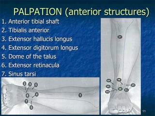
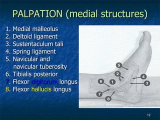
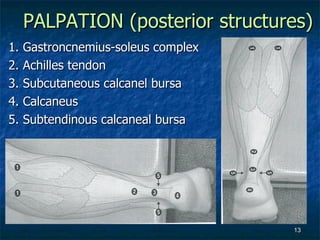
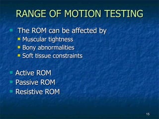
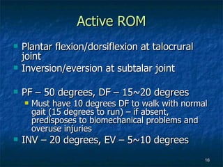
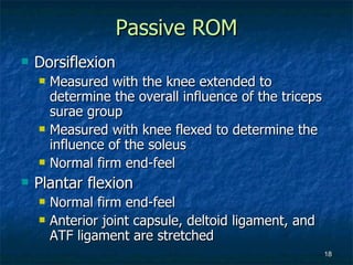
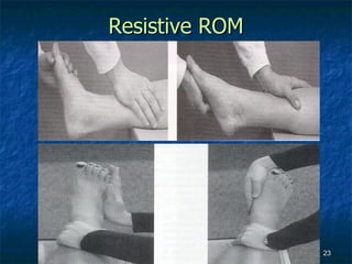
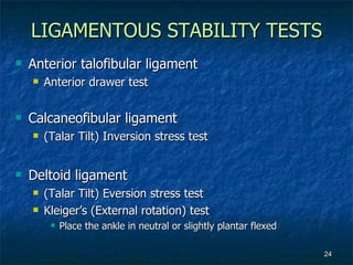
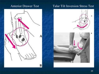
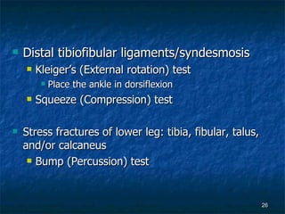
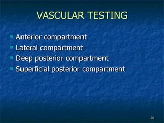
KIN 191A Advanced Assessment of  Lower Extremity Injuries ANKLE/LOWER LEG EVALUATION
INTRODUCTION HISTORY INSPECTION PALPATION ROM TESTING LIGAMENTOUS STABILITY TESTING NEUROLOGIC TESTING VASCULAR TESTING
Location of pain May be referred site for compartment syndrome, tarsal tunnel syndrome, peroneal/tibial nerve injury, sciatic nerve or lumbar nerve root impingement Nature/type of pain Aggravating/alleviating activities Effect of pain on activities HISTORY
Onset of pain Acute: trauma (sprain, strain, fracture, etc.) Gradual/insidious - overuse syndromes Mechanism of injury Macrotrauma - typically easy to identify tissues involved Microtrauma - typically involves more in depth questioning to arrive at cause of problem
Activity changes Duration, frequency, intensity, surface, shoes, orthotics, etc. Prior history Those with prior history of ankle injury may present with increased laxity and/or decreased proprioception vs. normal
INSPECTION General inspection Weight bearing status Gait pattern Swelling Obvious deformity Discoloration
Inspection of the lateral structures Peroneal muscle group Distal one third of the fibula Lateral malleolus Inspection of the anterior structures Appearance of the anterior lower leg Contour of the malleoli Talus Sinus tarsi Lateral aspect of the talus (anterior from the lateral malleolus)
Inspection of the medial structures Medial malleolus Medial longitudinal arch Inspection of the posterior structures Gastrocnemius-soleus complex Achilles tendon Bursae Subtendinous (Retrocalcaneal) bursa Subcutaneous calcaneal bursa Calcaneus
PALPATION (fibular structure) 1. Common peroneal nerve 2. Peroneus longus and brevis 3. Fibular shaft 4. Anterior and posterior tibiofibular ligaments 5. Interosseous  membrane 6. Superior peroneal  retinaculum
PALPATION (lateral ankle) 1. Lateral malleolus 2. Calcaneofibular ligament 3. Anterior talofibular ligament 4. Posterior talofibular ligament 5. Peroneal retinaculum 6. Peroneal tubercle 7. Cuboid 8. Base of the fifth metatarsal 9. Peroneus tertius
PALPATION (anterior structures) 1. Anterior tibial shaft 2. Tibialis anterior 3. Extensor hallucis longus 4. Extensor digitorum longus 5. Dome of the talus 6. Extensor retinacula 7. Sinus tarsi
PALPATION (medial structures) 1. Medial malleolus 2. Deltoid ligament 3. Sustentaculum tali 4. Spring ligament 5. Navicular and  navicular tuberosity 6. Tibialis posterior 7 . Flexor  digitorum  longus 8 . Flexor  hallucis  longus
PALPATION (posterior structures) 1. Gastroncnemius-soleus complex 2. Achilles tendon 3. Subcutaneous calcanel bursa 4. Calcaneus 5. Subtendinous calcaneal bursa
Palpation of Pulses Posterior tibial artery Between the flexor digitorum longus and flexor hallucis longus tendons as they pass behind the medial malleolus Dosalis pedis artery Between the extensor digitorum longus and extensor hallucis longus tendons as they pass over the cuneiforms
RANGE OF MOTION TESTING The ROM can be affected by Muscular tightness Bony abnormalities Soft tissue constraints Active ROM Passive ROM Resistive ROM
Active ROM Plantar flexion/dorsiflexion at talocrural joint  Inversion/eversion at subtalar joint PF – 50 degrees, DF – 15~20 degrees Must have 10 degrees DF to walk with normal gait (15 degrees to run) – if absent, predisposes to biomechanical problems and overuse injuries INV – 20 degrees, EV – 5~10 degrees
 
Passive ROM Dorsiflexion Measured with the knee extended to determine the overall influence of the triceps surae group Measured with knee flexed to determine the influence of the soleus Normal firm end-feel Plantar flexion Normal firm end-feel Anterior joint capsule, deltoid ligament, and ATF ligament are stretched
Inversion Should lie supine position Normal firm end-feel (especially the CF ligament stretch) and peroneal muscles stretch The tibia and fibular are stabilized to prevent hip or lower leg rotation Eversion A hard end-feel (because of the fibular’s striking the calcaneus)
Goniometry Plantar flexion/Dorsiflexion Sitting or lying with the knee flexed to at least 30 degrees Fulcrum Centered over the lateral malleolus Stationary arm Aligned with the long axis of the fibula Movement arm Parallel with the long axis of the 5 th  metatarsal
 
Goniometry Inversion/Eversion Sitting or lying with the knee flexed to at least 30 degrees Fulcrum Over the talocrural joint line, centered between the malleoli Stationary arm Centered over the long axis of the tibia Movement arm Over the 2 nd  metatarsal
Resistive ROM
Anterior talofibular ligament Anterior drawer test Calcaneofibular ligament (Talar Tilt) Inversion stress test Deltoid ligament (Talar Tilt) Eversion stress test Kleiger’s (External rotation) test Place the ankle in neutral or slightly plantar flexed LIGAMENTOUS STABILITY TESTS
Anterior Drawer Test Talar Tilt Inversion Stress Test
Distal tibiofibular ligaments/syndesmosis Kleiger’s (External rotation) test Place the ankle in dorsiflexion Squeeze (Compression) test Stress fractures of lower leg: tibia, fibular, talus, and/or calcaneus Bump (Percussion) test
Kleiger’s (External rotation) Test Squeeze (Compression) Test Bump (Percussion) Test
Homan’s Test Thompson Test For Achilles tendon rupture For Deep Vein Thrombosis
NEUROGIC TESTING L4 – S2 dermatomes/myotomes/reflex Tibial nerve Deep peroneal nerve Superficial peroneal nerve
VASCULAR TESTING Anterior compartment Lateral compartment Deep posterior compartment Superficial posterior compartment

Kin191 A. Ch.5. Ankle. Lower Leg. Evaluation. Fall 2007

  • 1.
    KIN 191A AdvancedAssessment of Lower Extremity Injuries ANKLE/LOWER LEG EVALUATION
  • 2.
    INTRODUCTION HISTORY INSPECTIONPALPATION ROM TESTING LIGAMENTOUS STABILITY TESTING NEUROLOGIC TESTING VASCULAR TESTING
  • 3.
    Location of painMay be referred site for compartment syndrome, tarsal tunnel syndrome, peroneal/tibial nerve injury, sciatic nerve or lumbar nerve root impingement Nature/type of pain Aggravating/alleviating activities Effect of pain on activities HISTORY
  • 4.
    Onset of painAcute: trauma (sprain, strain, fracture, etc.) Gradual/insidious - overuse syndromes Mechanism of injury Macrotrauma - typically easy to identify tissues involved Microtrauma - typically involves more in depth questioning to arrive at cause of problem
  • 5.
    Activity changes Duration,frequency, intensity, surface, shoes, orthotics, etc. Prior history Those with prior history of ankle injury may present with increased laxity and/or decreased proprioception vs. normal
  • 6.
    INSPECTION General inspectionWeight bearing status Gait pattern Swelling Obvious deformity Discoloration
  • 7.
    Inspection of thelateral structures Peroneal muscle group Distal one third of the fibula Lateral malleolus Inspection of the anterior structures Appearance of the anterior lower leg Contour of the malleoli Talus Sinus tarsi Lateral aspect of the talus (anterior from the lateral malleolus)
  • 8.
    Inspection of themedial structures Medial malleolus Medial longitudinal arch Inspection of the posterior structures Gastrocnemius-soleus complex Achilles tendon Bursae Subtendinous (Retrocalcaneal) bursa Subcutaneous calcaneal bursa Calcaneus
  • 9.
    PALPATION (fibular structure)1. Common peroneal nerve 2. Peroneus longus and brevis 3. Fibular shaft 4. Anterior and posterior tibiofibular ligaments 5. Interosseous membrane 6. Superior peroneal retinaculum
  • 10.
    PALPATION (lateral ankle)1. Lateral malleolus 2. Calcaneofibular ligament 3. Anterior talofibular ligament 4. Posterior talofibular ligament 5. Peroneal retinaculum 6. Peroneal tubercle 7. Cuboid 8. Base of the fifth metatarsal 9. Peroneus tertius
  • 11.
    PALPATION (anterior structures)1. Anterior tibial shaft 2. Tibialis anterior 3. Extensor hallucis longus 4. Extensor digitorum longus 5. Dome of the talus 6. Extensor retinacula 7. Sinus tarsi
  • 12.
    PALPATION (medial structures)1. Medial malleolus 2. Deltoid ligament 3. Sustentaculum tali 4. Spring ligament 5. Navicular and navicular tuberosity 6. Tibialis posterior 7 . Flexor digitorum longus 8 . Flexor hallucis longus
  • 13.
    PALPATION (posterior structures)1. Gastroncnemius-soleus complex 2. Achilles tendon 3. Subcutaneous calcanel bursa 4. Calcaneus 5. Subtendinous calcaneal bursa
  • 14.
    Palpation of PulsesPosterior tibial artery Between the flexor digitorum longus and flexor hallucis longus tendons as they pass behind the medial malleolus Dosalis pedis artery Between the extensor digitorum longus and extensor hallucis longus tendons as they pass over the cuneiforms
  • 15.
    RANGE OF MOTIONTESTING The ROM can be affected by Muscular tightness Bony abnormalities Soft tissue constraints Active ROM Passive ROM Resistive ROM
  • 16.
    Active ROM Plantarflexion/dorsiflexion at talocrural joint Inversion/eversion at subtalar joint PF – 50 degrees, DF – 15~20 degrees Must have 10 degrees DF to walk with normal gait (15 degrees to run) – if absent, predisposes to biomechanical problems and overuse injuries INV – 20 degrees, EV – 5~10 degrees
  • 17.
  • 18.
    Passive ROM DorsiflexionMeasured with the knee extended to determine the overall influence of the triceps surae group Measured with knee flexed to determine the influence of the soleus Normal firm end-feel Plantar flexion Normal firm end-feel Anterior joint capsule, deltoid ligament, and ATF ligament are stretched
  • 19.
    Inversion Should liesupine position Normal firm end-feel (especially the CF ligament stretch) and peroneal muscles stretch The tibia and fibular are stabilized to prevent hip or lower leg rotation Eversion A hard end-feel (because of the fibular’s striking the calcaneus)
  • 20.
    Goniometry Plantar flexion/DorsiflexionSitting or lying with the knee flexed to at least 30 degrees Fulcrum Centered over the lateral malleolus Stationary arm Aligned with the long axis of the fibula Movement arm Parallel with the long axis of the 5 th metatarsal
  • 21.
  • 22.
    Goniometry Inversion/Eversion Sittingor lying with the knee flexed to at least 30 degrees Fulcrum Over the talocrural joint line, centered between the malleoli Stationary arm Centered over the long axis of the tibia Movement arm Over the 2 nd metatarsal
  • 23.
  • 24.
    Anterior talofibular ligamentAnterior drawer test Calcaneofibular ligament (Talar Tilt) Inversion stress test Deltoid ligament (Talar Tilt) Eversion stress test Kleiger’s (External rotation) test Place the ankle in neutral or slightly plantar flexed LIGAMENTOUS STABILITY TESTS
  • 25.
    Anterior Drawer TestTalar Tilt Inversion Stress Test
  • 26.
    Distal tibiofibular ligaments/syndesmosisKleiger’s (External rotation) test Place the ankle in dorsiflexion Squeeze (Compression) test Stress fractures of lower leg: tibia, fibular, talus, and/or calcaneus Bump (Percussion) test
  • 27.
    Kleiger’s (External rotation)Test Squeeze (Compression) Test Bump (Percussion) Test
  • 28.
    Homan’s Test ThompsonTest For Achilles tendon rupture For Deep Vein Thrombosis
  • 29.
    NEUROGIC TESTING L4– S2 dermatomes/myotomes/reflex Tibial nerve Deep peroneal nerve Superficial peroneal nerve
  • 30.
    VASCULAR TESTING Anteriorcompartment Lateral compartment Deep posterior compartment Superficial posterior compartment