Download to read offline

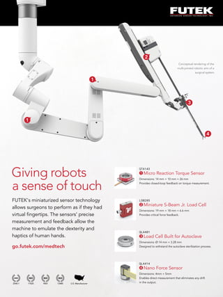
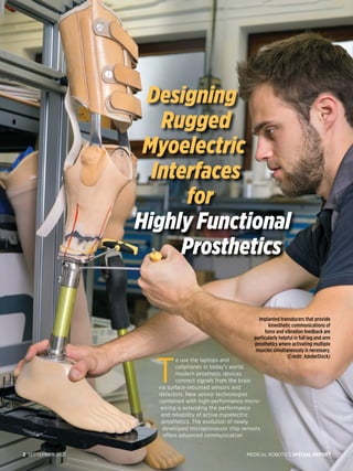
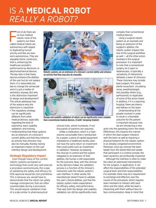
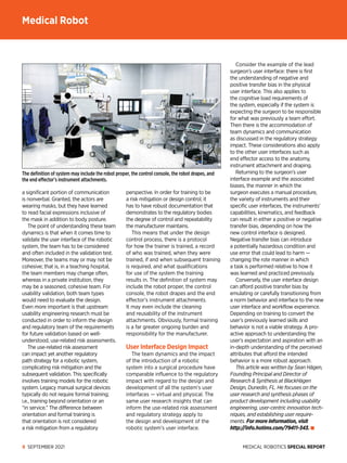


The special report on medical robotics highlights advancements in sensor technologies that enhance surgical procedures and prosthetics, allowing for precise force feedback and improved functionality. It discusses the complexity of integrating robotic systems into surgical workflows and the importance of usability validation in design, emphasizing that current medical robots are extensions of clinician abilities rather than fully autonomous machines. Additionally, the report outlines the need for formal training and robust documentation to ensure safe and effective use of robotic systems in medical settings.


![谷歌留痕技术 [ 𝙩𝙤𝙥 𝟮𝟯𝟯. 𝙘 𝙤𝙢 ]](https://cdn.slidesharecdn.com/ss_thumbnails/top233-260130174328-3833018c-thumbnail.jpg?width=640&height=640&fit=bounds)


